RESEARCH ARTICLE
An unusual case of childhood osteoarticular tuberculosis from the A ´rpa ´dian Age cemetery of Győrszentiva ´n-Re ´vhegyi tag (Győr-Moson- Sopron county, Hungary)
Olga SpekkerID1*, Luca Kis1, Andrea Dea´k2, Eszter Makai3, Gyo¨ rgy Pa´lfi1, Orsolya Anna Va´radi1,4, Erika Molna´r1
1 Department of Biological Anthropology, University of Szeged, Szeged, Hungary, 2 Ro´ mer Flo´ris Museum of Art and History, Győr, Hungary, 3 Department of Radiology, University of Szeged, Szeged, Hungary, 4 Department of Microbiology, University of Szeged, Szeged, Hungary
*olga.spekker@gmail.com
Abstract
Ancient human remains exhibiting bony changes consistent with osteoarticular tuberculosis (OATB) indicate that the disease has afflicted mankind for millennia. Nonetheless, not many pediatric OATB cases have been published in the paleopathological literature–from Hun- gary, only three cases have been described up to now. In our paper, we demonstrate a child (S0603) from the A´ rpa´dian Age cemetery of Győrszentiva´n-Re´vhegyi tag (northwestern Hungary), who represents a unique case of OATB regarding both the pattern and severity of the observed bony changes. During the macromorphological and radiological investigations, the most serious alterations were discovered in the upper thoracic spine–the development of osteolytic lesions led to severe bone loss and consequent collapse and fusion of several adjacent vertebrae. The pathological process terminated in a sharp, rigid angular kyphosis.
Disruption of the normal spine curvature resulted in consequent deformation of the whole thoracic wall–it became “rugby-ball-shaped”. The overall nature and pattern of the detected alterations, as well as their resemblance to those of described in previously published archaeological and modern cases from the pre-antibiotic era indicate that they are most con- sistent with OATB. Based on the severity and extent of the lesions, as well as on the evi- dence of secondary healing, S0603 suffered from TB for a long time prior to death. Besides body deformation, OATB resulted in consequent disability in daily activities, which would have required regular and significant care from others to survive. It implies that in the A´ rpa´- dian Age community of Győrszentiva´n-Re´vhegyi tag, there was a willingness to care for people in need. Detailed archaeological case studies can give us a unique insight into the natural history and different presentations of OATB. Furthermore, they can provide paleopa- thologists with a stronger basis for diagnosing TB and consequently, with a more sensitive means of assessing TB frequency in past populations.
PLOS ONE
a1111111111 a1111111111 a1111111111 a1111111111 a1111111111
OPEN ACCESS
Citation: Spekker O, Kis L, Dea´k A, Makai E, Pa´lfi G, Va´radi OA, et al. (2021) An unusual case of childhood osteoarticular tuberculosis from the A´rpa´dian Age cemetery of Győrszentiva´n-Re´vhegyi tag (Győr-Moson-Sopron county, Hungary). PLoS ONE 16(4): e0249939.https://doi.org/10.1371/
journal.pone.0249939
Editor: Mark Spigelman, Hebrew University, ISRAEL
Received: October 12, 2020 Accepted: March 27, 2021 Published: April 14, 2021
Peer Review History: PLOS recognizes the benefits of transparency in the peer review process; therefore, we enable the publication of all of the content of peer review and author responses alongside final, published articles. The editorial history of this article is available here:
https://doi.org/10.1371/journal.pone.0249939 Copyright:©2021 Spekker et al. This is an open access article distributed under the terms of the Creative Commons Attribution License, which permits unrestricted use, distribution, and reproduction in any medium, provided the original author and source are credited.
Data Availability Statement: All relevant data are within the manuscript and itsSupporting Informationfiles.
Introduction
Despite growing interest in the last few decades, pediatric tuberculosis (TB) as a main cause of morbidity and mortality, especially in TB-endemic regions, remains relatively neglected today [1–6]. However, particular attention should be paid to eliminate or at least control TB in the foreseeable future [1,2,5–7]. Childhood TB is an important TB control indicator, since it reflects recent and/or ongoing transmission in the community, as it is usually acquired postna- tally from an adult contact with clinically active TB disease [2,3,8–10]. Children and adoles- cents with latent TB infection represent reservoirs for future transmission following disease reactivation, often many years after the primary infection; and therefore, can provide the source of future epidemics [2,3,8]. According to the latest Global Tuberculosis Report from the World Health Organization (WHO), children under the age of 15 years accounted for approximately 11% of all TB cases and 13.8% of all TB deaths in 2018 –an estimated 1.1 million children became ill with TB and about 205,000 children died of TB worldwide [7]. Nevertheless, the actual global burden of pediatric TB is very likely higher as substantial challenges in diagnosis and surveillance compromise the quality of epidemiological data on the disease [1,3,4,7,9–11].
Once infected with TB bacteria (i.e., members of theMycobacterium tuberculosiscomplex;
MTBC), children are at particularly higher risk of rapid progression to clinically active TB dis- ease than adults, with the vast majority of them (>95%) developing it within the first 12 months after exposure [1,6,8–13]. Moreover, children are more prone to develop severe, extra-pulmonary forms of TB (e.g., tuberculous meningitis and miliary TB) that are associated with high morbidity and mortality [1,2,6,8,13–15]. Age-related differences in both the innate and adaptive immune responses to TB may play a crucial role in increasing the vulnerability of children to the disease compared to adults [1,13]. Based on pre-chemotherapy literature data from the first half of the 20thcentury, Marais and his co-workers [12] identified two high-risk periods of childhood for progression to clinically active TB disease following primary infec- tion: infancy (less than 2 years of age) and adolescence (more than 10 years of age).
Although in most pediatric cases (up to 80%), TB presents as pulmonary disease, other parts of the human body, including the skeletal system, can also be affected [6,15,16].
Osteoarticular or skeletal TB (i.e., tuberculous involvement of the bones and/or joints; OATB) is more frequent in children than in adults, accounting for approximately 10–35% of pediatric extra-pulmonary TB cases and about 5–7% of all pediatric TB cases [17–22]. OATB usually arises secondary to hematogenous seeding of TB bacteria from an often unknown primary site of infection outside the skeleton into the bone and/or synovial tissue during or shortly after the mycobacteremic phase of primary infection or late reactivation of the disease [21,23,24].
Less commonly, lymphogenous dissemination, contiguous spread from adjacent structures or direct inoculation of TB bacteria into a skeletal site can also occur [21,25,26]. Virtually any bone or joint of the human body can be affected by the disease–the three main forms of OATB are spinal TB (i.e., combination of tuberculous vertebral osteomyelitis and arthritis;*50%), tuberculous osteomyelitis of the extra-spinal bones (*11%), and tuberculous arthritis of the extra-spinal joints (*30%) [18,19,23,27]. Although the aforementioned forms of OATB are usually present alone, their concomitant occurrence can be observed in some cases [23]. Its rel- ative rarity and highly variable clinical and radiological presentations make pediatric OATB a diagnostic challenge in the modern medical practice [28,29]. However, early diagnosis is cru- cial to improve the clinical outcome, as OATB can be a debilitating medical condition with serious and potentially irreversible orthopedic and/or neurologic complications, even many years after the onset of the disease [18,19,30,31].
Recent evolutionary genetics studies on the age of the MTBC by Comas and his co-workers [32] revealed that the human-adapted members of the complex may have co-evolved with PLOS ONE A pediatric skeletal TB case from an A´ rpa´dian Age cemetery of northwestern Hungary
Funding: This work was funded by the University of Szeged Open Access Fund (5006) to OS. The National Research, Development and Innovation Office (Hungary) (K 125561) and the "A´rpa´d-ha´z Program" (39509/2018/KFSZ) of the Hungarian Ministry of Human Capacities provided funding for GP. The funders had no role in study design, data collection and analysis, decision to publish, or preparation of the manuscript.
Competing interests: The authors have declared that no competing interests exist.
their host to successfully infect, cause disease, and transmit over tens of thousands of years [33–36]. Besides genetic findings, ancient human skeletons and mummies exhibiting bony changes consistent with OATB [e.g.,37–41] also indicate that the disease has afflicted mankind for millennia [34,35,42]. Nonetheless, not many cases with OATB in children (less than 15 years of age) have been published in the paleopathological literature [e.g.,43–50]. The afore- mentioned studies reporting archaeological and modern pediatric OATB cases from the pre- antibiotic era have pointed out that the wide variety of manifestations of the disease observed in patients today were present in prehistoric and historic communities [49]. Besides meticu- lous descriptions from the literature from the first half of the 20thcentury (pre-chemotherapy era), detailed archaeological case studies also provide a unique insight into the natural history and different presentations of OATB in children. They help us in identifying non-pathogno- monic bony changes and/or patterns of lesions that can later be used as diagnostic criteria for OATB in both the modern medical and paleopathological practices. By scrutinizing these new diagnostic criteria in living patients with similar alterations, a more appropriate diagnosis could be established that may contribute to improving the clinical outcome of the disease in those patients. It should be noted that the bony changes observed in archaeological cases may differ from those detectable in living patients, due in part to the introduction of antibiotics in the management of TB from the second half of the 20thcentury [51]. However, in both devel- oping and developed countries, with the emergence of multidrug-resistant TB (in which the applied antibiotic therapy is often not effective enough), presentations of OATB that are simi- lar to those of discovered in archaeological cases from the pre-antibiotic era may become more common in the future. On the other hand, by providing physicians and paleopathologists with a stronger basis for diagnosing different manifestations of OATB, a more sensitive means of assessing the disease frequency in current and archaeological populations can be achieved.
In our paper, we demonstrate a child (i.e., S0603) from the A´ rpa´dian Age cemetery of Győrszentiva´n-Re´vhegyi tag (Győr-Moson-Sopron county, Hungary), who represents a unique case of pediatric OATB with multifocal involvement of the axial skeleton (i.e., spine and ribs) regarding both the pattern and severity of the observed bony changes, as well as the archaeological context.
Materials and methods
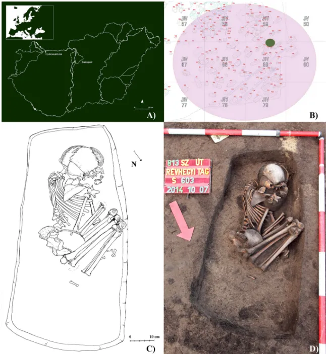
Between 2014 and 2015, prior to the construction works of main road #813 bypassing the city of Győr (Győr-Moson-Sopron county, northwestern Hungary), test and preventive excava- tions were carried out under the direction of Andrea Dea´k at the merged archaeological sites of Győr–Győrszentiva´n and Győr–Re´vhegyi tag, geographically located in close vicinity to the present-day city of Győrszentiva´n (Fig 1A). Remains of settlements from the Bronze Age, Iron Age, Roman period, and A´ rpa´dian Age, as well as of cemeteries from the Roman period and A´ rpa´dian Age were also discovered in the large excavation area that coincided with the trail of main road #813.
From the A´ rpa´dian Age cemetery of the archaeological site (Fig 1B), a total of 57 burials were unearthed that, based on the grave goods, can clearly be dated to the 11th–12thcenturies CE. It should be noted that the A´ rpa´dian Age cemetery is only partially excavated, and its exact extent is not known. The 57 burials can be divided into two main groups. The earlier (11thc. CE), western grave group consisted of 11 burials covering a semicircular area of the cemetery, whereas the later (12thc. CE), eastern grave group, which was contiguous with the eastern end of the first one, was comprised of rows of burials. In the uniformly orientated, rect- angular pit-graves, the individuals were buried in an extended supine position with the head at the southwestern end of the grave–consistent with the west-east grave orientation typical of PLOS ONE A pediatric skeletal TB case from an A´ rpa´dian Age cemetery of northwestern Hungary
A´ rpa´dian Age burials. Although the grave of S0603 was in one of the 12th-century-CE grave rows of the cemetery, the body of the deceased was buried in a flexed position, with the oppo- site orientation to that of the other individuals from the cemetery and without any grave goods (Fig 1B–1D).
Along with the remains of the 56 other individuals, the skeleton of S0603 is temporarily housed at the Department of Biological Anthropology, University of Szeged (Szeged, Hun- gary). Based on tooth formation and development [52], tooth eruption pattern [53], and diaphyseal length of long tubular bones [54], S0603 was around 12 years of age at the time of
Fig 1. A) Map of Hungary showing the location of the Győrszentiva´n-Re´vhegyi tag archaeological site; B) Plan drawing of the A´ rpa´dian Age cemetery of Győrszentiva´n-Re´vhegyi tag archaeological site with the location of burial S0603; C) Drawing of the burial of S0603in situ; and D) Photo of the burial of S0603in situ.
https://doi.org/10.1371/journal.pone.0249939.g001
PLOS ONE A pediatric skeletal TB case from an A´ rpa´dian Age cemetery of northwestern Hungary
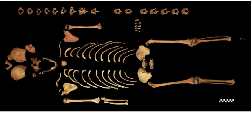
death. Both the qualitative and quantitative state of preservation of the child’s skeletal remains are quite good (Fig 2). The skeleton was subject to a detailed macromorphological investiga- tion, focusing on the detection of pathological bony changes. CT imaging of the T1–6 region of the spine was also performed to improve the diagnosis established on the basis of prior mac- roscopic observations–it was carried out with a Philips Brilliance iCT 256.
Ethics statement Specimen number: S0603.
The skeleton evaluated in the described study is housed in the Department of Biological Anthropology, University of Szeged, in Szeged, Hungary. Access to the specimen was granted by the Department of Biological Anthropology, University of Szeged (Szeged, Hungary) and the Ro´mer Flo´ris Museum of Art and History (Győr, Hungary).
No permits were required for the described study, complying with all relevant regulations.
Results
In the 12-year-old child’s skeletal remains, a number of severe pathological bony changes were macroscopically observed, predominantly in the axial skeleton (i.e., spine and ribs).
Skull
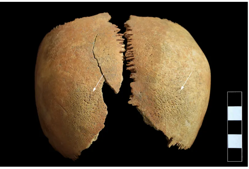
In the skull, both parietal bones exhibited signs ofcribra craniiin close vicinity to the joining point of the sagittal and lambdoid sutures (Fig 3). On the ectocranial surface of the occipital bone, pitting and slight cortical remodeling were observed on its basal and lateral parts;
the pathological process spared the articular surfaces (Fig 4). Similar pitting was detected on the inferior surface of the left temporal pyramid, adjacent to the affected occipital areas (Fig 4).
Fig 2. The skeleton of S0603.
https://doi.org/10.1371/journal.pone.0249939.g002
PLOS ONE A pediatric skeletal TB case from an A´ rpa´dian Age cemetery of northwestern Hungary
Spine
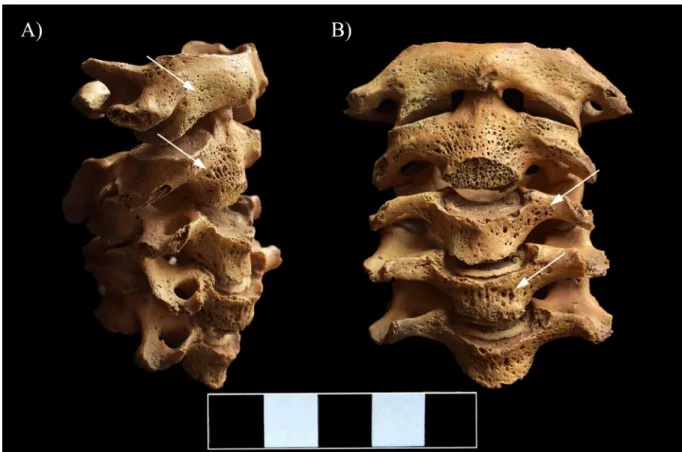
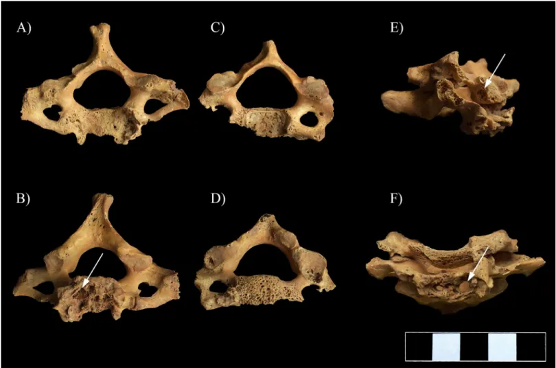
In the vertebral column, the C1–5 vertebral bodies showed signs of inflammation in form of pitting and slight cortical remodeling, particularly on their anterior aspect and costal processes (Fig 5). The original height of the C1–5 vertebral bodies was maintained (Fig 5). In the C6–7 vertebral bodies, the pathological process affected not only the cortical bone layers but also the trabeculae: multiple, well-circumscribed osteolytic lesions corresponding to round, oval or slightly lobulated necrotizing foci were detected (Fig 6A–6D and 6F). The presence of osteolytic lesions led to considerable bone loss, predominantly in the antero-inferior parts of the C6–7 vertebral bodies; because of the severe bone destruction, they became wedge-shaped (Fig 6E). In the C6–7 vertebral bodies, the pathological process extended towards the trans- verse processes: pitting and slight cortical remodeling were noted on both sides (Fig 6A, 6C and 6F).
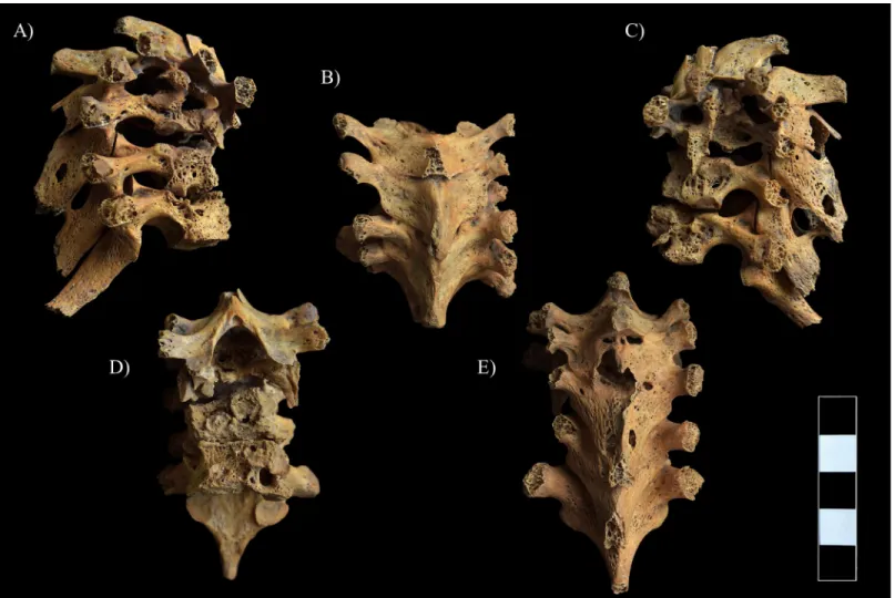
The most severe pathological bony changes were observed in the upper thoracic spine: the development of multiple, well-circumscribed osteolytic lesions resulted in almost complete destruction of the T1–5 vertebral bodies with disappearance of the intervening disc spaces and formation of a severe angular kyphosis in the cervicothoracic region of the spine (Fig 7andS1 Video). Subsequently, the small, wedge-shaped remnants of the T1–5 vertebral bodies fused
Fig 3. Signs ofcribra craniion the ectocranial surface of both parietal bones (white arrows).
https://doi.org/10.1371/journal.pone.0249939.g003
PLOS ONE A pediatric skeletal TB case from an A´ rpa´dian Age cemetery of northwestern Hungary
together. The anterior part of the T6–7 bodies was almost completely destroyed by the patho- logical process; however, the T5–6 and T6–7 intervening disc spaces and the body height of T6 and T7 were maintained (Fig 7A, 7C and 7D). The posterior part of the T6–7 vertebral bodies was also affected: abnormal porosity, multiple, variable-sized, well-circumscribed osteolytic lesions with thin sclerotic margins, pitting, and slight cortical remodeling were detected in their remnants (Fig 7A, 7C and 7D). The upper thoracic vertebral arci and their processes also showed signs of inflammation in form of pitting, slight cortical remodeling, and osteolytic lesions, especially in the T4–7 region (Fig 7A–7C and 7E). Ankylosis of the intervertebral joints and fusion of the laminae were also noted in the upper thoracic spine (T1–6) (Fig 7B, 7Eand S1 Video). Furthermore, the CT imaging of the T1–6 region revealed that the vertebral canal was slightly narrowed and altered in shape, especially at the apex of the severe angular kyphosis.
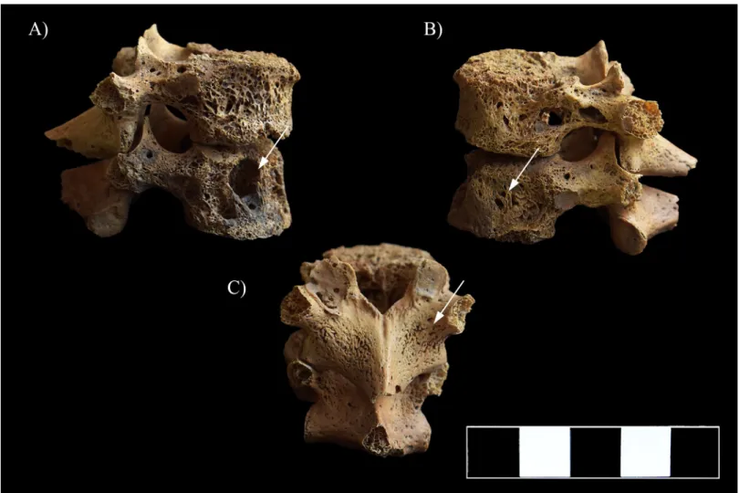
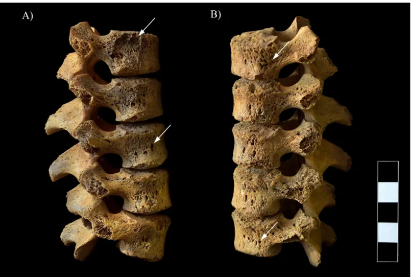
In the lower thoracic (Fig 8A and 8B) and lumbar spine (Fig 9A and 9B), multiple, well-cir- cumscribed osteolytic lesions, cortical remodeling, and signs of hypervascularization were recorded on the anterior and lateral aspects of all the preserved vertebral bodies. Moreover, they presented swelling. The lower thoracic and lumbar vertebral arci and their processes also exhibited pitting and slight remodeling of the cortical bone layers (Fig 8C).
Fig 4. Pitting and slight cortical remodeling on the ectocranial surface of the occipital and left temporal bones (white arrows).
https://doi.org/10.1371/journal.pone.0249939.g004
PLOS ONE A pediatric skeletal TB case from an A´ rpa´dian Age cemetery of northwestern Hungary
Ribs
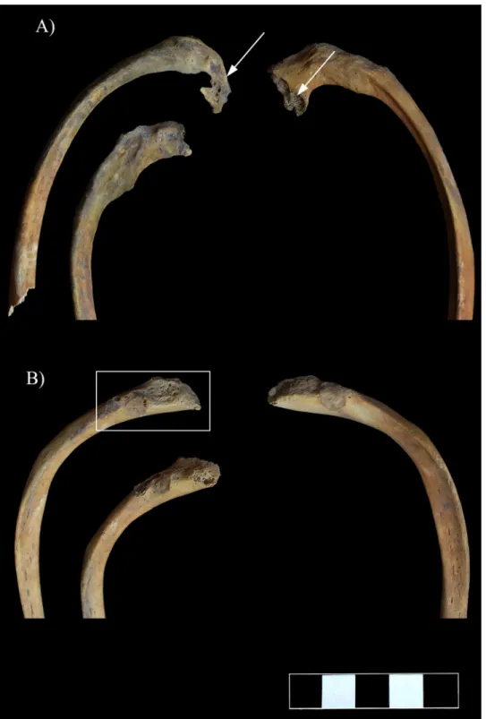
On both sides of the thoracic wall, almost all the preserved ribs showed pathological bony changes, predominantly on their vertebral end adjacent to the affected thoracic vertebral bod- ies. The vertebral ends of the first six ribs, especially from the 3rdto the 6th, were supero-inferi- orly flattened and thinned, and displayed an abnormal curvature (Fig 10A). An irregular morphology of the articular area was also observed from the 4thto the 6thribs (especially on the left side) (Fig 10A). From the 7thto the 11thribs, the vertebral ends presented swelling and pitting (the 12thribs are missingpost-mortem) (Fig 10B). Furthermore, they were supero-infe- riorly flattened and their original curvature changed (Fig 10B).
Clavicles
Although the clavicles exhibited no signs of inflammation, similar to the vertebral ends of the ribs, they showed an abnormal curvature on both the sternal and acromial ends (Fig 11).
Pelvic area
On the right iliac bone, reactive new bone formation covering the whole iliac fossa of the iliac wing was detected (Fig 12). The proximal end of the right femur displayed similar alterations:
periosteal new bone formation was noted on its anterior, medial, and posterior surfaces until the level of the lesser trochanter (Fig 13).
Fig 5. Pitting and slight cortical remodeling on the C1–5 vertebrae (white arrows). A) Lateral view (right side) and B) Anterior view.
https://doi.org/10.1371/journal.pone.0249939.g005
PLOS ONE A pediatric skeletal TB case from an A´ rpa´dian Age cemetery of northwestern Hungary
Discussion
The numerous pathological alterations (mainly of a lytic nature with very little bone forma- tion), which were observed in the skeletal remains of S0603, indicate that the 12-year-old child suffered from a systemic infectious disease. It affected various regions of the skeleton but the most remarkable bony changes were discovered predominantly in the spine–in form of well- circumscribed osteolytic lesions leading to severe bone loss and collapse and fusion of several adjacent vertebrae. The pathological process terminated in a localized sagittal spinal deformity with a sharp angulation (i.e., sharp, rigid angular kyphosis of the upper thoracic spine). Dis- ruption of the normal curvature of the vertebral column resulted in consequent deformation of the whole thoracic wall: besides the spinal changes, the altered shape of the clavicles and ribs (especially of their vertebral ends) gave the thoracic wall a “rugby-ball-shaped” appearance.
The signs of inflammation on the vertebral ends of ribs, as well as of an overlying paravertebral cold abscess in the pelvic area (i.e., new bone formation on the iliac fossa of the right iliac bone and on the proximal end of the right femur) are suggestive of direct extension of the infection from the vertebral column into the adjacent ribs and soft tissues. The overall nature and pat- tern of the detected bony changes, as well as their resemblance to those of described in
Fig 6. Multiple, well-circumscribed osteolytic lesions (white arrows) and consequent severe bone loss in the C6–7 vertebrae. A) Superior view of C7, B) Inferior view of C7, C) Superior view of C6, D) Inferior view of C6, E) Lateral view of C6–7 (right side), and F) Anterior view of C6–7.
https://doi.org/10.1371/journal.pone.0249939.g006
PLOS ONE A pediatric skeletal TB case from an A´ rpa´dian Age cemetery of northwestern Hungary
previously published archaeological and modern cases from the pre-antibiotic era [e.g., 45, 49, 50, 55, 56] indicate that they are most consistent with pediatric OATB.
In the vast majority of the cases (90–95%), spinal TB–also known as TB spondylitis or Pott’s disease–arises from the anterior subchondral (paradiscal) region of the vertebral body [19,21,23,57–59]. This area has a dense end-arteriolar network that makes it susceptible to bacterial seeding via the segmental arterial circulation [21,23,57,59,60]. Lodgment of TB bac- teria into the anterior subchondral region triggers the onset of granulomatous inflammation of the cancellous bone with tubercle formation in the red bone marrow [21,23,61]. The devel- opment and caseous necrosis of the gradually expanding and coalescing tubercles induce the growth of the initial intra-vertebral abscess and the establishment of additional intra-vertebral abscesses within the affected vertebral body [23,61,62]. Furthermore, TB involvement of the segmental artery branches terminating in the anterior subchondral region generates depriva- tion of the blood supply to the cancellous bone [62]. Any or all of the aforementioned pro- cesses result in necrosis and consequent resorption of the bone trabeculae that ultimately lead to the formation of osteolytic lesions in the anterior subchondral region, with subsequent involvement of the entire vertebral body and occasionally of the posterior vertebral elements [23,30,57,59,60,62]. As the pathological process progresses, not only the trabeculae but also the cortical bone layers of the affected vertebral body can become destroyed; and thus, the
Fig 7. Pott’s gibbus in the T1–7 region. A) Lateral view (right side), B) Superior view, C) Lateral view (left side), D) Anterior view, and E) Posterior view.
https://doi.org/10.1371/journal.pone.0249939.g007
PLOS ONE A pediatric skeletal TB case from an A´ rpa´dian Age cemetery of northwestern Hungary
intra-vertebral abscess can extend towards the sub-ligamentous space, the adjoining interver- tebral discs or the adjacent soft tissues (e.g., ligaments and muscles) [23,30,57,59,60].
The anterior longitudinal ligament (i.e., a fibrous structure that covers the anterior and lat- eral surfaces of the vertebral bodies), at least temporarily, resists the horizontal progression of the infection [62]. Thus, the TB mass extending into the sub-ligamentous space (i.e., an extra- vertebral abscess) can spread only vertically (upwards or downwards) beneath the ligament from the initially affected vertebral body into a similar location at one or more contiguous ver- tebrae or beyond [62]. The sub-ligamentous extension of the extra-vertebral abscess results in stripping of the periosteum and anterior longitudinal ligament from the anterior and lateral vertebral surfaces [62]. This generates deprivation of the periosteal blood supply to the affected vertebral bodies with consequent ischemia [28,61,62]. The combination of pressure and ischemic effects caused by the presence and spread of the extra-vertebral abscess in the sub-lig- amentous space results in shallow cortical erosion on the anterior and lateral vertebral surfaces that gives them a scalloped appearance (i.e., anterior gouge defect) [23,28,62]. Besides cortical erosion, reactive new bone formation can also occur on the vertebral surfaces underlying the extra-vertebral abscess [23,63]. As the pathological process progresses, it can involve not only
Fig 8. Multiple, well-circumscribed osteolytic lesions, cortical remodeling, and signs of hypervascularization in the T11–12 vertebrae (white arrows). A) Lateral view (right side), B) Lateral view (left side), and C) Posterior view.
https://doi.org/10.1371/journal.pone.0249939.g008
PLOS ONE A pediatric skeletal TB case from an A´ rpa´dian Age cemetery of northwestern Hungary
the cortical bone layers of the affected vertebral bodies but also their trabeculae, since avascular vertebrae are more susceptible to infection [28,63].
Until more advanced stages of spinal TB, the intervertebral disc is relatively spared by the infection–most likely due to the lack of proteolytic enzymes in TB bacteria [18,19,23,58,59, 61]. However, the progressive destruction of the subchondral region of two adjacent vertebrae can compromise the nutrition of the adjoining intervertebral disc [57]. This can lead to disc degeneration and/or result in disc herniation into the weakened adjoining vertebral bodies, with gradual diminution or eventual loss of the intervening disc space [64]. Degenerated and/
or herniated discs are more prone to seeding by TB bacteria from the subchondral cancellous bone either via sub-ligamentous dissemination or contiguous spread; and therefore, to be sec- ondarily involved by the infection [57]. In children, not only the vertebral bodies but also the intervertebral discs can represent the initial site of infection due to their vascularized nature [59,61,65].
Extension of the intra-vertebral abscess into the adjacent soft tissues can result in the forma- tion of an extra-vertebral cold abscess (i.e., a slowly progressive abscess without characteristic signs of inflammation (such as heat, erythema or tenderness), which can become encapsulated and calcified over time) [59,60,63,66,67]. In spinal TB, the development of an extra-vertebral
Fig 9. Multiple, well-circumscribed osteolytic lesions, cortical remodeling, and signs of hypervascularization in the L1–5 vertebrae (white arrows). A) Lateral view (right side) and B) Lateral view (left side).
https://doi.org/10.1371/journal.pone.0249939.g009
PLOS ONE A pediatric skeletal TB case from an A´ rpa´dian Age cemetery of northwestern Hungary
cold abscess is a common complication–it occurs in about two-thirds of the cases [60,66]. The TB mass can accumulate within the prevertebral and/or paravertebral spaces with the forma- tion of prevertebral or paravertebral cold abscesses that are commonly associated with fistulae
Fig 10. A) Supero-inferior flattening, thinning, and destructive articular changes (white arrows) and B) swelling and pitting (white rectangle) on the vertebral end of some ribs.
https://doi.org/10.1371/journal.pone.0249939.g010
PLOS ONE A pediatric skeletal TB case from an A´ rpa´dian Age cemetery of northwestern Hungary
[58,63,67]. Although the extra-vertebral cold abscess can remain localized at the initial site of infection, in most cases, it extends vertically (usually downwards) beneath the anterior longitu- dinal ligament or along the fascial planes [63,67]. In response to an overlying cold abscess, erosive cortical bone destruction and/or reactive new bone formation can occur on the adja- cent bone surfaces (e.g., vertebrae, hip bones, and femora) [63].
The weakening of the affected vertebral bodies due to the formation of osteolytic lesions can result in their consequent collapse under the weight of the trunk [23,62]. This is character- ized by a wedge-shaped appearance, since the cavitation affects predominantly the anterior portion of the vertebral body, with intact or less destroyed posterior vertebral elements [62, 63]. Depending on the spinal location, the collapse of one or more contiguous vertebral bodies may lead to the development of different spinal deformities, most frequently of a sharp angular kyphosis in the thoracic spine (i.e., Pott’s gibbus) [57,60,63,67]. The progressive bone destruction and consequent deformity can result in mechanical instability of the spine [59,60, 66,67]. To stabilize the vertebral column, subsequent bony fusion of the remnants of the col- lapsed vertebral bodies and posterior vertebral elements, bony ankylosis of the intervertebral joints, ossification of the intervertebral ligaments, and formation of bony extensions intercon- necting the adjacent vertebrae can occur [59,63].
Fig 11. Abnormal curvature of the clavicles.
https://doi.org/10.1371/journal.pone.0249939.g011
PLOS ONE A pediatric skeletal TB case from an A´ rpa´dian Age cemetery of northwestern Hungary
Although any part of the vertebral column can be affected by TB, the lower thoracic (40–
50%) and upper lumbar (35–40%) spine represent the most commonly involved regions in adults [58,60,65]. Nonetheless, besides the thoracolumbar junction, the cervicothoracic spine can also become affected quite frequently in children [17,68,69]. Spinal TB can be more aggressive and involve more vertebrae in children than in adults; therefore, following vertebral destruction and collapse, they are at particular risk of rapid and severe deformity progression [17,18,67,69,70]. Moreover, in pediatric spinal TB cases, progression of the deformity even after healing of the disease is not uncommon due to the growing nature of the vertebral col- umn in childhood [31,57,65,67,71].
In the skeleton of S0603, based on the severity and extent of the observed bony changes, the initial site of TB infection could be the upper thoracic spine (T1–5). From this region, the infection could spread (upwards and downwards) beneath the anterior longitudinal ligament–
first to the adjacent cervical and thoracic vertebral bodies (C6–7 and T6–7) and later to all true vertebrae (upwards even to the skull base). In later stages of the disease, not only the spine, but the adjacent ribs and soft tissues (e.g., the iliopsoas muscle) could also become affected by direct extension of the infection. TB involvement of the right iliopsoas muscle could result in the development of a paravertebral cold abscess. Later, the TB mass could extend downwards following the course of the right psoas fascia to its tendinous insertion at the lesser trochanter
Fig 12. Reactive new bone formation on the iliac fossa of the right iliac wing (white arrows).
https://doi.org/10.1371/journal.pone.0249939.g012
PLOS ONE A pediatric skeletal TB case from an A´ rpa´dian Age cemetery of northwestern Hungary
of the right femur. The 12-year-old child’s case shows a number of similarities to those of pre- viously described in the paleopathological literature [e.g., 45, 49, 50, 55, 56].
Sparacello and his co-workers [49] reported a probable case of pediatric multifocal OATB (i.e., PO21) from Middle Neolithic Italy. Similarities with S0603 include involvement of the
Fig 13. Reactive new bone formation on the anterior surface of the right femur (proximal end) (white arrows).
https://doi.org/10.1371/journal.pone.0249939.g013
PLOS ONE A pediatric skeletal TB case from an A´ rpa´dian Age cemetery of northwestern Hungary
spine, ribs, and pelvic area. In the 5-year-old child’s vertebral column, the cervical, thoracic, lumbar, and sacral regions were concomitantly affected; however, in contrast to S0603, only one or two non-contiguous vertebrae were involved in each region, and the spine did not dis- play angular kyphosis. Similar to S0603, bony changes (i.e., pitting, bone resorption with cavi- tation, and periosteal new bone formation), presumably infectious in nature, were detected in four right-side ribs of PO21; however, they were located not on the vertebral ends but on the sternal ones. Moreover, the shape of the affected ribs was not altered. Although both S0603 and PO21 exhibited signs of an overlying paravertebral cold abscess (i.e., an iliopsoas abscess) in the pelvic area, in P021, the lesions were bilateral and particularly of a lytic/erosive nature.
In contrast to S0603, some of the upper limb bones of PO21 (left humerus and right scapula) were also affected by the pathological process.
In a 10-year-old child’s skeleton (i.e., L/74) from Roman period Hungary, Hlavenkova´ and her co-workers [55] identified a number of bony changes that are suggestive of OATB. Similar to S0603, the most remarkable alterations were discovered in the spine and ribs of L/74. In the 10-year-old child’s vertebral column, the sharp angular kyphosis developed in the thoracolum- bar region with involvement of five adjacent vertebrae (T9–L1). Due topost-mortemdamages, no signs of cortical remodeling or reactive new bone formation could be observed on the affected vertebral bodies; nevertheless, ankylosis of the T11–12 intervertebral joints was noted.
In contrast to S0603, the disease affected only one region of the vertebral column (i.e., thoraco- lumbar region) and no signs of an overlying paravertebral cold abscess could be detected on the skeletal remains of L/74. Although all the preserved ribs of L/74 presented pathological alterations (i.e., slight or moderate periosteal new bone formation on the visceral surface), their appearance was quite different from those of detected in S0603. Only one left-side rib of L/74 showed osteolytic lesions, but not on its vertebral end. In contrast to S0603, some of the lower limb bones of L/74 (right femur and both tibiae) exhibited signs of periostitis on the pos- terior surface of the shaft.
Another probable case of pediatric OATB from Sarmatian period Hungary was described by Marcsik and Kuja´ni [56]. Similar to S0603, the cervicothoracic spine of a 5–6-year-old child (i.e., grave no. 178) was affected by the disease–destruction and collapse of the C4–T2 vertebral bodies and subsequent fusion of their remnants resulted in the formation of a Pott’s gibbus. Furthermore, the unequal destruction of the C6–7 vertebral bodies led to scoliosis of the affected spinal region. No other bony changes probably related to TB have been mentioned by the authors.
Matos and his co-workers [45] reported the case of a 12-year-old child (i.e., skeleton no. 8) from medieval Portugal, whose remains exhibited numerous lesions, particularly in the axial skeleton (i.e., spine and ribs), that are consistent with multifocal OATB. Similar to S0603, the most severe alterations were observed in the spine. The T3–7 vertebral bodies were completely destroyed, whereas the T2 and T8–10 vertebral bodies displayed flattening, extensive bone resorption, and cavitation. In the T8–L3 region, the posterior vertebral elements were also involved by TB–they demonstrated pitting and osteolytic lesions. Furthermore, ankylosis of some of the intervertebral joints and ossification of the interspinous ligaments between the T7 and T8 vertebrae were noted. The pathological process terminated in a sharp angular kyphosis of the thoracic spine with moderate scoliosis. Similar to S0603, disruption of the normal curva- ture of the 12-year-old child’s vertebral column resulted in consequent deformation of the whole thoracic wall–the shape of both clavicles and the 3rd–8thribs (especially of their vertebral ends) changed. Moreover, direct extension of the infection from the spine to the vertebral end of ribs was also observed in form of destruction, reactive new bone formation, and swelling. In contrast to S0603, no signs of an overlying paravertebral cold abscess could be detected on the remains of skeleton no. 8.
PLOS ONE A pediatric skeletal TB case from an A´ rpa´dian Age cemetery of northwestern Hungary
In a 9-year-old child’s remains from the skeletal reference collection of Lisbon (National Museum of Natural History and Science, Lisbon, Portugal), Gooderham and her co-workers [50] observed bony changes presumably resulted from TB involvement of the skeletal system.
Similar to S0603, the spine and ribs were most severely affected by the disease. In the T4–12 region, the vertebral bodies exhibited numerous osteolytic lesions that led to considerable bone loss or even complete body destruction (T8–11) with subsequent development of a sharp angular kyphosis and scoliosis. In the same thoracic region, the pathological process extended from the vertebral bodies towards the posterior vertebral elements. Furthermore, the L1–3 ver- tebral bodies presented swelling. Besides the thoracic and lumbar vertebrae, four right-side ribs (8th–12th) and three left-side ribs (7th, 9th, and 10th) were also affected by TB in form of osteolytic lesions on their visceral surface near the vertebral end. In contrast to S0603, no signs of an overlying paravertebral cold abscess could be detected on the skeletal remains of the 9-year-old child. Gooderham and her colleagues [50] noticed that the prolonged disease dura- tion resulted in growth deficit in their case. This could also happen to S0603. Therefore, it can- not be excluded that the chronological age of S0603 was higher than the biological one we could estimate based on the observable skeletal remains.
It should be noted that a number of infectious conditions can lead to the development of vertebral osteolytic lesions; and thus, to the destruction of the affected vertebrae with subse- quent formation of an abnormal kyphosis in the spine [58]. In the modern medical practice, it can be very difficult to determine, which particular etiology is indicated by the observed bony changes, due in part to the overlap in manifestations of the aforementioned pathological con- ditions [41,45,49]. Nevertheless, in archaeological cases, it can be even more challenging to establish an appropriate diagnosis, as a lot of diagnostic technologies, which can be used to diagnose living patients (e.g., anamnesis, serological testing, and soft tissue analysis), cannot be applied in the paleopathological practice [45]. Although, based on the modern medical and paleopathological literature, pediatric OATB seems to be the most likely underlying cause of the pathological alterations detected in the skeleton of S0603, other infectious etiologies should also be considered in the differential diagnosis. The most relevant ones are granulomatous spi- nal infections other than TB (fungal infections–e.g., candidiasis, aspergillosis, coccidioidosis, and blastomycosis, and bacterial infections–e.g., actinomycosis and brucellosis) and pyogenic spinal infections [45,49,57,60].
Although in recent years, there has been an increase in incidence of fungal spondylitis, especially in immunocompromised patients, it is still a rare medical condition [72–74]. The most frequent fungal granulomatous infections with potential spinal involvement are candidi- asis and aspergillosis that can be found throughout the world [73,74]. Most cases withCan- didaspondylitis have been reported in adults [75,76], with the lower thoracic and lumbar spine representing the most commonly affected regions (*95%); involvement of the cervical spine seems to be uncommon [72–74]. Similar toCandidaspondylitis, vertebral osteomyelitis due toAspergillusspecies occurs predominantly in the thoracic and lumbar regions; the disease scarcely affects the cervical spine [72,74,77,78]. Both candidiasis and aspergillosis can cause vertebral body destruction and collapse with subsequent kyphosis formation [62]; and there- fore, they cannot be completely rejected as diagnostic options in S0603. However, based on their age and localization preference, as well as of their rarity, they seem to be less likely to be responsible for the development of bony changes observed in the spine of S0603.
In contrast to candidiasis and aspergillosis, some fungal granulomatous infections with potential spinal involvement are limited to particular geographic areas of the world–among these diseases, coccidioidosis and blastomycosis are the most common ones [72,74]. Cocci- dioidosis is endemic to parts of North, Central, and South America [72,74,79,80], whereas blastomycosis is endemic to North America, but there have been increasing reports of the PLOS ONE A pediatric skeletal TB case from an A´ rpa´dian Age cemetery of northwestern Hungary
disease from other parts of the world in the last few decades (e.g., Central and South America, Africa, and Asia) [72,81–83]. If we assume that about one thousand years ago the geographic distribution of coccidioidosis and blastomycosis was similar to that of today, both diseases can be excluded in the differential diagnosis of S0603.
Besides TB, other bacterial granulomatous infections, such as actinomycosis, can cause ver- tebral osteomyelitis; however, actinomycosis is a rare medical condition, and actinomycotic involvement of the spine is even more uncommon [62,84–87]. Actinomycotic spondylitis occurs mainly in adults, and is usually secondary to direct extension of adjacent soft tissue infection rather than to hematogenous spread ofActinomycesbacteria into the vertebrae [62, 84,88]. In actinomycotic spondylitis, the cervical and thoracic regions represent the most fre- quently involved sites, with reactive new bone formation and osteolytic lesions in the affected vertebrae; the intervertebral discs are usually spared [62,85,86]. The disease tends to involve the posterior vertebral elements rather than the vertebral bodies–it scarcely results in vertebral body collapse; and thus, consequent kyphosis formation [62,85]. Based on the above, actino- mycosis can be ruled out with high certainty as a diagnostic option in S0603.
Similar to actinomycosis, brucellosis is a bacterial granulomatous infection with worldwide distribution [62,89,90]. Brucellar spondylitis, most frequently located in the lumbar region, usually affects adults, particularly in their fifth decade of life [62,90–93]. In brucellar involve- ment of the spine, destructive and reparative processes occur concurrently, with eventual osteophyte-like reactive new bone formation on the anterior aspect of the affected vertebral bodies [62,92]. Severe vertebral body destruction and collapse, as well as paravertebral abscess formation are uncommon features in brucellar spondylitis [62,89,92,93]. Therefore, it is unlikely that an infection withBrucellaspp. resulted in the development of bony changes dis- covered in the vertebral column of S0603.
Pyogenic spinal infection is a common cause of vertebral osteomyelitis, especially in adults over 50 years of age [94–97]. In most cases with pyogenic spondylitis, the lumbar vertebrae are affected, with typical involvement of two contiguous vertebrae and the adjoining intervertebral disc [94–97]. Pyogenic spondylitis usually affects the anterior portion of the vertebral body–
destruction of more than half of it, as well as consequent collapse and kyphosis formation are not characteristic features of the disease [95]. Furthermore, in pyogenic spondylitis, involve- ment of the posterior vertebral elements is relatively rare [97]. Although pyogenic spondylitis cannot be completely excluded in the differential diagnosis of S0603, the overall nature and pattern of the observed bony changes indicate that they are more consistent with pediatric OATB.
Conclusions
Based on the severity and extent of the lesions, as well as on the evidence of secondary healing in the skeleton of S0603, the 12-year-old child suffered from TB for a long time prior to death.
In S0603, OATB resulted in severe body deformation and consequent disability in daily activi- ties, which would have required regular and significant care from others to survive. This implies that in the A´ rpa´dian Age community of Győrszentiva´n-Re´vhegyi tag, there was a will- ingness to care for people in need.
From the present-day territory of Hungary, only a few cases with pediatric OATB have been published in the paleopathological literature up to now–one case from the Roman period [55] and two other cases [56] from the Sarmatian period. S0603 is the first reported case from the A´ rpa´dian Age. Since children usually acquire TB from an adult contact with clinically active disease, the presence of a childhood OATB case in the A´ rpa´dian Age community of Győrszentiva´n-Re´vhegyi tag indicates that other individuals also lived with TB in this historic PLOS ONE A pediatric skeletal TB case from an A´ rpa´dian Age cemetery of northwestern Hungary
population. Moreover, different bony changes probably related to TB infection (e.g., signs of rib periostitis, endocranial alterations, vertebral hypervascularization, and diffuse long bone periostitis) were discovered in a number of other sub-adult and adult skeletons from the A´ rpa´- dian Age cemetery of Győrszentiva´n-Re´vhegyi tag, which also imply that TB was endemic in the community–increased population density and unsanitary living conditions could play a crucial role in the transmission of the disease.
Besides meticulous descriptions from the literature from the first half of the 20thcentury (pre-chemotherapy era), detailed archaeological case studies–like S0603 –can give us a unique insight into the natural history and different presentations of OATB in children. The establish- ment of a more reliable and accurate TB diagnosis and the assessment of a more relevant TB frequency in past populations require excessive scientific knowledge on the macromorphologi- cal diagnostics of TB. Archaeological case studies, like S0603, help us in identifying non- pathognomonic bony changes and/or patterns of lesions that can later be used as macromor- phological diagnostic criteria for TB in the paleopathological practice. By providing paleopa- thologists with a stronger basis for diagnosing TB in ancient human skeletal remains that reveal bony changes resembling that of S0603 or other published archaeological case studies, a more sensitive means of assessing TB frequency in past populations can be achieved.
Supporting information
S1 Video. 3D reconstruction of the severe angular kyphosis (Pott’s gibbus) in the T1–6 region of the spine.
(MP4)
Author Contributions Conceptualization: Olga Spekker.
Data curation: Olga Spekker, Andrea Dea´k.
Formal analysis: Eszter Makai.
Funding acquisition: Olga Spekker, Gyo¨rgy Pa´lfi.
Investigation: Olga Spekker, Andrea Dea´k, Eszter Makai, Orsolya Anna Va´radi, Erika Molna´r.
Project administration: Olga Spekker.
Resources: Gyo¨rgy Pa´lfi.
Supervision: Erika Molna´r.
Visualization: Luca Kis, Andrea Dea´k, Eszter Makai.
Writing – original draft: Olga Spekker.
Writing – review & editing: Olga Spekker.
References
1. Brent AJ. Childhood TB surveillance: Bridging the knowledge gap to inform policy. J Trop Med. 2012;
2012: a865436.https://doi.org/10.1155/2012/865436PMID:22518169
2. Tsai K-S, Chang H-L, Chien S-T, Chen K-L, Chen K-H, Mai M-H, et al. Childhood tuberculosis: Epidemi- ology, diagnosis, treatment, and vaccination. Pediatr Neonatol. 2013; 54(5): 295–302.https://doi.org/
10.1016/j.pedneo.2013.01.019PMID:23597517
PLOS ONE A pediatric skeletal TB case from an A´ rpa´dian Age cemetery of northwestern Hungary
3. Hamzaoui A, Yaalaoui S, Cherif FT, Saidi LS, Berraies A. Childhood tuberculosis: A concern of the modern world. Eur Respir Rev. 2014; 23(133): 278–291.https://doi.org/10.1183/09059180.00005314 PMID:25176964
4. Jenkins HE, Yuen CM, Rodriguez CA, Nathavitharana RR, McLaughlin MM, Donald P, et al. Mortality in children diagnosed with tuberculosis: A systematic review and meta-analysis. Lancet Infect Dis. 2017;
17(3): 285–295.https://doi.org/10.1016/S1473-3099(16)30474-1PMID:27964822
5. Kendall EA. Tuberculosis in children: Under-counted and under-treated. Lancet Glob Health 2017; 5(9):
e845–e846.https://doi.org/10.1016/S2214-109X(17)30305-4PMID:28807170
6. Carvalho ACC, Cardoso CAA, Martire TC, Migliori GB, Sant’Anna CC. Epidemiological aspects, clinical manifestations, and prevention of pediatric tuberculosis from the perspective of the End TB Strategy. J Bras Pneumol. 2018; 44(2): 134–144.https://doi.org/10.1590/s1806-37562017000000461PMID:
29791553
7. World Health Organization. TB disease burden. In: Global tuberculosis report 2019. Geneva, Switzer- land: WHO; 2019. pp. 27–70.
8. Hoskyns W. Paediatric tuberculosis. Postgrad Med J. 2003; 79(931): 272–278.https://doi.org/10.1136/
pmj.79.931.272PMID:12782773
9. Marais BJ. Childhood tuberculosis: Epidemiology and natural history of disease. Indian J Pediatr. 2011;
78(3): 321–327.https://doi.org/10.1007/s12098-010-0353-1PMID:21213073
10. Perez-Velez CM, Marais BJ. Tuberculosis in children. N Engl J Med. 2012; 367(4): 348–361.https://doi.
org/10.1056/NEJMra1008049PMID:22830465
11. Marais BJ, Schaaf HS. Tuberculosis in children. CSH Perspect Med. 2014; 4(9): a017855.https://doi.
org/10.1101/cshperspect.a017855PMID:25037105
12. Marais BJ, Gie RP, Schaaf HS, Hesseling AC, Obihara CC, Starke JJ, et al. The natural history of child- hood intra-thoracic tuberculosis: A critical review of literature from the pre-chemotherapy era. Int J Tuberc Lung Dis. 2004; 8(4): 392–402. PMID:15141729
13. Lewinson DA, Lewinson DM. Immunologic susceptibility of young children to Mycobacterium tuberculo- sis. Pediatr Res. 2008; 63(2): 115.https://doi.org/10.1203/PDR.0b013e3181652085PMID:18209663 14. Driessche KV, Persson A, Marais BJ, Fink PJ, Urdahl KB. Immune vulnerability of infants to tuberculo-
sis. Clin Dev Immunol. 2013; 2013: 781320.https://doi.org/10.1155/2013/781320PMID:23762096 15. Dawani A, Gupta AK, Jana M. Imaging in pediatric extra-pulmonary tuberculosis. Indian J Pediatr.
2019; 86(5): 459–467.https://doi.org/10.1007/s12098-019-02858-yPMID:30697676
16. Lotfian F, Iotfian G, Bolursaz MR, Tabarsi P, Velayati A. Comparison between pulmonary and extrapul- monary tuberculosis in adolescents. Arch Pediatr Infect Dis. 2017; 5(3): e57253.https://doi.org/10.
5812/pedinfect.57253
17. Kiymaz N, Yilmaz N, Demir O¨ . Spinal cord compression from spinal tuberculosis in a child. Pediatr Neu- rosurg. 2006; 42(3): 180–182.https://doi.org/10.1159/000091864PMID:16636622
18. Piccini P, Chiappini E, Tortoli E, de Martino M, Galli L. Clinical peculiarities of tuberculosis. BMC Infect Dis. 2014; 14: S4.https://doi.org/10.1186/1471-2334-14-S1-S4PMID:24564419
19. Kritsaneepaiboon S. Skeletal involvement in pediatric tuberculosis. Radiologic imaging manifestations.
TB Corner 2016; 2(2): 1–6.
20. Held M, Bruins M-F, Castelein S, Laubscher M, Dunn R, Hoppe S. A neglected infection in literature:
Childhood musculoskeletal tuberculosis–A bibliometric analysis of the most influential papers. Int J Mycobacteriol. 2017; 6(3): 229–238.https://doi.org/10.4103/ijmy.ijmy_99_17PMID:28776520 21. Procopie I, Popescu EL, Huplea V, Pleşea RM, GhelaseŞM, Stoica GA, et al. Osteoarticular tuberculo-
sis–Brief review of clinical morphological and therapeutic profiles. Curr Health Sci J. 2017; 43(3): 171–
190.https://doi.org/10.12865/CHSJ.43.03.01PMID:30595874
22. Agarwal A. Paediatric osteoarticular tuberculosis: A review. J Clin Orthop Trauma 2020; 11(2): 202–
207.https://doi.org/10.1016/j.jcot.2020.01.005PMID:32099280
23. Teo HEL, Peh WCG. Skeletal tuberculosis in children. Pediatr Radiol. 2004; 34(11): 853–860.https://
doi.org/10.1007/s00247-004-1223-7PMID:15278319
24. Spiegel DA, Singh GK, Banskota AK. Tuberculosis of the musculoskeletal system. Tech Orthop. 2005;
20(2): 167–178.https://doi.org/10.1097/01.bto.0000167745.24646.dc
25. Hamijoyo L. Tuberculous arthritis: An overview. Indonesian Journal of Rheumatology 2010; 2(2): 10–
14.https://doi.org/10.37275/ijr.v2i3.77
26. Patel R, Gannamani V, Shay E, Alcid D. Spinal tuberculosis and cold abscess without known primary disease: Case report and review of the literature. Case Rep Infect Dis. 2016.https://doi.org/10.1155/
2016/1780153PMID:28070429
PLOS ONE A pediatric skeletal TB case from an A´ rpa´dian Age cemetery of northwestern Hungary
27. Carender CN, Akoh CC, Kowalski HR. Mycobacterium tuberculosis monoarthritis of the knee in chil- dren: A case report. Iowa Orthop J. 2018; 38: 17–23. PMID:30104920
28. Kalane UD, Kulkarni S. Spinal tuberculosis in young children: Is it really unusual presentation in endemic countries like India?. J Pediatr Neurol. 2011; 9(1): 69–73.https://doi.org/10.3233/
JPN20100444
29. Dura´n PMJ, Sanz-Gadea MB, Raves RT, Peña MMJ, Baquero-Artigao F. Osteoarticular tuberculosis in paediatrics: A review of 20 years of cases in a tertiary hospital. An Pediatr. 2017; 87(5): 291–292.
https://doi.org/10.1016/j.anpede.2017.01.006
30. Seddon JA, Donald PR, Vlok GJ, Schaaf HS. Multidrug-resistant tuberculosis of the spine in children–
Characteristics from a high burden setting. J Trop Pediatr. 2012; 58(5): 341–347.https://doi.org/10.
1093/tropej/fmr104PMID:22170512
31. Moon M-S, Kim S-J, Kim M-S, Kim D-S. Most reliable time in predicting residual kyphosis and stability:
Pediatric spinal tuberculosis. Asian Spine J. 2018; 12(6): 1069–1077.https://doi.org/10.31616/asj.
2018.12.6.1069PMID:30322249
32. Comas I, Coscolla M, Luo T, Borrell S, Holt KE, Kato-Maeda M, et al. Out-of-Africa migration and Neo- lithic coexpansion of Mycobacterium tuberculosis with modern humans. Nat Genet. 2013; 45(10):
1176–1182.https://doi.org/10.1038/ng.2744PMID:23995134
33. Bañuls A-L, Sanou A, Nguyen TVA, Godreuil S. Mycobacterium tuberculosis: Ecology and evolution of a human bacterium. J Med Microbiol. 2015; 64(11): 1261–1269.https://doi.org/10.1099/jmm.0.000171 PMID:26385049
34. Brites D, Gagneux S. Co-evolution of Mycobacterium tuberculosis and Homo sapiens. Immunol Rev.
2015; 264(1): 6–24.https://doi.org/10.1111/imr.12264PMID:25703549
35. Ecology Gagneux S. and evolution of Mycobacterium tuberculosis. Nat Rev Microbiol. 2018; 16(4):
202–213.https://doi.org/10.1038/nrmicro.2018.8PMID:29456241
36. Buzic I, Giuffra V. The paleopathological evidence on the origins of human tuberculosis: A review. J Prev Med Hyg. 2020; 61(Suppl. 1): e3–e38.https://doi.org/10.15167/2421-4248/jpmh2020.61.1s1.
1379PMID:32529097
37. Nerlich AG, Haas CJ, Zink A, Szeimies U, Hagedorn HG. Molecular evidence for tuberculosis in an ancient Egyptian mummy. Lancet 1997; 350(9088): 1404.https://doi.org/10.1016/S0140-6736(05) 65185-9
38. Suzuki T, Inoue T. Earliest evidence of spinal tuberculosis from the Aneolithic Yayoi period in Japan. Int J Osteoarchaeol. 2007; 17(4): 392–402.https://doi.org/10.1002/oa.871
39. Ko¨hler K, Pa´lfi Gy, Molna´r E, Zalai-Gaa´ l I, Oszta´s A, Ba´nffy E, et al. A late Neolithic case of Pott’s dis- ease from Hungary. Int J Osteoarchaeol. 2014; 24(6): 697–703.https://doi.org/10.1002/oa.2254 40. Baker O, Lee OY-C, Wu HHT, Besra GS, Minnikin DE, Llewellyn G, et al. Human tuberculosis predates
domestication in ancient Syria. Tuberculosis 2015; 95(Suppl. 1): S4–S12.https://doi.org/10.1016/j.
tube.2015.02.001PMID:25819157
41. Okazaki K, Takamuku H, Yonemoto S, Itahashi Y, Gakuhari T, Yoneda M, et al. A paleopathological approach to early human adaptation for wet-rice agriculture: The first case of Neolithic spinal tuberculo- sis at the Yangtze River Delta of China. Int J Paleopathol. 2019; 24: 236–244.https://doi.org/10.1016/j.
ijpp.2019.01.002PMID:30660048
42. Human Perrin P. and tuberculosis co-evolution: An integrative view. Tuberculosis 2015; 95(Suppl. 1):
S112–S116.https://doi.org/10.1016/j.tube.2015.02.016PMID:25841342
43. Dabernat H, Crube´ zy E´ . Multiple bone tuberculosis in a child from pre-dynastic Upper Egypt (3200 BC).
Int J Osteoarchaeol. 2010; 20(6): 719–730.https://doi.org/10.1002/oa.1082
44. Klaus HD, Wilbur AK, Temple DH, Buikstra JE, Stone AC, Fernandez M, et al. Tuberculosis on the north coast of Peru: Skeletal and molecular paleopathology of late pre-Hispanic and postcontact myco- bacterial disease. J Archaeol Sci. 2010; 37(10): 2587–2597.https://doi.org/10.1016/j.jas.2010.05.019 45. Matos V, Marques C, Lopes C. Severe vertebral collapse in a juvenile from the graveyard (13th/14th–
19thcenturies) of the São Miguel Church (Castelo Branco, Portugal): Differential palaeopathological diagnosis. Int J Osteoarchaeol. 2011; 21(2): 208–217.https://doi.org/10.1002/oa.1125
46. Dawson H, Brown KR. Childhood tuberculosis: A probable case from late medieval Somerset, England.
Int J Paleopathol. 2012; 2(1): 31–35.https://doi.org/10.1016/j.ijpp.2012.04.001PMID:29539350 47. Colombo A, Saint-Pierre C, Naji S, Panuel M, Coqueugniot H, Dutour O. Langerhans cell histiocystosis
or tuberculosis on a medieval child (Oppidum de la Granède, Millau, France– 10th–11thcenturies AD).
Tuberculosis 2015; 95(Suppl. 1): S42–S50.https://doi.org/10.1016/j.tube.2015.02.003PMID:
25747815
PLOS ONE A pediatric skeletal TB case from an A´ rpa´dian Age cemetery of northwestern Hungary
48. Cieślik AI. Evidence of tuberculosis among children in medieval (13th–15thcentury) Wrocław: A case study of hip joint tuberculosis in a juvenile skeleton excavated from the crypt of the St. Elizabeth church.
Anthropol Rev. 2017; 80(2): 219–231.https://doi.org/10.1515/anre-2017-0014
49. Sparacello VS, Roberts CA, Kerudin A, Mu¨ller R. A 6500-year-old Middle Neolithic child from Pollera Cave (Liguria, Italy) with probable multifocal osteoarticular tuberculosis. Int J Paleopathol. 2017; 17:
67–74.https://doi.org/10.1016/j.ijpp.2017.01.004PMID:28521913
50. Gooderham E, Marinho L, Spake L, Fisk S, Prates C, Sousa S, et al. Severe skeletal lesions, osteope- nia and growth deficit in a child with pulmonary tuberculosis (mid-20thcentury, Portugal). Int J Paleo- pathol. 2020; 30: 47–56.https://doi.org/10.1016/j.ijpp.2020.03.002PMID:32464525
51. Holloway KL, Link K, Ru¨hli F, Henneberg M. Skeletal lesions in human tuberculosis may sometimes heal: An aid to palaeopathological diagnoses. PLOS ONE 2013; 8(4): e62798.https://doi.org/10.1371/
journal.pone.0062798PMID:23638146
52. Schour I, Massler M. The development of the human dentition. J Am Dent Assoc. 1941; 28: 1153–1160.
53. Ubelaker DH. Human skeletal remains: Excavation, analysis, interpretation. Washington, DC, USA:
Taraxacum. 1984.
54. Stloukal M, Hanaˆkovaˆ H. Die La¨nge der La¨ngsknochen altslawischer Bevo¨lkerungen unter besonderer Beru¨cksichtigung von Wachstumsfragen. HOMO 1978; 29(1): 53–69.
55. Hlavenkova´ L, Teasdale MD, Ga´bor O, Nagy G, Beňusˇ R, Marcsik A, et al. Childhood bone tuberculosis from Roman Pe´cs, Hungary. HOMO 2015; 66(1): 27–37.https://doi.org/10.1016/j.jchb.2014.10.001 PMID:25456143
56. Marcsik A, Kuja´ni I. Analysis of Sarmatian period human’s skeletal remains at Apa´tfalva-Nagyu´ t-dűlő lelőhely (M43 site 43). In: Ba´rka´nyi I, F. Lajko´ O, editors. Yearbook of the Mo´ra Ferenc Museum. Sze- ged, Hungary: Mo´ra Ferenc Museum; 2015. pp. 139–155.
57. Agrawal A, Timothy J, Shetty MS, Shetty L, Shetty JP. Paediatric perspective of spinal tuberculosis.
Paediatr Today 2006; 9(1): 25–34.
58. Acharya J, Gibbs WN. Imaging spinal infection. Radiol Infect Dis. 2016; 3(2): 84–91.https://doi.org/10.
1016/j.jrid.2016.03.001
59. Esteves S, Catarino I, Lopes D, Sousa C. Spinal tuberculosis: Rethinking an old disease. J Spine 2017;
6(1): 358.https://doi.org/10.4172/2165-7939.1000358
60. Garg RK, Somvanshi DS. Spinal tuberculosis: A review. J Spinal Cord Med. 2011; 34(5): 440–454.
https://doi.org/10.1179/2045772311Y.0000000023PMID:22118251
61. Onuminya JE, Morgan E, Shobode MA. Spinal tuberculosis–Current management approach. Nigerian Journal of Orthopaedics and Trauma 2019; 18(2): 35–43.https://doi.org/10.4103/njot.njot_25_19 62. Aufderheide AC, Rodrı´guez-Martı´n C. The Cambridge encyclopedia of human paleopathology. Cam-
bridge, UK: Cambridge University Press; 1998. pp. 118–141.
63. Ortner DJ. Infectious diseases: Tuberculosis and leprosy. In: Ortner DJ, editor. Identification of patho- logical conditions in human skeletal remains. San Diego, CA, USA: Academic Press; 2003. pp. 227–
271.
64. Palmer PES. Tuberculosis of bone. In: The imaging of tuberculosis: With epidemiological, pathological, and clinical correlation. Berlin, Germany: Springer Science & Business Media; 2002. pp. 84–103.
65. Rasouli MR, Mirkoohi M, Vaccaro AR, Yarandi KK, Rahimi-Movaghar V. Spinal tuberculosis: Diagnosis and management. Asian Spine J. 2012; 6(4): 294–308.https://doi.org/10.4184/asj.2012.6.4.294PMID:
23275816
66. Batirel A, Erdem H, Sengoz G, Pehlivanoglu F, Ramosaco E, Gu¨lsu¨n S, et al. The course of spinal tuberculosis (Pott disease): Results of the multinational, multicentre Backbone-2 study. Clin Microbiol Infect. 2015; 21(11): 1008e9–1008e18.https://doi.org/10.1016/j.cmi.2015.07.013PMID:26232534 67. Rajasekaran S, Soundararajan DC, Shetty AP, Kanna RM. Spinal tuberculosis: Current concepts.
Global Spine J. 2018; 8(Suppl. 4): S96–S108.https://doi.org/10.1177/2192568218769053PMID:
30574444
68. Tuli SM, Srivastava TP, Varma BP, Sinha GP. Tuberculosis of spine. Acta Orthop Scand. 1967; 38(1–
4): 445–458.https://doi.org/10.3109/17453676708989653PMID:5596124
69. Xing X, Yuan H. Imaging and differential diagnosis of pediatric spinal tuberculosis. Radiol Infect Dis.
2015; 1(2): 78–82.https://doi.org/10.1016/j.jrid.2015.02.005
70. Zhang HQ, Wang Y-X, Guo C, Zhao D, Deng A, Wu J-H, et al. One-stage posterior focus debridement, fusion, and instrumentation in the surgical treatment of cervicothoracic spinal tuberculosis with kyphosis in children: A preliminary report. Childs Nerv Syst. 2011; 27(5): 735–742.https://doi.org/10.1007/
s00381-010-1319-3PMID:21057955
PLOS ONE A pediatric skeletal TB case from an A´ rpa´dian Age cemetery of northwestern Hungary