Screw-Mechanical Basis of Protoplasmic Movement
ROBERT JAROSCH
Biological Research Division, Austrian Nitrate Works, Linz/Donau, Austria
During the last few decades, most biologists seem to have agreed that the basic mechanism of biological movements is contraction. The term "contraction" is generally understood to be an active shortening of contractile elements, whatever these may be. This concept alone, however, is insufficient to explain the great variety of biological move- ments. I should like to outline in this paper a different view of the process of contraction, but first I would like briefly to review the theory of contraction from a historical viewpoint.
Historical Sketch of Contraction Hypotheses
As early as 1854, Schultze (1854, 1863) spoke of protoplasm as a
"contractile substance." Afterward there followed a period during which a wide variety of speculations on the nature of the contractile process were put forward, which, however, did not lead to any progress in understanding. Progress did not, in fact, become possible until 1902 when Fischer (1906a, b) elucidated the primary molecular structure of proteins, and until the secondary structure was studied by X-ray diffrac- tion (Herzog and Jancke, 1920). T h e α-β transformation of the poly- peptide chain (Astbury and Street, 1931) appeared to biologists to hold the first clue to the explanation of contractility, and several hypotheses were advanced. Finally, a little over a decade ago, Pauling et al. (1951) pointed out an important aspect of protein structure: many proteins contain polypeptide chain regions that are in the relatively rigid α-helix form. More modern explanations of contraction have thus tended to look for explanations of the contraction process in structural aspects of proteins more complex than the polypeptide chain sequence itself.
The Basic Structure of the α-Proteins
The α-helix (Fig. 1) behaves as a rigid, elastic screw, since the hy- drogen bonds between adjacent gyres do not allow the monomers to rotate freely around the axis of the polypeptide chain. T h e helix has
599
of polypeptide chain is inhibited.
600
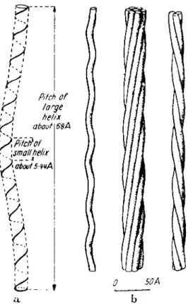
Screw-Mechanical Basis of Protoplasmic Movement 601 constant dimensions: pitch about 5.4 A, diameter about 11.0 A, pitch
angle about 26°. However, Corey and Pauling (1953) and Pauling and Corey (1954) pointed out the possibility of slight variations both in the length of the hydrogen bonds (2.68-2.92 A) and in the bond angle at the α-carbon atom (108.9-110.8°). This means a possible maximal variation of about 7% in the pitch. "The changes in the nature of the side-chain
a b
FIG. 2. T h e axis of the α-helix also takes a spiral course (helix of the second order) in keratin so that three or seven spirals form a strand. (After Pauling and Corey, 1953.)
groups might well cause the hydrogen bond distance to vary, either directly through the interaction of side chains with the carbonyl and the imino groups, or indirectly by steric hindrance or van der Waals' attraction"
(Corey and Pauling, 1955).
Crick (1952), and independently Pauling and Corey (1953) came to the further conclusion that in keratin, the axes of the α-helices are wound in a screwlike manner (Fig. 2), due, in part, to special sequences of amino acids. "Let us consider an α-helix composed of a polypeptide
in which a unit of four amino acid residues of different types is continu- ally repeated. Two of the hydrogen bonds might be longer than the other two, by about 0.2 A. This difference in length would cause a curva- ture of the axis of the α-helix" (Pauling and Corey, 1953). If we speak of the α-helix as a screw of the first order, then we can call wound helices,
"second-order screws." Thus, in considering the structures involved in movement, we may have to consider screws of the second, third, fourth, and even higher orders.
The existence of higher-order screws in cells appears to be demon- strated by the major and minor coils in chromosomes [where possibly as many as seven orders may be observed (Amano et al., 1956; Bopp-Hassen- kamp, 1959; Howanitz, 1953)] and in the bacterial flagellum (cf. Figs. 7 and 8) and, occasionally, in spirochetes, where two higher orders are apparent.
Since one aspect of the basic structure of α-proteins seems to be that they are elastic screws, we would expect among their dynamic functions not only simple shortening by contraction, but also other processes that are connected with the special mechanics of coiled structures. Various aspects of the mechanics and dynamics of coils can be studied in model experiments using helices made of steel wire (Jarosch, 1963a,b).
Since much of what we shall say concerning coiled structures is sug- gested directly by certain phenomena occurring in bacterial flagellae and in the behavior of filaments and bundles of filaments in extruded drop- lets of plant cytoplasm, we shall discuss both model experiments and ob- servations simultaneously. In this presentation, we shall first discuss processes occurring in single helical structures and then those occurring in multiple helices. It should be pointed out that phenomena seen in the model experiments occur in steel-wire screws and, thus, these phe- nomena are independent of the ultimate nature of the elastic material of which the helices are composed. However, since proteins involved in protoplasmic movement undoubtedly possess α-helical regions, much of what we say can probably be referred back to this configuration, as the one from which higher-order helices are built.
The Motive Principle in Protein Helices
If the pitch of an elastic screw is changed, a torsional force is built up in the screw. This force, in turn, causes a rotation. In a revolving screw, the single gyres progress as "apparent waves" and may displace the screw relative to its environment, as, for example a corkscrew moving relative to a cork. This principle, which was already known to Archi- medes, is the basis for the wide technological use of the screw. In an
Screw-Mechanical Basis of Protoplasmic Movement 603
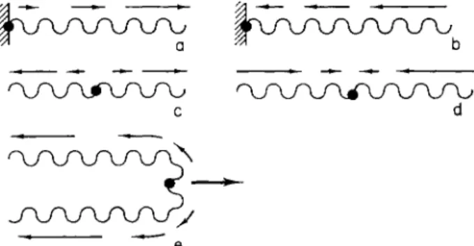
FIG. 3. Direction of the progress of apparent waves (small arrows) in the screw e
resulting from rotation caused by changes in pitch. T h e fixed position is shown with a black spot. With an increase in the pitch (a,c,e), the apparent waves progress away from the fixed end. With a decrease in the pitch (b,d) they proceed toward the fixed end. (e) Diagram of a chromosome during an anaphase movement.
rotations occur. From Pauling and Corey's data on α-helices, the number of residues per turn can vary from 3.60 to 3.67. The difference of 0.07 of a residue between these two extremes for a given α-helix is 1.9% of one turn. Thus, an α-helix 1 μ long will change length by approximately 190 A for this pitch change, and since the pitch of the α-helix is about 5.4 A, this represents about 190/5.4 or 35.2 rotations. However, in a real fluid medium, the torsion resulting in rotation will cause movement of the gyres against the medium, resulting in frictional forces. Thus, one would not expect to find the full 35.2 rotations actually occurring. In our work we, therefore, use the value 17.5 derived from model experi- ments, although this value is undoubtedly too small, since some of the torsion introduced into the model is dissipated in plastic deformation of the steel wire—a phenomenon we would not expect in elastic protein helices.
Together with the α-helices, the helices of higher order will also rotate, whereas the rigidity of the screws decreases with increasingly higher orders. This means that screws of the higher orders may tend to elastic screw, decreasing the pitch always results in a rotation, in which the gyres travel as apparent waves toward the fixed end of the screw (Fig. 3b,d). An increase in the pitch, by contrast, always causes the apparent waves to travel away from the fixed end (Fig. 3a,c,e). These rules apply regardless of the coiling-sense of the screw.
In a coil of steel wire, which has dimensions proportional to those of an α-helix 1 μ in length and with a pitch angle of 26°, experiments show that a change in pitch of 7% (caused by stretching) results in about 17.5 revolutions of the free end of the screw. In a longer screw, more
bend rather than participate in rotation around the long axis, the latter behavior being the main response of helices of lower order. If we ignore this tendency to bend, then we may derive an expression for the number of rotations R of a superhelix composed of wound α-helices corresponding to the maximum number of rotations of the lower-order helix of which it is composed. For a helix of third order this expression is
sin a2 sin a3
where / is the actual length of the axis of the third-order helix in microns, 17.5 is the number of rotations of an α-helix 1 μ in length if it undergoes a maximally allowable change in pitch (as discussed previ- ously), and a2 and a3 are the inclination angles of the second- and third- order helices, respectively.
The displacement, S, of a cytoplasmic structure is mainly dependent on the pitch, P, of the highest-order screw that rotates about its axis.
Under the most favorable conditions, that is, if the environment behaves as a firm body,
S = PR (2) Frequently, however, especially in higher-order screws, the displace-
ment will be less than this value [cf. Hancock (1953) and Taylor (1951, 1952) for discussion of wave propagation in screwlike structures] because the properties of the environment become more similar to a fluid at the microscopic and macroscopic levels.
The idea I wish to develop is that the cause of biological movements may be torsion pressure within protein helices. The basic process that produces this tension—i.e., the change in pitch of the helix—occurs be- fore the screw rotates. This may explain the fact that after interruption of some biological movements by injury, the movement takes some time to decay (e.g., cf. Metzner, 1920). Under certain circumstances a change of pitch in protein helices may be possible even in dead cells. This, at least, is one possible interpretation of the results of Hoffmann-Berling (1953, 1955).
Now I should like to discuss two possible applications of the mechan- ics of screws in nature.
Muscular Contraction
According to the recent work of Huxley's group (Hanson and Huxley, 1955; Huxley, 1956, 1957), contraction and relaxation of striated muscle are accompanied by a sliding motion of protofibrils relative to one another. The cause of this motion remains unexplained. If, however, one
Screw-Mechanical Basis of Protoplasmic Movement 605 assumes that the actin protofibrils are screws which are attached on one
side of the Z-membrane, then contraction can be explained as caused by a decrease in pitch (Figs. 3b and 4a), whereas relaxation might result from an increase in pitch (Figs. 3a and 4b). This concept is supported by older electron-microscopic observations which indicate that the axial
J A J
FIG. 4. A diagram showing the movement in muscle, (a) Condition at the begin- ning of contraction after a decrease in pitch of actin screw (cf. Fig. 3b). (b) Condition at the beginning of relaxation phase after an increase in pitch (cf. Fig. 3a).
period (which apparently corresponds to the pitch of the actin helices) is less in the contracted than in the relaxed state [250 versus 450 A, according to Draper and Hodge (1949)].
Movement of Chromosomes
The movements of chromosomes may also be related to changes in their coiling pitch. It is known that chromosomes exhibit a cycle of spiralization which reaches a peak in the condensed metaphase state, after which the chromosomes begin to elongate again. If the coils of both arms, which are fixed at the centromere, rotate when their pitch
increases on uncoiling, then the centromere would be pushed ahead, in a manner similar to the screw action in propelling a ship. It is possible that such considerations are important in anaphase movements (Fig.
3c and e).
Temperature Dependence of Cytoplasmic Streaming If the cause of movement is torsional force in protein helices, then during the relaxation of this torsion, the resulting apparent waves will encounter resistance in the surrounding aqueous medium. The viscosity of water, and, therefore, this resistive force are both temperature depen- dent. It is probably for this reason that cytoplasmic movements are proportional to temperature. On the other hand, the motive force [which we interpret as the expression of the torsional force and which in slime molds has a torsional component (Kamiya and Seifriz, 1954; Kamiya, 1959)] is not temperature dependent. This has been experimentally dem- onstrated by Kamiya (1953, 1959), in the slime mold and in Characeae by Hayashi (1960), who used the balance-centrifugal acceleration method of Kamiya and Kuroda (1958) to measure the motive force for cytoplasmic rotation. Hayashi's data strongly suggest that the effect of temperature on the velocity of cytoplasmic streaming is basically an effect on cytoplasmic viscosity.
If the change in pitch always occurs when the torsion tension has decreased to a certain minimal value, then the frequency of the change in pitch must be temperature dependent, because an increasing of tem- perature will cause a quicker release by quicker rotations. This explains the shortening in the motion period, e.g., of shuttle streaming (Kamiya, 1953, 1959) and gliding movements, as, for example, of Bacillaria para- doxa (Jarosch, 1958b) when the temperature is increased.
The ability to take up torsional forces will arise proportionally with the length of a screw. T h e previously mentioned motion period will, therefore, also arise proportionally with the screw length. The periods, e.g., of the "shunting movements" in gliding diatoms and Oscillatoriacae
(cf. Jarosch, 1962) seem, therefore, to be dependent on the organism's length.
Helices of Different Pitch Wound Together into a Superhelix So far we have dealt mostly with the behavior of a single helix. If two coils with slightly different pitch are wound together (let the pitches be px and p2, respectively, p2 < pi), a superhelix is obtained with a con- siderably larger pitch, Ρ (Fig. 5). The same is true for a superhelix made
Screw-Mechanical Basis of Protoplasmic Movement 607 of three (Fig. 6) or more single helices. T h e superhelix can be thought of
as the resultant of the superposition of single helices. An exact mathe- matical treatment of these combinations has not yet been possible.
Many model experiments with wound steel-wire helices have, how-
FIGS. 5 to 9. Bacterial flagella and their imitation with a screw model. Figure 5—a superscrew with a pitch Ρ is formed when two screws of pitches pl and slightly smaller, p2, are wound together; a, a pitch angle. Figure 6—a model of a super- screw, which consists of three screws in its left and right portions and of two screws in the middle portion. There are only two different pitches (p1 and smaller, p^).
Figure 7—a bacterium (Brucella bronchiseptica) with flagella (superscrews). Scale: 0.5 μ.
(From Labaw and Mosley, 1955.) Figure 8—the same flagella as shown in Fig. 7 enlarged. Each flagellum consists of three fibrils wound around one another. Scale:
0.5 μ. (From Labaw and Mosley, 1955.) Figure 9—string model demonstrating the course of the fibrils. Top, two fibrils; bottom, three fibrils.
ever, yielded the following empirical approximation for a superhelix formed from two separate helices:
Ρ — 2p l.bK
(P1/P2) · + 3.1 (3)
(Pi
+
P2)where p = - , the average pitch of the two helices.
Graphs of Ρ against p were made for helices with different pitches, and Eq. (3) is an empirical formula which best fits these data. It is valid only within the limits px/p2 < 2 and P/p1 < 60 (Jarosch, 1963b).
The value of Κ is dependent on the average of the pitch angles, a, and is given in Table I (based on empirical measurements).
T A B L E I
VALUES OF Κ
a Κ α Κ
10° 2.14 35° 0.30
15° 1.30 40° 0.23
20° 0.84 45° 0.20
25° 0.50 50° 0.18
30° 0.33 60° 0.15
Whereas protein helices of second order may originate from certain specific amino acid sequences in a single helix (Pauling and Corey, 1953), higher-order helices may more reasonably be assumed to be the product of two or more helices wound together. The possible variations in pitch of such helices increases with the order of the helix. Thus, the α-helix may vary only by about 7% in pitch whereas Pauling and Corey (1953) show that a pitch decrease of 5.5% in an α-helix may cause the pitch of a second-order helix to double. Thus, helices of higher order may have a nearly unlimited variability in pitch (e.g., compare the chromo- nena helices of chromosomes, which may be completely extended or completely coiled).
Since coiled structures of a variety of sizes are common in many plants, it might be expected that the protoplasm of plants would show more striking differences in the pitch of its protein helices than would the proteins of animals. Detailed analyses have been presented so far only for bacteria.
The flagella of bacteria (Fig. 7) behave in a manner analogous to steel wires in the model experiments (Fig. 5). T h e pitch of the bacterial flagellum (P) is characteristic for each species (Reichert, 1909; Pijper and Abraham, 1954; Pijper, 1957). The two or three intertwined helical
FIGS. 10 to 15. Imitation of phenomena displayed by bacterial flagella with a screw model. Figure 10—detached bacterial flagella with big arcs. (From Houwink and van Iterson, 1950.) Figure 11—the screw model in Fig. 5 can easily be laid in a plane making it form an arc such as is shown in Fig. 10. Figure 12—"biplicity"
in two bundles of flagella in a bacterium. (From Pijper, 1957.) Figure 13—imita- tion of the biplicity with one and the same screw model. Biplicity with opposite wind- ing direction (bottom) is brought about by torsion in winding direction at the primary condition. Slight pull brings back the primary condition (top). Figure 14—biplicity of one and the same flagellar bundle (From Pijper, 1957.) Figure 15—imitation of the biplicity in Fig. 14 with the screw model.
609
fibrillae have been demonstrated most clearly by Starr and Williams (1952) and by Labaw and Mosley (1954, 1955) (cf. Fig. 8). These fibrillae are apparently helices of the third order. The helix of the flagellum itself would consequently be of the fourth order. A model demonstrates the course of the two (Fig. 9, upper) or three (Fig. 9, lower) fibrillae more clearly. The difference between the model of Fig. 5 and the bacterial flagellum would seem to be primarily the proportionately larger diameter of the fibrils relative to the diameter of their constituent helices.
If values for Ρ and α are taken from the paper by Labaw and Mosley (1954) and introduced into Eq. (3), a difference in pitch of 3.5% is ob- tained for the two fibrillar helices of the flagellum. It can probably be taken as evidence for the actual existence of pitch differences in com- ponent helices of the flagellum that certain characteristic properties of the bacterial flagellum can be imitated in a wire coil model. These properties are: (1) the arc which is visible frequently at the end of a flagellum (Figs. 10 and 11), and (2) the phenomenon of "biplicity," that is, the sudden appearance of one-half the usual pitch (Fig. 12). The latter can be seen in the same flagellum (Fig. 14) and can easily be imitated in the coil model by application of torsion in the direction of the coil winding (Figs. 13, 15).
Dynamics of a Stable Superhelix
In working with steel-wire models, such as that in Fig. 5, it is difficult to produce torsional forces by a pitch change, as they probably are pro- duced in reality, and we have therefore done our experiments by twist- ing the model to produce internal torsional forces. There are two ex- treme cases in superhelices: those in which twisting the first-order helix either with or against its coiling sense will not reverse the sense of the superhelix; and those in which it will. Let us consider the latter case first. If we produce a torsional force by twisting the first-order helices against the direction in which they are wound, apparent waves of the superhelix will travel away from the fixed end of the screw. If we pro- duce such a force by twisting in the direction of winding of the first- order coil, it may suddenly change the direction of coiling of the second- ary helix (as can easily be seen in model experiments), and again the waves travel away from the fixed point. Thus, in this case, the direction of apparent wave motion is independent of the direction of torsional force. The primary superhelix (defined here as a superhelix wound in the same sense as the individual helices of which it is constituted) changes into a secondary superhelix (one in which the sense of winding is opposite to the sense of the separate helices). In the "biplicity" phe-
Screw-Mechanical Basis of Protoplasmic Movement 611 nomenon of bacterial flagella, the flagellum with the doubled pitch ap-
pears to be a stable secondary superhelix. This behavior means that a periodic change in pitch can cause a movement which goes always in the same direction, i.e., away from the fixed end of the helix. This, apparently, is the situation in flagella: continuous movement in the
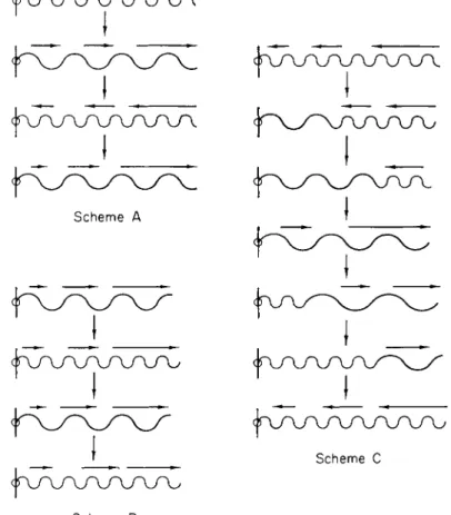
Scheme Β
FIG. 1 6 . Possible schemes for shifting of protein helices with periodic increases and decreases in pitch. For further details see the text.
direction toward the fixed end of the helix is mechanically impossible.
The first case where the superhelix does not reverse was considered earlier (cf. Fig. 3a,b).
If there is a common mechanism to protoplasmic movements, we believe that it is based on changes in the pitch of protein helices. Figure 16 shows three possible propulsion schemes based on this simple idea.
In scheme A, there is no change in the sense of the superhelix, so that
the direction of propulsion reverses when the pitch increases and de- creases. This applies to muscular contraction. In scheme Β there is a change in the direction of coiling, so that propulsion is always in the same direction (away from the point of fixation). While scheme Β ap- pears to fit the facts of flagellar movement, both schemes A and Β prob- ably apply to the motions observed in the cytoplasm of plant cells; in addition, there are probably transitions between these schemes. For ex- ample, if the change in pitch occurs very slowly (scheme C), then the conditions in the scheme Β may not be fulfilled and the primary super- helices will respond during the transition by rotations. Because of these rotations, the torsion tension required for reversal of the sense cannot be achieved. This might well be the case in the gliding movements of diatoms, blue-green algae (Jarosch, 1962), and similar organisms. Exactly which scheme is operative depends not only on the structure of the helix but also on the energetics of the change in pitch.
If these considerations are correct, then there does not seem to be any fundamental difference between flagellar and gliding movement. T h e superhelix of the bacterial flagellum, when wound around the cell mass, becomes a gliding organ if it cannot rotate around the axis of the high- est helical order, but instead bends in a flexible manner around the bacterium. In the eubacteria and spirochetes, this rotation proceeds in the direction opposite to body rotation and causes swimming. In the filose bacteria and Cyanophyceae the rotation is no longer observed; we assume in these cases that gliding is caused by rotation of helices of the next lower order.
If superhelices, which are under high torsional forces, have free play, certain characteristic phenomena are observed in model experiments with wire. (1) If the superhelix is fixed at both ends, a braidlike con- figuration of the individual helices may form and grow in extent; forma- tions of this sort may occur in accidental intertwining of blue-green algae and in filamentous bacteria. (2) If the superhelix is fixed only at one end and if there occurs a sudden increase in torsional force, the helix may snap back toward the fixation point and twist around it. During the change of direction of certain gliding movements and during bacterial movements, these phenomena, especially in flagella, occur widely. A more detailed discussion of these phenomena will not be given here.
Behavior of Unstable Bundles of Helices
In contrast to flagella, which are stable bundles of helices, the cyto- plasm of many cells apparently contains unstable bundles of helices. T h e behavior of these is very complicated and variable. The combination of single helices into bundles seems to be a physical phenomenon common
Screw-Mechanical Basis of Protoplasmic Movement 613 to all active (i.e., rotating) single helices which, because of the rotation,
will twist around one another. This leads to the formation of helices of higher order. Parallel rotating superhelices (e.g., the single flagella of a bacterium) must become entrained with respect to phase (cf. Gray 1951;
Taylor 1951). This in turn leads to a real wave motion of the bundle (helical waves), whereas the rotating single helices exhibit only apparent wave motion.
If the protoplasmic fibrils of Characeae discussed earlier in this Symposium by Dr. Kuroda and previously by us (Jarosch, 1956a, 1957, 1958a, 1960; cf. Yotsuyanagi, 1953 and Kamiya, 1959), can be considered as bundles of helices, their properties can be explained rather simply in terms of the mechanics of helices. In drops of cytoplasm freshly squeezed from living cells, the bundles of fibrils first appear bent and flexible (Fig. 18). In this state the fibrils themselves are hardly visible, but they can be recognized by small attached particles. These fibrillar bundles may form rings, which, however, soon exhibit straight stiffened regions, with the result that polygonal shapes are formed. The sides and corners of these fibrillar bundles move in a wavelike fashion, whereas the par- ticles attached to the ring do not participate in the movement (Fig. 20).
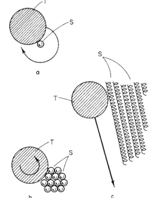
In older preparations, the polygons come to rest, showing diffuse regions of brightness in dark-field illumination and birefringence in polarized light (Fig. 19). Particle movements along the fibers and bundles of fibers are complex but may also be interpreted in terms of rotating helices. A particle attached to a single rotating helix will take part in this rotation (Fig. 17a). However, one which is attached to a bundle of rotating helices may either be pushed parallel to the axis of the bundle, or else exhibit some rotation without translation (Fig. 17b and c). T h e motions parallel to the axis of the bundle occur as a kind of reaction movement similar to those in, e.g., bundles of bacterial flagella, on the surfaces of gliding or- ganisms, and in protoplasmic filaments. However, it is at first difficult to understand how the fibrils may exhibit strong active propulsion in a lengthwise direction, yet small attached particles may move only slightly.
This can be explained if we assume that the propulsive activity of the fibrils is located in a higher-order coil than that to which the particle is attached (see Fig. 22). A particle which becomes detached correspond- ingly obtains an impulse counter to the direction of fibrillar movement, because, by being detached it may enter the counter-moving streamlet.
Coil models can imitate certain aspects of behavior of bundles of fila- ments described previously. For example, if a normally wound wire coil
is bent, it appears to be curved smoothly (Fig. 21, outer coil). However, a maximally contracted wire coil (i.e., α equals nearly zero) shows straight stiffened regions and, at the site of the bend, a rather sharp arc (Fig. 21,
inner coil). This sharp arc can often be observed clearly in completely stiffened fibrillar bundles in cytoplasmic droplets. In Fig. 21 the two coils are illuminated by a kind of dark-field illumination; it can be seen that the normally (outer) wound coil does not shine so brightly as the tightly wound one. It seems reasonable to assume, then, that in cyto- plasmic droplets the straight stiffening of the fibrils is due to some kind of "supercontraction" of the superhelices of higher orders. It can be
b c
FIG. 1 7 . T h e behavior of a particle (T) attaching to rotating screws (S). (a) At- tached to a single screw and (b, c) to a bundle of screws.
thought of as the mechanical consequence of a persisting tendency to contract, even when the single turns are closely apposed. T h e stiffening force will probably increase with the internal tension.
Thus* in cytoplasm squeezed from Characeae the single helices prob- ably mostly decrease in pitch resulting in increased stiffness. Over an extended period (some hours) it can be observed that the wave motion becomes progressively slower. At about the time that the light scattering of the polygons increases very slight wave motion of the corners of the polygons sometimes still persists. Here again the existing torsional force appears to run out slowly.
Screw-Mechanical Basis of Protoplasmic Movement 615
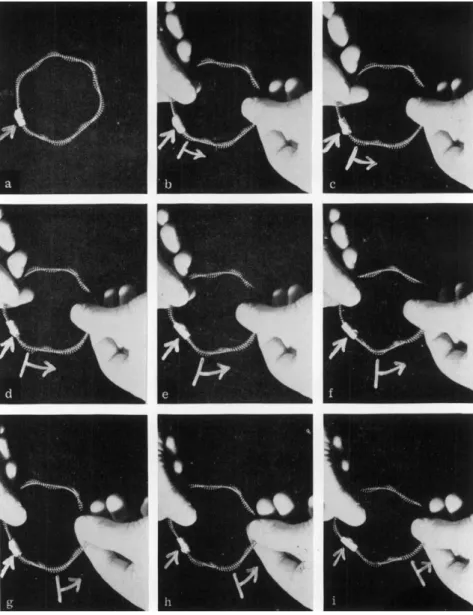
FIGS. 18 to 21. Imitation of the phenomena shown by protoplasmic fibrils of Characeae with a screw model. Figure 18—a ring of fibril bundle undulating in the form of arc in the freshly squeezed out protoplasmic drop. Figure 19—Linearly stiffened fibril bundles in an old plasma drop. Figure 20—wavelike propagation of a corner in a ring of fibril bundle. A particle imbedded in the ring, to which the arrow points, scarcely changes its position. T h e nine cine pictures were taken at intervals of 0.25 sec. Figure 21—imitation of the rectilinear stiffening of fibrils with a "supercontracted" coil.
FIG. 22. Imitation of the undulatory motion (see Fig. 20) by revolving a ringlike superhelix model around the helical axis. T h e simple arrow marks a gummed tape as a "particle." T h e arrow with cross beam marks a moving "wave."
The wave motion of the rings of fibrillar bundles in characeous cyto- plasm can be imitated in a striking manner if a superhelix model similar to that shown in Fig. 5 is made into a ring by joining the ends and is made to revolve around the helical axis. The similarity is so striking that
Screw-Mechanical Basis of Protoplasmic Movement 617 it is not difficult to become convinced that there are helices in protoplasm which rotate and give rise to protoplasmic streaming phenomena. Fig- ures 22a-i are successive photos of such a coiled model and show the wave motion of the sides and corners and fixed position of the "particle."
It is unfortunate that the phenomena which I have tried to explain in terms of screw-mechanical principles lie mostly at or just below the limit of resolution of the light microscope. Although observations on the organization of protein structures at this level must be regarded with caution, it should be mentioned that Strugger (1956, 1957; Strugger and Lindner, 1959) has claimed the existence of submicroscopic helices as protoplasmic structural elements on the basis of electron-microscopic observations on plant cells. Even if his analysis of the ribosomes as cross sections through these helices seems to be incorrect, the value of his observations themselves should be acknowledged (cf. also Weissenfels,
1957, 1958).
We have been assuming that much of plant cytoplasm streaming may be related to rotating superhelices and that the organization of such streaming is due to superhelix formation. For example, the transition from agitation to order by streaming in cells (Jarosch, 1956b; Kamiya,
1959) may very well be due to the organization of previously randomly arranged, rotating helices into bundles of superhelices, in a manner analogous to that discussed for fibril and polygon formation. If so, it may be of some value to calculate the number of resolutions per second to be expected of superhelices. Starting with the equation for the velo- city of wave motion;
Velocity — frequency χ wavelength
we can write for a rotating submicroscopic helix V = rP. In this ex- pression the displacement velocity, V, corresponds to the streaming velocity of the protoplasm; r designates the number of rotations per second. The pitch, P, can be measured in electron micrographs. Its value, if we accept Strugger's results for the purposes of this argument, averages about 300 A (z= 0.03 μ). Protoplasmic streaming at a rate of 2 μ/sec, therefore, yields for r a value of 2/0.03 or 67 rotations/sec.
Though this rotation velocity is high, it might well be the basis for the
"dynamical organization" of the protoplasm, as described, for instance, for the amoeba cell by Abé (1961, 1962). What we are suggesting can be demonstrated by rotating the model of a superhelix, of the kind shown in Fig. 5, at high speed around its long axis, for instance by connecting it to an electric motor. If this is done, standing waves will result, the distances of their nodes correspond to the repeat distances of the rotating superscrew (Fig. 23a, b, c). Using the proper repeat distances, varying
patterns of standing waves may be produced. Frequently these standing waves are not quite stationary. Then their movement resembles the wave- like motions observed at protoplasmic surfaces. They are quite unstable, as each mechanical alteration changes the state of equilibrium (Fig. 23c).
FIG. 23. (a) Resting superhelix similar to that shown in Fig. 5. (b) "Standing waves" on a quickly rotating superhelix (about 17 rotations/sec). T h e distances of neighboring nodes correspond to the pitch, (c) "Disturbed standing waves" caused by touching the rotating superhelix with the fingers.
Screw-Mechanical Basis of Protoplasmic Movement 619 If such rotating helices do exist in cells, then these standing waves may set up fixed patterns. These patterns may even play a decisive role in cell morphogenesis determining, for instance, in growing plant cells the site of addition of cell wall material (compare Kiermayer and Jarosch, 1962). It may also be that a pattern of this kind determines the localiza- tion of the pseudopods in amebae. Perhaps high-speed movies of cells would reveal some of these phenomena.
In summary, a general theory has been developed to suggest that torsional forces within protein helices giving rise to alterations in pitch may explain a variety of motions in living organisms from flagellated and gliding bacteria to the striated muscle of higher organisms. Although the idea is new and therefore untested, it has the advantage that it is generally applicable to a wide variety of so far unexplained motions which we associate with life.
ACKNOWLEDGMENT
I am indebted to Dr. R. D. Allen and Dr. L. Rebhun for bringing this article to its present form. I am also very grateful to Mr. K. Bachmann for the translation of the manuscript.
REFERENCES Abé, T . H. (1961). Cytologia 26, 378.
Abé, T. H. (1962). Cytologia 27, 111.
Amano, S., Dohi, S., Tanaka, H., Uchino, F., and Hanaoka, M. (1956). Cytologia 21, 241.
Astbury, W. T., and Street, A. (1931). Phil Trans. Roy. Soc. A230, 75.
Bopp-Hassenkamp, G. (1959). Protoplasma 50, 243.
Corey, R. Β., and Pauling, L. (1953). Proc. Roy. Soc. B141, 10.
Corey, R. B., and Pauling, L. (1955). Rend. Ist. Lombardo Sei. Lettere B89, 10.
Crick, F. H. C. (1952). Nature 170, 882.
Draper, M. H., and Hodge, A. J . (1949). Australian J. Exptl. Biol. Med. Sei. 27, 465.
Fischer, E. (1906a). Ber. Deut. Chem. Ges. 39, 453, 2893.
Fischer, E. (1906b). "Untersuchungen über Aminosäuren, Polpeptide und Proteine."
Springer, Berlin.
Gray, J . (1951). Nature 168, 929.
Hancock, G. J . (1953). Proc. Roy. Soc. A217, 96.
Hanson, J . , and Huxley, Η. Ε. (1955). Symp. Soc. Exptl. Biol. 9, 228.
Hayashi, T. (1960). Sei. Papers Coll. Gen. Educ. Univ. Tokyo 10, 245.
Herzog, R. O., and Jancke, W. (1920). Ber. Deut. Chem. Ges. 53, 2162.
Hoffmann-Berling, H. (1953). Biochim. Biophys. Acta 10, 629.
Hoffmann-Berling, H. (1955). Biochim. Biophys. Acta 16, 146.
Houwink, A. L., and van Iterson, W. (1950). Biochim. Biophys. Acta 5, 10.
Howanitz, W. (1953). Wasman J. Biol. 11, 1.
Huxley, Η. Ε. (1956). Endeavour 15, 177.
Huxley, Η. Ε. (1957). Biophys. Biochem. Cytology 3, 631.
Jarosch, R. (1956a). Phyton (Buenos Aires) 6, 87.
Jarosch, R. (1956b). Protoplasma 47, 478.
Jarosch, R. (1957). Biochim. Biophys. Acta 25, 204.
Jarosch, R. (1958a). Protoplasma 50, 93.
Jarosch, R. (1958b). Protoplasma 50, 277.
Jarosch, R. (1960) Phyton (Buenos Aires) 15, 43.
Jarosch, R. (1962). In "Physiology and Biochemistry of Algae" (R. A. Lewin, ed.), Academic Press, New York.
Jarosch, R. (1963a). Protoplasma: Höfler-Festschrift. 57, 448.
Jarosch, R. (1963b). / . Theoret. Biol. In press.
Kamiya, N. (1953). Ann. Kept. Sei. Works Fac. Sei. Osaka Univ. 1 , 53.
Kamiya, N. (1959). Protoplasmatologia 8 (3a).
Kamiya, N., and Kuroda, S. (1958). Protoplasma 50, 144.
Kamiya, N., and Seifriz, W. (1954). Exptl. Cell Res. 6, 1.
Kendrew, J . C. (1961). Set. Am. 205, 96.
Kiermayer, O., and Jarosch, R. (1962). Protoplasma 54, 382.
Labaw, L . W., and Mosley, V. M. (1954). Biochim. Biophys. Acta 15, 325.
Labaw, L. W., and Mosley, V. M. (1955). Biochim. Biophys. Acta 17, 322.
Metzner, P. (1920). Jahrb. Wiss. Botan. 59, 325.
Pauling, L., Corey, R. B., and Branson, H. H. (1951). Proc. Natl. Acad. Set. U.S.
37, 205.
Pauling, L., and Corey, R. B. (1953). Nature 171, 59.
Pauling, L., and Corey, R. B. (1954). Fortschr. Chem. Org. Naturstoffe 8, 310.
Pijper, A. (1957). Ergeb. Mikrobiol. Immunitätsforsch. Exptl. Therap. 30, 37.
Pijper, Α., and Abraham, G. (1954). / . Gen. Microbiol. 10, 425.
Reichert, Κ. (1909). Zentr. Bakteriol. Parasitenk. Abt. 1, Orig. 5 1 , 14.
Schultze, M. (1854). "Über den Organismus der Polythalamien." Leipzig, Germany.
Schultze, M. (1863). "Das Protoplasma der Rhizopoden und der Pflanzenzellen." Leip- zig, Germany.
Starr, M. P., and Williams, R. C. (1952). / . Bacteriol. 63, 701.
Strugger, S. (1956). Naturwissenschaften 43, 451.
Strugger, S. (1957). Ber. Deut. Botan. Ges. 70, 91.
Strugger, S., and Lindner, H. (1959). Protoplasma 50, 607.
Taylor, G. (1951). Proc. Roy. Soc. A209, 447.
Taylor, G. (1952). Proc. Roy. Soc. A 2 1 1 , 225.
Weissenfels, N. (1957). Naturwissenschaften 44, 241.
Weissenfels, N. (1958). Z. Naturforsch. 13b, 182.
Yotsuyanagi, Y. (1953). Cytologia 18, 146, 202.
DISCUSSION
DR. REBHUN: I would like to point out that there are really two elements in what you have discussed. One is that you developed a certain number of models and phenomena for elastic helices. T h e second is that in your paper you tried to link these with processes that occur in the α-helix.
I think that these two things are quite separate, because, while I think your mechanical models are extremely interesting, I feel there could be considerable dis- agreement as to whether your application of these to the biological systems in terms of the α-helix is proper. For example, if the thin filaments in muscle are actually actin filaments, there is considerable evidence that they do not consist of α-helices elongated throughout the length of the fibril, but of F-actin composed, at least from recent microscopic work, of globular elements. I think, therefore, that your analysis might refer to the higher order of helix without necessarily having to relate it to an α-helix.
Screw-Mechanical Basis of Protoplasmic Movement 621
DR. JAROSCH: This work describes mechanics which might apply to helices of higher order, even if the smallest order is not an α-helix but any kind of screw.
But certainly the values described for the changes in the proportion of α-helix show a remarkable agreement with the values demanded by the screw-mechanical concept.
DR. ANDREW G. SZENT-GYÖRGYI: I must comment on some of the experimental data on muscle which Dr. Jarosch mentioned. First, actin is perhaps the only fibrous muscle protein that does not show the 5.1 A periodicity. It is very hard to consider it a fully coiled α-helix. Second, the changes in the periodicity were observed at an early stage of electron microscopy at a time when effects of sectioning artifacts and shrinkage were not clearly realized and taken into account. It really should be done over before one could accept it without reservations. If the observation is true, it would be one of the most important pieces of information concerning changes in the structure of contracting muscle.
DR. JAROSCH: T h e computations which were made here using these data taken from the literature are not supposed to be more than an example, using some values and obtaining a result which is of the right order of magnitude.
DR. HOFFMANN-BERLING: I would like to apply your models to the movements of bacterial flagella. It is an unsolved problem whether bacterial flagella contract throughout their whole length as the flagella of higher organisms do. However, if they do, energy is required to produce or to reverse the mechanical alterations at the flagellar tip. T h a t energy must be transported from the bulk of the cytoplasm over the length of the flagellum to the tip. T h e flagella of higher organisms are surrounded by a membrane, and the transport can be imagined to occur inside the flagellum. However, bacterial flagella are extremely narrow and do not seem to have membranes.
What I ask is: Do your models offer some explanation for how energy could be transported mechanically from one end of the flagellum to the other? Do you see what I mean? In your models, you twist a screw, fixed at both ends. Can you imag- ine a different model, where one end of the screw is unfixed and where, by twisting, you can make waves travel to the unfixed end?
DR. JAROSCH: Concerning the energy that transports along the flagellum, there is a problem, not only in the explanation with screw models but in any other ex- planation. Dr. Gray has shown that the energy content is the same along the whole flagellum.
DR. INOUÉ: I would like to answer part of Dr. Hoffmann-Berling's comments about introducing mechanical distortions in the middle of a flagellum. W e have done similar experiments with protozoan axostyles by altering their mechanical prop- erties simply by irradiating with a microbeam of blue light. We can change the wavelengths and the wave form traveling down the axis, which is ordinarily a saw- tooth wave. Then one can watch what happens at the irradiated spot and one, indeed, finds the waves altered; as soon as the waves come to a normal part again, they get transmitted in the same way as before, getting exactly the type of deformation that Dr. Jarosch is proposing, namely, a differential contraction wave or stiffening wave which is traveling down the length. There is no time to present the data in detail, but the observations do agree with Dr. Jarosch's model and Miss Kuroda's observation.
DR. ALLEN: I just wanted to say that I am very glad that Dr. Jarosch decided to give his presentation on the screw-mechanical basis of protoplasmic movement rather than on the topic he was originally invited to discuss, namely, the movements in isolated droplets of Nitella cytoplasm. There are many motions in living material which have spiral components to them. T o remind ourselves of just a few, I might mention the helical waves in flagella, the twisting of slime mold strands that Dr.
Kamiya showed in his film, the spiral path of the ameba (Schaeffer), the spiral con- traction of the Vorticella stalk, the twisting during contraction of acephaline gre- garines (which Christopher Watters has shown us in his film) and many other phenomena.
So far as I know, no general theory of contraction that has ever been proposed has ever taken these twisting motions into account. I think that Dr. Jarosch's model, whether it is exactly correct or only in the right direction, is a very important thing for us to think about when considering general mechanisms of contractility.