IX./2.: Phoniatrics
dr. Zoltán Fent
IX./2.1.: The physiology of phonation
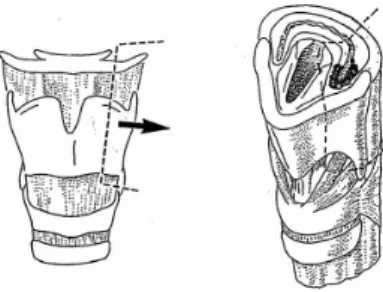
The three main factors of human voice phonation are: the air column created by
exhaling, the phonational configuration, tonicity and vibration of the vocal folds and the extension tube as vocal tract resonator space. The vocal fold vibration caused by the air column flowing through the glottal aperture can be characterised by three main features:
(fig.1.)
Frequency- the number of oscillations define voice pitch, Amplitude size- is a factor that influences volume,
The comparative ratio of the opening, closing and finishing phases of vocal fold vibration influence voice tone colour.
Figure 1.: healthy larynx in inhaling and phonation position
The multiple resonance frequencies of the primary voice, (fundamental frequency F0) will be filtered in the extension tube into singing or speaking voice. (fig.2.).
Figure 2.: Vocal fold movements during phonation (frontal section) a) closed glottal aperture b) increasing subglottal pressure c) due to increased subglottal pressure the vocal folds start to open up d) due to decreased
pressure the glottal aperture is flexibly closing e) repetitive cycle
What define tone of voice?
The various spaces of the extension tube: the supraglottis, pharynx, oral cavity and nasal cavity act as resonators. (fig.3.). These resonator spaces enhance the primary voice of the larynx, damp some of the harmonics, while other groups of harmonics are
highlighted. This way they influence the intensity and tonicity of the perceived sound.
Due to the resonance some groups of harmonics are especially amplified. These amplified clusters of harmonics are called formants. The location and intensity of formants define the speech sounds of a particular language.
Figure 3.: Soft matter extension tube and resonator spaces. The formation places of sounds
The frequency range within which the fundamental voice traces the speech tune is called range of voice in phonetics. The phonetic range of voice for men is between 70-170 Hz, for women 120-260 Hz.
IX./2.2.: The most frequent phoniatric examinations in clinical practice.
IX./2.2.1.: Anamnesis, auditive examination of voice
Instrumental examinations do not make interviewing the patients to learn historical data and complaints, observing the voice, superfluous.
During anamnesis the following needs to be considered:
family and social circumstances former illnesses
smoking medication
who and when noticed the voice production disorder present complaints
length of workplace voice demand (hour/week) singing?
Degree of noise pollution disturbance
With children – circumstances of birth, mental development
How does hoarseness develop?
During the auditive examination of the voice we observe the tone colouring of the voice, the ability of raising the loudness of voice, the ability of voice-holding, (maximum phonation time) the kind of voice starting and interrupting, and the tessitura, the spoken voice range. We establish the voice range and voice class.
The occurrence of noise elements in the primary laryngeal voice is called hoarseness.
Two factors contribute to the development of hoarseness. The first one is pathological voice fold vibration, caused by change in the mass and flexibility of the vocal folds, the second are the turbulences appearing in the air column flowing through the glottal aperture. In the case of insufficient glottal closure the voice-tone is breathy. When the glottal aperture is forcibly closed the tone becomes raspy, croaky, grating.
The normal length of the ability of voice holding, maximum phonation time, for men is 25s (pathological under l5s), for women l7s (pathological under l4s). With experienced singers it can even reach 30-50 seconds.
The voice effect appearing at the same time as the phonation configuration of the vocal folds is called voice initiation. Considering its character voice initiation can be breathy, soft, strained, hard, and pressed.
The voice effect appearing when the phonation configuration of the vocal folds ceases is called voice interruption.
Tessitura or voice range is that range of voice frequency which the fundamental voice during speech occupies from the full range.
The normal pitch of the speaking voice is generally in the lower third of the voice range.
Keeping spontaneous speech range at indifferent pitch ensures long non-tiring speech with the least energy consumption.
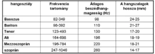
Physiological voice range is the sound frequency range which embraces that group of sounds that the person examined is able phonate at all. Within that can be found the musical voice range, constituted by the purely phonated singing voices that the singer can produce without exertion. According to voice range, body build, the size of the larynx and the vocal folds, the shapes of the resonating cavities singers are classified into voice ranges: (Table1.)
Table 1.:Voice ranges (singing and speech) and the characteristic frequency range
IX./2.2.2.: Indirect examination of the larynx with a mirror
Basic otorhinolaryngological examination
IX./2.2.3.: Videostroboscopy
Examination using a fiber-optic laryngoscope (fig.4. ) or rigid laryngoscope (fig.5.).
Stroboscopic light deviates from the vibration frequency of the vocal folds by 2 Hz, so the light illuminates sequences of voice fold waves at places that hardly deviate within the periods. Every image is preserved on the retina for 0.2 second and this way we perceive the effect of a slowed down video shoot (fig.6.).
Figure 4.: Flexible endoscope for diagnostic purposes Figure 5.: Rigid laryngoscope
Figure 6.: The basic principle of stroboscopy.
Stroboscopic light illuminates certain parts of series of periods. The eye puts the illuminated points into a slow motion movement. When the stroboscope is synchronic with the vocal fold vibrations (A), the same points of the periods will be illuminated and so in the registered shot the vocal folds will be motionless. When, however, the light is slightly desyinchronised (B), illumination falls on hardly deviating points in the periods following each other, this way creating the effect of a slow-motion film. (Hirano, 1981)
IX./2.2.4.: High speed video recording
The high speed video equipment records 2000-4000 black and white pictures per second. As speed increases the quality of the pictures slightly deteriorates. The length of the recording can be freely adjusted, generally 2 seconds are sufficient.
IX./2.2.5.: Acoustic examination of the voice, voice analysis
The examined parameters:
Maximum Phonation Time frequency range of phonation
the speaker’s fundamental frequency (f0)
frequency-intensity profile, the Voice Range Profile, can be charted by recording the maximum and minimum sound levels of phonation as a function of the fundamental frequency. The resultant figure is the voice field, the phonetogram.
During the acoustic examination of speech certain parameters can be easily quantified, expressed numerically, and examined.
Spectographs represent sounds visually. In representing the acoustic sign the horizontal axis of the diagram shows the time (t), the vertical axis the frequency (Hz) and the third dimension, the intensity measured at the various frequencies which is indicated by shades of colour. (fig.7.).
Figure 7.: spectogram of the word: „január”
Beside the spectogram we can measure fundamental frequency, frequency-fluctuation (jitter), amplitude-fluctuation (shimmer) and Harmonics-to-Noise Ratio. (fig.8. ).
Figure 8.: Jitter and shimmer (norm values: jitter - max. 1.04%, or 83,2 µs, shimmer - max.3.81%, or 0,35dB)
IX./2.3.: Partial and complete larynx removals. Voice rehabilitation
In general in Hungary on average 600-700 male patients die of laryngeal cancer annually. The prognosis of laryngeal cancers depends on the location and extension of the tumour. Based on location three regions can be distinguished:
1) supraglottic region – more than 50% of laryngeal cancers originate from here;
2) glottic region – these tumours constitute more than 40% of cases;
3) subglottic region – here the ratio of cancerous diseases hardly reaches 1-2%.
What do we is call junctional tumour?
The location of the cancer also indicates the frequency of lymph node metastasis.
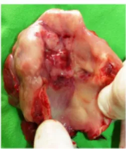
Tumours of glottic origin (fig. 9.) relatively rarely metastatize into regional lymph nodes, it is more frequent with tumours of supraglottic origin. (fig.10.) Subglottic tumours (fig.11.) will metastatize with high probability. The so-called junctional tumour is a separate clinico-pathological entity, which originates from the borderline of the true and the false vocal folds, from the so-called junctional epithelial zone. The distinction is justified because junctional tumours spreading into the paraglottic space deeply infiltrate the larynx, worsening the prognosis of this disease. (Répássy 1988).
Figure 9.: Laryngeal tumour of glottic origin (operative preparation – complete larynx)
Figure 10.: Laryngeal tumour of Supraglottic origin (operative preparation – complete larynx)
Figure 11.: Laryngeal tumour of subglottic origin (operative preparation – complete larynx)
The curative treatment of the tumour aims at residue-free complete removal of the tumour and restoration of pre-disease state, rehabilitation: regaining lost physical abilities, ability to work, and successful reintegration into society.
The surgical therapy of laryngeal cancer involves either complete or partial removal of the larynx. In certain types of tumours partial resection is enough, this way the
irreversibly mutilating nature of complete larynx ectomy can be avoided. The type of surgical intervention, the possibility of partial larynx resection are chosen primarily on the basis of the place and mass of the tumour, the presence or exclusion of neck or distant metastases and the general health status of the patient.
Figure 12.: Extensive laryngeal tumour (operative preparation – complete larynx)
IX./2.3.1.: Consequences of complete larynx removal
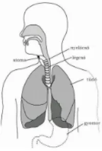
The essence of complete larynx removal is that the larynx must be removed in one piece from the tongue root to the windpipe, if necessary together with part of the tongue, the trachea, the pharynx and the thyroid gland. The trachea is sewn to the skin of the neck, creating a permanent tracheostoma and the remaining sub-pharyngeal defect will be
closed with sutures, sealing off the pathway of nourishment from the pathway of air.
Figure 13.: Complete larynx removal and postoperative state
What can the consequences be of
complete larynx removal?
Swallowing is generally undisturbed but with the loss of the larynx the patients face a completely new and unknown situation. This new state involves the loss of the ability of speech and serious restriction of communication compared to the past. Apart from the loss of voice there are other consequences too. Due to the loss of airflow in the nose sensing smell and the aroma of food deteriorates and the sense of taste changes. As the air is no longer filtered for pollutants, nor is it warmed and humidified it may dry the mucous membrane of the trachea and the ciliated cylindrical epithelium can turn into metaplasic flattened epithelium. Inhaling polluted air often leads to the appearance of crust and ample discharge.
After complete larynx removal voice rehabilitation is possible by learning esophageal speech and with the help of electrolarynx- or voice-prosthesis.
Esophageal speech
The air column necessary for phonation is provided by the air stream inhaled into the esophagus and then released. The function of the vocal folds is taken over by the so called pseudoglottis, which is a mucous membrane-muscle fold in the area of the upper esophageal sphincter or in the hypopharynx.
One third of laryngectomy patients are able to acquire esophageal speech, another third reaches an acceptable level. Learning esophageal speech is a long process that requires great perseverance and regular practice.
The teaching of esopharingeal speech is most successful in small therapeutic
communities, in the frame of self-help circles, under the supervision of a well trained speech therapist, in our part of the world (Central Europe) this person is called a
logopede. During club-like meetings new and old laryngectomy patients help each other and together improve, perfect their ability of esophageal speaking.
Esophageal speakers, due to their anatomical and functional situation, have much less air at their disposal for the pronunciation of sentences, consequently they always have to supplement air quantity.
Electrolarynx
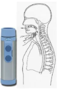
After complete larynxectomy, lacking voice creating space, the generation of primary sound is done with the help of a small instrument whose membrane part is attached to the skin of the neck, and the induced voice vibrations are then passed on through the soft tissues into the resonating space, and then using normal articulation the sound can be turned into speech. (fig.14.).
Figure 14.: Electrolarynx and its use
Electrolarynx aided speech used to be considered monotonous, mechanical, lacking in dynamism, and expressionless. In order to approach the natural speaking voice as much as possible on the new instruments there is a possibility to regulate volume and pitch in a wide range. The use and application of electolarynx requires 8-10 days of learning and practice. A drawback of these instruments is that in themselves they cannot be used in the case of X-ray scarred necks. Such patients can use the instrument with the help of an oral adapter. There is a manual operation free version too, triggered by the
electromyographic activity of the neck muscles.
Voice prostheses
How does voice prosthesis work?
The operation principle of the voice prosthesis in use since the 1980’s, and ever since continuously perfected, is that the air necessary for phonation comes from the lungs from the direction of the trachea and through a tracheo-oesophageal fistula created by the operation reaches the eosophagus, where spare vocal folds, developed from pharyngeal musculature and the mucous membrane-fold take on the phonation role of the larynx. Articulation, as originally, is done by the oral cavity. The created fistula has a valve built into it which allows only one direction of air flow and also prevents eventual aspiration.
Figure 15.: functional perspective of voice prosthesis
The operation principle of voice prosthesis
How does voice prosthesis work?
The voice prosthesis (fig.16.) is mostly implanted simultaneously with the larynxectomy, but the implantation can take place during a second operation too. During teaching phonation the patient must be taught the step by step procedure of inhaling - closing the stoma – insufflation - voicing - articulation, which they have to practice under
supervision until the procedure becomes a reflex.
Figure 16.: Provox prosthesis
Excellent quality of sound is an undisputed advantage of voice prostheses. Depending on type, however, they need to be changed every 3-5 months due to unsatisfactory work or because of a fistula appearing between the oesophagus and the trachea.
IX./2.3.2.: About some partial larynx operation
Hemilaryngectomy
It is indicated when the tumour infiltrates the cartilage, reaches the anterior commissure, causing limitation of movement, but it does not cross the middle line in the back part of the pharynx, and does not reach the articulation between the cricoid and the arytenoid cartilage. (fig.17. ).
Figure 17.: Resecatum during hemilaryngectomy
Supraglottic horizontal larynxresection
In the case of what tumour localisation can we perform supraglottal horizontal laryngeal resection?
It is indicated when the tumours of the lingual surface of the epiglottis reach the aryepiglottic fold, and the vallecula glosso-epiglottica, but they do not infiltrate the latter, and also tumours which originate from the aryepiglottic fold. It is counter- indicated when the localisation of the tumour deviates from what is described above, for example infiltration of the arycartilage or the sinus piriformis.
During the operation the whole preepiglottic pouch, the hyoidean, the epiglottis, the aryepiglottic folds, false vocal folds, Morgagni’s pouches and the upper third of the thyroid cartilage with its inner perichondrium will be removed. (figs.18., 19. ).
Figure 18.: Supraglottic horizontal larynx resection Stripes indicate the specimen to be removed
Figure 19.: Fiberoscopic image of post horizontal supraglottic larynxresection with open and closed
configurations of the vocal folds.
Tumours of which anatomical region are suitable for the surgical solution of supracricoid lateral larynx hiatus resection?
Lateral supracricoid larynxresection
Répássy et al. performed this operation for the first time in Hungary in 1994, and gave an account in 2000 about the lateral supracricoid larynxresection as a possible way of treatment of recessus piriformis tumours. The authors consider the operation procedure described as a suitable function preserving treatment in selected cases .
Figure 20.: Large tumour of the aryepiglottic fold Figure 21.: Removed specimen from lateral supracricoid larynx resection
During the operation the affected side of the lamella of the thyroid gland together with its back horn will be removed with a 2.5 cm incision leading from the posterior edge towards the medial. Depending on the extension of the tumour on the affected side we remove half of the hyoidean together with the same side ary-region and the back part of the vocal folds and as needed a larger portion of lateral pharyngeal wall along with the sinus piriformis. ( fig.21.). During reconstruction the remaining pharyngeal wall is stitched to the remaining thyroid sheet and then the pharyngeal defect will be closed. For post operative fiberoscopic picture see (fig.22).
Figure 22.: Post right side supracricoid lateral larynxremoval state with open and closed glottic aperture
Being aware of the spreading of the recessus piriformis tumour, best professional
practice needs to preserve laryngeal functions because the patient’s quality of life is significantly better than in the case of complete larynx removal. In the case of recessus piriformis tumour operations also the successful surgical therapy depends on early diagnosis as in an early stage the larynx may be mostly tumour free and can be saved.
(Répássy 2000).
Supracricoid horizontal larynx resection
During the operation of extensive supraglottic and glottic tumours and transglottic tumours that involve several regions of the larynx in selected cases the opening
attachment of the trachea into the neck, making a permanent neck stoma can be avoided by preserving the cricoid, the hyoidean and perhaps a part of the flap covering the larynx and in some of these cases even voice rehabilitation results can be good.
This operation was first described by Mayer and Rieder in 1959. About modifying the operation technique and its application results Laccourreye and Chevalier have
published a number of studies. In Hungary supracricoid horizontal larynx removal was first performed by Répássy in 1997. He summed up the modified operation technique and experience with the operation in a publication in 2000. (Répássy 2000).
The laryngeal parts to be removed can be seen in (fig. 23).
Figure 23.: Part of the larynx to be removed in supracricoid horizontal larynx resection
Following the removal of the tumour we pull the cricoid and the hyoidean together with frontal cross stitching suture. (fig.24.). Two forms of this operation can be distinguished:
1. Cricohyoidopexia (CHP) – if we have removed the whole epiglottis then we sew the arch of the cricoid cartilage to the hyoidean.
2. Cricohyoido-epiglottopexia (CHEP) – if the upper 2/3 of the epiglottis can be preserved after than the hyoidean stitch we cross stitch the lower edge of the epiglottis, then comes the cross stitching of the arch of the cricoid cartilage and the suture will fix the epiglottis forward, to the cricoid cartilage.
Figure 24.: Reconstruction after supracricoid horizontal larynx resection
The area of operation indications:
Tumour of supraglottic origin with Morgagni’s pouch infiltration aryregion infiltration
anterior commissure infiltration paraglottic area infiltration
vocal fold cancer with transglottic spreading
particular selected cases of T4 supraglottic and glottic cancers
The
alternative of which larynx operation could the horizontal supracricoid larynx resection?
Counter indications:
infiltration of the hyoidean and the vallecula glosso-epiglottica tongue root infiltration
vocal folds fixation
subglottic spreading towards to cricoid interarytenoid infiltration
tumours treatable with horizontal supraglottic resection
Répássy et al. have found that generally for the preservation of laryngeal functions in a smaller selected group of patients who qualify for complete pharynx removal
supracricoid horizontal laryngectomy proves to be a good alternative solution.
According to their experience, however, after the operation regular swallowing and the removal of the cannula are problematic, in some cases the removal of the cannula is not possible. (Répássy 2000).
Figure 25.: Phonation after SCH-CHEP
The ary-region is marked with an asterisk, which during phonation closes to the inner surface of the epiglottis.