Synthesis of TiO
2nanofibers by electrospinning using water-soluble Ti- precursor
Odhiambo Vincent Otieno1•Edina Csa´ki1• Orsolya Ke´ri1•La´szlo´ Simon2•Istva´n Endre Luka´cs3• Katalin Me´sza´ros Sze´cse´nyi4•Imre Miklo´s Szila´gyi1
Received: 4 May 2019 / Accepted: 18 May 2019 / Published online: 29 May 2019 ÓThe Author(s) 2019
Abstract
A new electrospinning process was developed for preparing TiO2 nanofibers using a water-soluble Ti-precursor, [bis(kappa1O-hydroxo)(bis(kappa2O,O0-lactato)titanium(IV)] commonly known as titanium(IV) bis (ammonium lactato) dihydroxide (TiBALDH). The importance of the study is justified by the fact that Ti-precursors used for electrospinning, sol–gel, hydrothermal and other fiber synthesis processes are mostly non-water soluble. Accordingly, anatase TiO2
nanofibers of diameter between 20 and 140 nm were synthesized by electrospinning and annealing. Polyvinylpyrrolidone (PVP) and different concentrations of TiBALDH were dissolved in a mixture of water, ethyl alcohol and acetic acid to optimize the electrospinning conditions. The thermal decomposition and fragmentation of PVP, TiBALDH and the fibers with 50% mass fraction of TiBALDH were studied by TGA-MS measurements. The fibers were then annealed at 1°C min-1until 600°C. The TiO2fibers were characterized using SEM–EDX, FTIR and XRD
Keywords TiBALDHPVP ElectrospinningAnnealingTiO2TGA-MSSEM–EDXFTIR and XRD
Introduction
Titanium dioxide is used in a number of applications including self-cleaning, antimicrobial thin film coatings, photocatalysis, gas sensing and dye-sensitized solar cells [1–5]. This has led to a lot of research in the preparation of
TiO2 nanofibers with well-controlled morphology [6–10].
Many methods of synthesis of TiO2nanofibers have been developed including sol–gel, hydrothermal and electro- spinning [11–13]. Electrospinning as a method of synthesis of nanofibers has been widely reported [14–17]. The technique is a versatile and efficient method for synthe- sizing uniform fibers with large specific surface area [18–21]. In electrospinning, high static voltage is applied to a polymer solution or melt, which can contain a precursor salt of metal oxide in a syringe. The solution or melt is ejected from the needle tip, accelerated by electric field and is collected on a grounded substrate in form of thin con- tinuous fibers [17,22–24]. The nanofiber properties can be controlled to offer more flexibility in surface functionalities of the end product. Many studies use Ti-alkoxides and Ti- halides as precursors for the synthesis of TiO2; however, these are insoluble in water [25]. There is a need to prepare TiO2nanofibers from water-soluble precursors. This would enable coupling TiO2 with other metal oxides having water-soluble precursors. In this study, [bis(kappa1O-hy- droxo)(bis(kappa2O,O0-lactato)titanium(IV)] commonly known as titanium(IV) bis (ammonium lactato) dihydrox- ide (TiBALDH) was used as the precursor in preparing Electronic supplementary material The online version of this
article (https://doi.org/10.1007/s10973-019-08398-z) con- tains supplementary material, which is available to autho- rized users.
& Odhiambo Vincent Otieno
vincent.odhiambo@mail.bme.hu
1 Department of Inorganic and Analytical Chemistry, Budapest University of Technology and Economics, Szent Gelle´rt te´r 4., Budapest 1111, Hungary
2 Department of Organic Chemistry and Technology, Budapest University of Technology and Economics, Budafoki u´t 8., Budapest 1111, Hungary
3 Research Institute for Technical Physics and Materials Science, Hungarian Academy of Sciences, Konkoly Thege M. u´t 29-33., Budapest 1121, Hungary
4 Department of Chemistry, Biochemistry and Environmental Protection, University of Novi Sad, Trg Dositeja Obradovica 3, 21000 Novi Sad, Serbia
https://doi.org/10.1007/s10973-019-08398-z(0123456789().,-volV)(0123456789().,-volV)
TiO2nanofibers. The structure of TiBALDH is shown in Fig.1.
TiBALDH is a water-soluble titanium precursor with low reactivity [26, 27]. It has been used to make various titania nanomaterials. Mockel et al. reported that TiBALDH was utilized to produce almost monodispersed anatase nanocrystals by thermohydrolysis [28].
Mayya et al. [29] reported a nanoscale coating of gold nanoparticles with titania based on TiBALDH. Lee et al.
[30] prepared anatase TiO2 nanoparticles coupled with carbon nanotubes (CNTs) by controlled hydrolysis of TiBALDH in CNTs containing aqueous media.
Hongzhi et al. [31] prepared TiO2 nanocrystals by hydrolysis and hydrothermal treatment of TiBALDH.
There is only one report in which a water-soluble TiO2 precursor was used to synthesize TiO2 nanofibers by electrospinning. Nakane et al. [32] prepared hybrid nano- fibers of poly(vinyl-alcohol) and titanium lactate by elec- trospinning. In their study, Nakale et al did not optimize the
concentration for the precursors and only one set of con- centration was published. However, no studies have been reported about the use of TiBALDH as a precursor in the synthesis of TiO2nanofibers by electrospinning. There are also no reports on thermal analysis of TiBALDH which is important since to obtain crystalline TiO2, an annealing step is often needed.
In this study, TiO2 nanofibers were prepared by elec- trospinning a mixture of alcoholic and aqueous solutions of polyvinylpyrrolidone (PVP) and TiBALDH. We studied how the properties of the electrospun fibers could be con- trolled by using different concentrations of the precursor.
The thermal decomposition and fragmentation of PVP, TiBALDH and the fiber with 50% mass fraction of TiBALDH were studied by TGA-MS measurements. The thermal properties of the electrospun fibers were investi- gated in nitrogen by simultaneous thermogravimetry/dif- ferential thermal analysis (TG/DTA). The electrospun fibers were annealed at 600°C to remove the polymer component, decompose the precursor and obtain TiO2 nanofibers. The electrospun and annealed nanofibers were characterized by scanning electron microscopy (SEM) and energy-dispersive X-ray spectroscopy (EDX). The fibers, PVP and TiBALDH were studied by Fourier transform infrared spectroscopy (FTIR). The annealed fibers were investigated by X-ray diffraction (XRD).
2NH4+ H3C
HO
CH3
O O
O
O O
O Ti OH
2–
Fig. 1 Structure of TiBALDH
67 °C
66 °C
72 °C
192 °C 225 °C
285 °C 285 °C
340 °C 386 °C
468 °C
0.004%/°C
0.01%/°C
0.01%/°C 416 °C
434 °C
100 200 300 400 500 600
Temperature/°C
Deriv.mass/% °C–1
0.5
%/°C
PVP TiBALDH TiBALDH-PVP
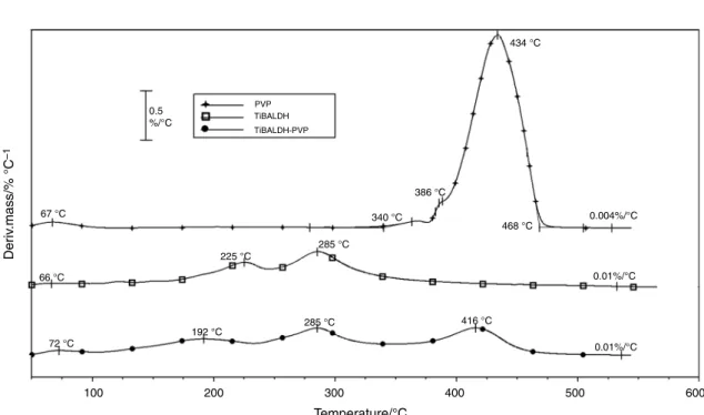
Fig. 2 DTG curves of PVP, TiBALDH and PVP-TiBALDH mixture in argon
100 200 300 400 500 600 35
30 25 20 15 10 4 3 2 1 0
100 200 300 400 500 600
Temperature/°C
Temperature/°C Temperature/°C
m/z 14.00 m/z 15.00 m/z 16.00 m/z 17.00 m/z 18.00
Signal intensity/a.u.
DTG 0
– 1
– 2
– 3
– 4
DTG/% °C–1
100 200 300 400 500 600
6
4 3 2 5
0.1
100 200 300 400 500 600
Temperature/°C
Temperature/°C
m/z 36.00 m/z 37.00 m/z 38.00 m/z 39.00 m/z 41.00
Signal intensity/a.u.
0
– 1
– 2 0.0
m/z 42.00 m/z 43.00 m/z 44.00
100 200 300 400 500
– 0.5 – 0.6 – 0.7 1
4 3 2 5
0
100 200 300 400 500
– 0.2 Temperature/°C
Temperature/°C
m/z 29.00 m/z 30.00 m/z 31.00 m/z 32.00
m/z 55.00
Signal intensity/a.u.
– 0.1
– 0.3 – 0.4 0.0
m/z 42.00 m/z 43.00 m/z 46.00
100 200 300 400 500
– 0.5 – 0.6 – 0.7 100
140 180
4 8
2 6
0
100 200 300 400 500
– 0.2
Temperature/°C
Signal intensity/a.u.
– 0.1
– 0.3 – 0.4 0.0
m/z 14.00 m/z 15.00 m/z 16.00 m/z 17.00 m/z 18.00 60
100 200 300 400 500 600
Temperature/°C
– 0.5 – 0.6 – 0.2 – 0.1
– 0.3
– 0.4 0.0
DTG DTG
4 2
0 10 14 18 22 26
100 200 300 400 500
Temperature/°C
600 m/z 14.00 m/z 15.00 m/z 16.00 m/z 17.00 m/z 18.00 DTG
DTG
m/z 30.00 m/z 32.00
m/z 55.00 m/z 42.00 m/z 45.00
Signal intensity/a.u. Signal intensity/a.u.
100 200 300 400 500
Temperature/°C
100 200 300 400 500
Temperature/°C
– 0.5 – 0.6 – 0.7 – 0.2 – 0.1
– 0.3 – 0.4 0.0
0.0 0.1 0.2 0.5 1.0 1.5 2.0
(a) (b)
(c) (d)
(e) (f)
DTG/% °C–1 DTG/% °C–1 DTG/% °C–1DTG/% °C–1 DTG/% °C–1
Fig. 3 DTG-MS curves of PVP (a,b) TiBALDH (c,d) and PVP-TIBALDH mixture (e,f) in argon
Experimental
MaterialsAll materials were analytical grade and used as received.
Polyvinylpyrrolidone [PVP, (C6H9NO)n, K-90] and tita- nium(IV) bis(ammonium lactato)dihydroxide [(C6H18N2 O8Ti) TiBALDH, 50 mass% in water)] were obtained from Sigma-Aldrich.
Preparation and characterization of TiO2fibers
The polymer solution contained 20 mass% PVP dissolved in 1:1 mixture of acetic acid and ethyl alcohol. 2 mL of the polymer solution was mixed with 2 mL of an aqueous solution containing 50, 30, 25 and 10 mass% TiBALDH, respectively. The mixture was stirred for 6 h at room temperature. The mixed solution was loaded into a plastic syringe equipped with a needle for electrospinning. The feeding rate was 1 mL h-1, while the applied voltage was 25 kV. The fibers were collected on an Al foil screen covered by a polyethylene sheet.
For the thermal measurements, a 50 mass% solution of TiBALDH was carefully1 dried. TG/DTA–MS measure- ments for TiBALDH, PVP and as-spun fiber of 50 mass%
TiBALDH were carried out using the TA Instruments’
Q600 simultaneous TG/DSC setup coupled to a Hiden Analytical HPR-20/QIC mass spectrometer. The measure- ments were carried out in flowing argon (flow rate = 50 cm3 min-1) in an alumina crucible and empty crucible as a reference. The sample mass was ca. 7 mg.
Selected ions between m/z= 1–125 were monitored in
multiple ion detection mode (MID) at a heating rate of 10°C min-1.
The electrospun fibers were annealed in air to remove the polymer and decompose the precursor. The annealing was done at a rate of 1°C min–1up to 600°C. The thermal decomposition of the electrospun fibers in nitrogen was investigated in an STD 2960 simultaneous DTA/TGA (TA Instruments Inc.) thermal analyzer. The samples were heated up to 600°C using a heating rate of 10 °C min–1in nitrogen.
The morphology of the as-spun fibers was studied by scanning electron microscopy (SEM) in a JEOL JSM- 5500LV scanning electron microscope in a high vacuum mode at 20 kV. For the annealed samples, the SEM images were observed by a LEO 1440 XB electron microscope in a high vacuum mode with secondary electron detector. The EDX analysis of the annealed fibers was done using JEOL JSM-5500LV electron microscope. Before the measure- ment, the nanofibers were coated with a thin Au/Pd layer in a sputter coater. Fourier transform infrared spectroscopy (FTIR) measurements of carefully dried 50 mass%
TiBALDH, PVP, electrospun and annealed nanofibers were recorded with a Nicolet 6700 apparatus in the 400–4000 cm-1 domain in transmittance mode. The sen- sitivity of measurements was 4 cm-1, and 64 scans were accumulated per spectrum. The XRD patterns were recor- ded by a PANalytical X’pert Pro MPD X-ray diffrac- tometer using Cu Kairradiation.
Results and discussion
TG/DTA–MS measurements were carried out to study how the TiO2precursor, TiBALDH, affects the decomposition of PVP and the formation of TiO2. Figure2shows that the
3.2 2.4 1.6 0.8
Signal intensity/a.u. Signal intensity/a.u.
0.4
0.2
0.0
100 200 300
Temperature/°C Temperature/°C
400 500 100 200 300 400 500
0.020
0.020
0.010
0.005 0.000 m/z 1.00
m/z 2.00
m/z 36.00 m/z 68.00
m/z 112.00 m/z 56.00
(a) (b)
Fig. 4 MS curve for other characteristic fragments of PVP decomposition in argon
1 According to the Safety Information the hazard statement of TiBALDH is H226-H319.
decomposition of TiBALDH is continuous showing two DTG maxima to 550°C, while the decomposition of PVP takes place in practically one step between 340 and 468°C without residue [33, 34]. The decomposition of the fiber with 50 mass% TiBALDH and 20% PVP is also continuous with three DTG maxima up to 550°C. The decomposition
of the fiber and both of its components is practically fin- ished around 550°C. In the temperature range up to about 100 °C, the evaporation of the solvents is expected.
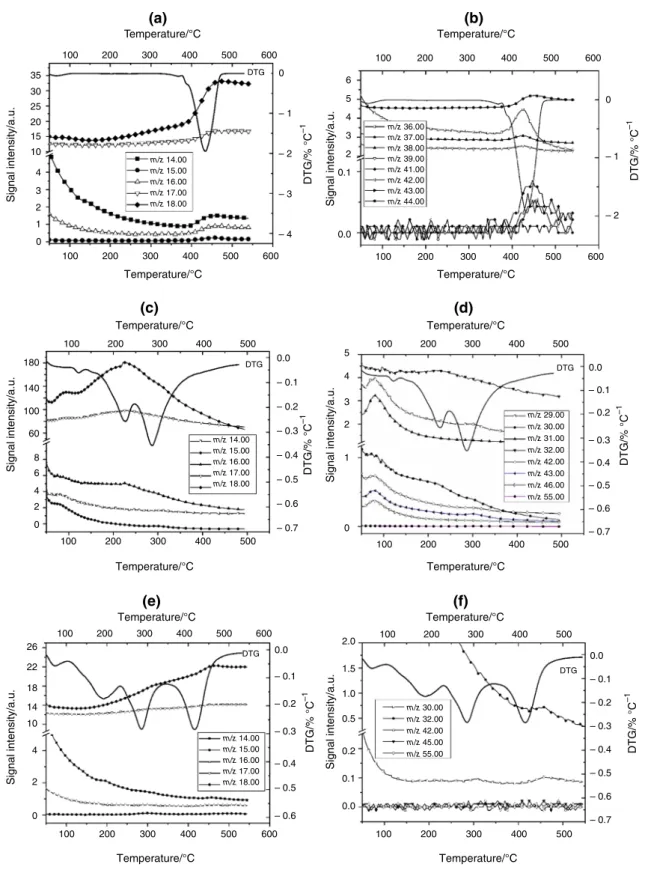
The DTG–MS data are shown in Fig.3a–f. In the samples of PVP (Fig.3a) and the fiber of PVP/50 mass%
TiBALDH (Fig.3e), at first, water evaporation was
100
91.5%
70.3%
152.8 °C 52.4 °C
287.2 °C
430.1 °C 524.3 °C
147.1 °C 61.5 °C
283.8 °C
434.7 °C 557.8 °C
168.7 °C 62.6 °C
294.6 °C
440.9 °C 562.3 °C 153.3 °C
51.2 °C
287.4 °C
436.4 °C 557.2 °C 45.7% 44.2%
92.5%
72.1%
43.3% 40.5%
95.8%
80.8%
39.9% 38.5%
95.9%
87.7%
38.3%
36.5%
50
0
100
50
0
100
50
0
100
50
0
0 100 200 300
Temperature/°C
Endo down Endo down
Endo down Endo down
Mass/% Mass/%Mass/%
Mass/% DTA/a.u.DTA/a.u. DTA/a.u.DTA/a.u.
TGTG TGTG
400 500 600
0 100 200 300
Temperature/°C
400 500 600 0 100 200 300
Temperature/°C
400 500 600
0 100 200 300
Temperature/°C
400 500 600
(a) (b)
(c) (d)
Fig. 5 TG/DTA of as-spun fibers in nitrogen a 50 mass% TiBALDH,b 30 mass% TiBALDH,c25 mass% TiBALDH and d10 mass%
TiBALDH
detected. In addition, in TiBALDH, the appearance of fragments with 14–18 and 29–46m/z ratio indicates a partial decomposition of the TiO2 precursor even below 100°C (Fig.3c, d). This is not surprising taking into account the hazard statements of TiBALDH. Above 100°C, in the DTG curve of the composite fiber, the peaks characteristic for both of PVP and of TiBALDH was observed. However, the DTG maxima appeared at lower temperatures than in the pure components.
The most intense MS signals of PVP are those withm/
z= 18 and 17. The intensity ratio of the peaks below 100°C agrees with that of water. At higher temperatures, the intensity ratio changes as a result of the evolution of fragments NH3?and NH4?. The intensity of the signals of
the fragments withm/z= 14, 15 and 16 is about ten times less than of fragmentsm/z= 18 and 17. Besides these, low- intensity signals with a higherm/zratio were also detected (Fig.3b). The courses of the m/z signals follow well the course of the DTG signal. Since the decomposition was recorded in flowing argon using TG/DTA-MS, relative intense signals of H? and H2?(m/z= 1, 2) and C3?(m/
z= 36) were detected due to the reduction of the polymer (Fig.4a). The molecular fragment of the PVP monomer with m/z= 111 was not found. The fragment with the highest m/z was detected at 112 (Fig.4b) which most probably originates from the recombination of the fragment withm/z= 56 (see Fig.4b). Besides, a low-intensity signal in PVP was detected atm/z= 68. The appearance of these
Table 1 As-spun and annealed fiber diameters and composition of annealed fibers
TiBALDH/mass% As-spun fibersd/nm Annealed fibersd/nm Atomic/%
Ti O
50 617–800 131–168 35.8 64.2
30 487–608 61–82 38.9 61.1
25 425–453 41–68 35.5 64.5
10 309–375 20–57 34.5 65.5
Fig. 6 SEM images ofaas-spun fibers 50 mass% TiBALDH,bas- spun fibers 30 mass% TiBALDH, c as-spun fibers 25 mass%
TiBALDH,das-spun fibers 10 mass% TiBALDH,eannealed fibers
50 mass% TiBALDH, e annealed fibers 30 mass% TiBALDH, fannealed fibers 25 mass% TiBALDH,gannealed fibers 10 mass%
TiBALDH
signals is most probably a result of the fragmentation of the pyrrolidone ring (e.g., C2H2NO and C3H2NO).
During the decomposition of TiBALDH, only signals up to m/z= 46 ratio were detected (Fig.3c, d). Fragments with m/z= 18, 29 and 31 had the most intense signals.
These could be attributed to H2O?, C2H5?
and C2H2OH? formed during the decomposition of TiBALDH (C6H18N2O8Ti).
The difference in the fragmentation of the as-spun fiber compared to the fragmentation of the components used for its preparation is that in the mixture all the detected signals were below m/z= 32 (Fig.3f). This means that in the preparation of the TiO2 fibers TiBALDH catalyzes the decomposition of PVP.
Due to the very small differences in the molar masses of the expected CO2?, N2O?, NOx?fragments, their identifi- cation by this way was not possible. Based on the TGA measurements, the as-spun fibers should be annealed to 600°C to obtain TiO2fibers.
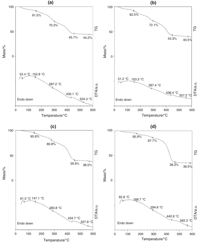
Figure5 shows the comparison of various composite fibers in nitrogen and that the decomposition is continuous.
The endothermic peaks in the DTA peaks are not sharp, because the precursor and the polymer components of the fibers decomposed without combustion. The decomposition occurred in three stages as discussed earlier. The mass of the residues was consistent with the increasing concentra- tion of the precursor. The percentage yield for the TiO2 fibers varied depending on the concentration of the pre- cursor, 50 mass% TiBALDH was 13.1%, 30 mass%
TiBALDH was 10.7%, 25 mass% TiBALDH was 10.4%
and 10 mass% TiBALDH was 9.4%.
The as-spun PVP/TiBALDH fibers had diameters between 310 and 800 nm depending on the concentration of TiBALDH. The fibers were annealed in air at a heating rate of 1°C min-1up to 600 °C. The slow heating rate was used to avoid disintegration of the oxide fibers [22]. The diameter of the as-spun and annealed fibers was larger when the concentration of TiBALDH was higher. After annealing, diameter of the fibers decreased significantly after annealing to 20–170 nm, as shown in Table1.
From the SEM image of annealed fibers shown in Fig.6, the fibers formed from 50 mass% and 30 mass% TiBALDH were smooth, while the fibers from 25 mass% and 10 mass% TiBALDH had some beads.
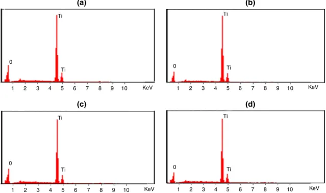
Results of EDX analysis are shown in Fig.7. They confirmed the presence of titanium and oxygen in the annealed fibers. Ti peak was observed at 4.5 kV [35,36].
The elemental composition in the annealed fibers is as shown in Table1.
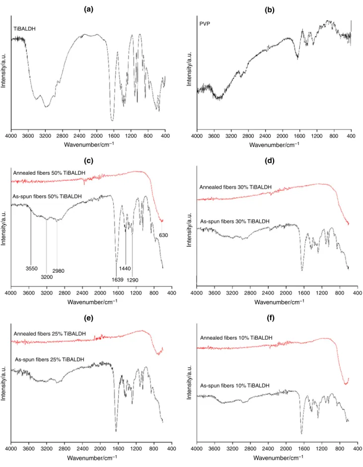
The results for the FTIR measurements of pure TiBALDH, PVP, as-spun and annealed composite fibers are shown in Fig. 8. The broadband around 3600 cm-1can be assigned to O–H stretching vibration while the peak around and 3200 cm-1 can be due to N–H stretching vibrations in TiBALDH. The C–H asymmetric vibrations of the methyl group in TiBALDH and PVP were observed around 2980 cm-1. The sharp absorption bands around 1639 cm-1can be assigned to C=O in the amide group in
0 0 Ti
Ti
Ti Ti
Ti Ti
(a) (b)
(c) (d)
Ti Ti
0 0
1 2 3 4 5 6 7 8 9 10
1 2 3 4 5 6 7 8 9 10
KeV
KeV 1 2 3 4 5 6 7 8 9 10 KeV
1 2 3 4 5 6 7 8 9 10 KeV
Fig. 7 EDX spectra of annealed fibersa50 mass% TiBALDH,b30 mass% TiBALDH,c25 mass% TiBALDH andd10 mass% TiBALDH
4000 3600 TiBALDH
(a) (b)
(c) (d)
(e) (f)
PVP
3200
Annealed fibers 50% TiBALDH
As-spun fibers 50% TiBALDH
Annealed fibers 30% TiBALDH
As-spun fibers 30% TiBALDH
Annealed fibers 25% TiBALDH
As-spun fibers 25% TiBALDH
Annealed fibers 10% TiBALDH
As-spun fibers 10% TiBALDH 2800 2400
Wavenumber/cm–1 Wavenumber/cm–1
Wavenumber/cm–1 Wavenumber/cm–1
Wavenumber/cm–1
Intensity/a.u. Intensity/a.u.
Intensity/a.u. Intensity/a.u.
Intensity/a.u. Intensity/a.u.
Wavenumber/cm–1
2000 1600 1200 800 400 4000 3600 3200 2800 2400 2000 1600 1200 800 400
4000 3600 3550
3200 2980
1639 1290 1440
630
3200 2800 2400 2000 1600 1200 800 400 4000 3600 3200 2800 2400 2000 1600 1200 800 400
4000 3600 3200 2800 2400 2000 1600 1200 800 400 4000 3600 3200 2800 2400 2000 1600 1200 800 400
Fig. 8 FTIR spectra of as-spun and annealed fibersa TiBALDH, b PVP, b50 mass% TiBALDH, c 30 mass% TiBALDH,d 25 mass%
TiBALDH ande10 mass% TiBALDH
PVP. The peak around 1440 cm-1can be assigned to O–H bending vibrations. The C–N stretching vibration absorp- tion peaks in PVP were observed around 1290 cm-1 [33,37,38]. These absorption bands were also observed in the as-spun fibers. For the annealed samples, the absorption band around 630 cm-1 can be assigned to the Ti–O–Ti bonds [37]. The FTIR measurements of the annealed fibers confirmed that the polymer and the precursor were decomposed during annealing.
Figure9shows the XRD pattern of the annealed fibers.
The fibers were crystalline with tetragonal structures. The XRD patterns exhibited strong diffraction peaks at 25°and 48°indicating TiO2in anatase phase [35,39]. The annealed fibers were indexed to ICDD 04-016-2837.
Conclusions
Anatase TiO2nanofibers of diameter between 20–170 nm were synthesized by electrospinning using a water-soluble Ti-precursor. Polyvinylpyrrolidone and different concen- trations of TiBALDH were dissolved in a mixture of water, ethyl alcohol and acetic acid followed by electrospinning at 20 kV to obtain nanofibers. The as-spun fibers were studied by TG/DTA-MS to establish annealing temperatures. The data of TGA-MS measurements revealed that TiBALDH catalyzes the decomposition of the as-spun fibers. During its decomposition, only fragments withm/z\32 evolved.
The fibers were annealed at 1°C min-1 until 600°C to form anatase TiO2 nanofibers. 50 mass% and 30 mass%
TiBALDH concentrations formed smooth fibers.
Acknowledgements Open access funding provided by Budapest University of Technology and Economics (BME). I. M. Szila´gyi
thanks for a Ja´nos Bolyai Research Fellowship of the Hungarian Academy of Sciences and an U´ NKP-18-4-BME-238 New National Excellence Program of the Ministry of Human Capacities, Hungary.
A GINOP-2.2.1-15-2017-00084, an NRDI K 124212 and an NRDI TNN_16 123631 grants are acknowledged. The research within pro- ject No. VEKOP-2.3.2-16-2017-00013 was supported by the Euro- pean Union and the State of Hungary, co-financed by the European Regional Development Fund. The research reported in this paper was supported by the Higher Education Excellence Program of the Min- istry of Human Capacities in the frame of Nanotechnology and Materials Science research area of Budapest University of Technol- ogy (BME FIKP-NAT) and Stipendium Hungaricum scholarship grant. Katalin Me´sza´ros Sze´cse´nyi thanks the Ministry of Education, Science and Technological Development, Serbia (Project Number:
ON172014)
Open Access This article is distributed under the terms of the Creative Commons Attribution 4.0 International License (http://creative commons.org/licenses/by/4.0/), which permits unrestricted use, dis- tribution, and reproduction in any medium, provided you give appropriate credit to the original author(s) and the source, provide a link to the Creative Commons license, and indicate if changes were made.
References
1. Liu S, Liu B, Nakata K, Ochiai T, Murakami T, Fujishima A.
Electrospinning preparation and photocatalytic activity of porous TiO2nanofibers. J Nanomater. 2012;2012:1–5.
2. Ke´ri O, Ko´cs L, Ho´rvo¨lgyi Z, Baji Z, La´szlo´ K, Taka´ts V, et al.
Photocatalytically active amorphous and crystalline TiO2 pre- pared by atomic layer deposition. Period Polytech Chem Eng.
2019;63(3):378–87.
3. Justh N, Mikula GJ, Bakos LP, Nagy B, La´szlo´ K, Parditka B, et al. Photocatalytic properties of TiO2@polymer and TiO2@- carbon aerogel composites prepared by atomic layer deposition.
Carbon N Y. 2019;147:476–82.https://doi.org/10.1016/j.carbon.
2019.02.076.
4. Sylwia W. Titanium dioxide doped with vanadium as effective catalyst for selective oxidation of diphenyl sulfide to diphenyl sulfonate. J Therm Anal Calorim. 2018;9:1471–80.
5. Gonzalez-Calderon JA, Pe´rez-Pe´rez C, Pe´rez Rodrı´guez RY, Fierro-Gonza´lez JC, Vallejo-Montesinos J. Silanization of di-n- octyldichlorosilane as a route to improve the integration of tita- nium dioxide in polypropylene. J Therm Anal Calorim.
2019;0123456789.https://doi.org/10.1007/s10973-019-08159-y.
6. Albetran H, Dong Y, Low IM. Characterization and optimization of electrospun TiO2 /PVP nanofibers using Taguchi design of experiment method. J Asian Ceram Soc. 2015;3:292–300.
7. Ke´ri O, Ba´rdos P, Boyadjiev S, Igricz T, Nagy ZK, Szila´gyi IM.
Thermal properties of electrospun polyvinylpyrrolidone/titanium tetraisopropoxide composite nanofibers. J Therm Anal Calorim.
2019.https://doi.org/10.1007/s10973-019-08030-0.
8. Boyadzhiev S, Georgieva V, Rassovska M. QCM gas sensor characterization of ALD-grown very thin TiO2films QCM gas sensor characterization of ALD-grown very thin. Journal of Physics Conference Series. 2018.
9. Choi HS, Kim T, Im JH, Park CR. Preparation and electro- chemical performance of hyper-networked Li4Ti5O12 / carbon hybrid nanofiber sheets for a battery–supercapacitor hybrid sys- tem. Nanotechnology. 2011;22:405402.
10. Keshavarz M, Zebarjad SM, Daneshmanesh H, Moghim MH. On the role of TiO2nanoparticles on thermal behavior of flexible 5 10 15 20 25 30 35
2 /°θ
Intensity/a.u.
40 45
Annealed 10% TiBALDH Annealed 25% TiBALDH Annealed 30% TiBALDH Annealed 50% TiBALDH
50 55 60 65
Fig. 9 XRD patterns of annealed fibers
polyurethane foam sandwich panels. J Therm Anal Calorim.
2017;127:2037–48.
11. D. A. Matolygina, A. E. Baranchikov VKI. Synthesis of superfine titania via high temperature hydrolysis of titanium(IV) bis(am- monium lactato) dihydroxide. [cited 2018 Dec 9]; https://link.
springer.com/content/pdf/10.1134/S0012500811120019.pdf.
12. C. Tekmen, A. Suslu UC. Titania nanofibers prepared by elec- trospinning.[cited 2019 Jan 10];http://hifyber.com/images/white Papers/2008_Titania-nanofibers-prepared-by-electrospinning.pdf.
13. Szila´gyi IM, Nagy D. Review on one-dimensional nanostructures prepared by electrospinning and atomic layer deposition. In:
Journal Physics Conference Series. 2014; vol 559.
14. Obradovic N, Pavlovic V. Thermal, morphological, and mechanical properties of ethyl vanillin immobilized in polyvinyl alcohol by electrospinning process. J Therm Anal Calorim. 2014;118:661–8.
15. Awal A, Sain M, Chowdhury M. Thermal analysis and spectro- scopic studies of electrospun nano-scale composite fibers.
J Therm Anal Calorim. 2011;107:1237–42.
16. Noori M, Ravari F, Ehsani M. Preparation of PVA nanofibers reinforced with magnetic graphene by electrospinning method and investigation of their degradation kinetics using master plot analyses on solid state. J Therm Anal Calorim.
2018;132:397–406.https://doi.org/10.1007/s10973-017-6927-7.
17. Li Y, Yang H, Hong Y, Yang Y, Cheng Y. Electrospun nanofiber- based nanoboron/nitrocellulose composite and their reactive properties. J Therm Anal Calorim. 2017;130:1063–8.
18. Carata˜o B, Carneiro E, Sa´ P, Almeida B, Carvalho S. Properties of electrospun TiO2 nanofibers. J Nanotechnol. Hindawi; 2014 [cited 2018 May 23];2014:1–5. http://www.hindawi.com/jour nals/jnt/2014/472132/.
19. Su Y, Wang B, Liu L, Wang G, Qi H, Li Z, et al. Electrospinning of a porous silica fiber-confined titanium dioxide catalyst for the degradation of methyl orange. J Nanoparticle Res. 2018;20:280.
https://doi.org/10.1007/s11051-018-4377-1.
20. Barakat NAM, Abadir MF, Sheikh FA, Kanjwal MA, Park SJ, Kim HY. Polymeric nanofibers containing solid nanoparticles prepared by electrospinning and their applications. Chem Eng J.
2010;156:487–95.
21. Castellano M, Alloisio M, Darawish R, Dodero A, Vicini S.
Electrospun composite mats of alginate with embedded silver nanoparticles. J Therm Anal Calorim. 2019;0123456789.https://
doi.org/10.1007/s10973-018-7979-z.
22. Szila´gyi IM, Santala E, Heikkila¨ M, Pore V, Kemell M, Nikitin T, et al. Photocatalytic properties of WO3/TiO2 core/shell nanofi- bers prepared by electrospinning and atomic layer deposition.
Chem Vap Depos. 2013;19:149–55.
23. Watthanaarun J, Pavarajarn V, Supaphol P. Science and tech- nology of advanced materials. 2005 [cited 2018 May 23];http://
iopscience.iop.org/article/10.1016/j.stam.2005.02.002/pdf.
24. Jia L, Qin XH. The effect of different surfactants on the elec- trospinning poly (vinyl alcohol)(PVA) nanofibers. J Therm Anal Calorim. 2013;112:595–605.
25. Katsumata KI, Ohno Y, Tomita K, Sakai M, Nakajima A, Kak- ihana M, et al. Preparation of TiO2thin films using water-soluble
titanium complexes and their photoinduced properties. Pho- tochem Photobiol. 2011;87:988–94.
26. Zhang Z, Brown S, Goodall JBM, Weng X, Thompson K, Gong K, et al. Direct continuous hydrothermal synthesis of high surface area nanosized titania. J Alloys Compd. 2009;476:451–6.
27. Kim J-H, Fujita S, Shiratori S. Fabrication and characterization of TiO2 thin film prepared by a layer-by-layer self-assembly method. Thin Solid Films. 2006;499:83–9.
28. Mo¨ckel H, Giersig M, Willig F. Formation of uniform size ana- tase nanocrystals from bis(ammonium lactato)titanium dihy- droxide by thermohydrolysis. J Mater Chem. 1999;9:3051–6.
29. Mayya KS, Gittins DI, Caruso F. Gold-titania core-shell nanoparticles by polyelectrolyte complexation with a titania pre- cursor the nanoscale coating of colloid particles with materials of different composition is currently an active area proaches widely used for the surface modifi. Chem Mater. 2001;13:3833–6.
30. Lee S, Sigmund WM. Formation of anatase TiO2nanoparticles on carbon nanotubes. Chem Commun. 2003;9(6):780–1.
31. Hao Y, Rui Y, Li Y, Zhang Q, Wang H. Size-tunable TiO2 nanocrystals from titanium (IV) bis (ammonium lactato) dihy- droxide and towards enhance the performance of dye-sensitized solar cells. Electrochim Acta. 2014;117:268–75.
32. Nakane K, Yasuda K, Ogihara T, Ogata N, Yamaguchi S. Formation of poly(vinyl alcohol)-titanium lactate hybrid nanofibers and prop- erties of TiO2nanofibers obtained by calcination of the hybrids.
J Appl Polym Sci. 2007;104:1232–5. https://doi.org/10.1002/app.
25769.
33. Song C, Dong X. Preparation and characterization of tricompo- nent SiO2/SnO2/TiO2composite nanofibers by electrospinning.
Optoelectron Adv Mater. 2012;6:225–9.
34. Santala E, Heikkila M, Miklo I, Kemell M, Nikitin T, Khri- achtchev L, et al. Thermal study on electrospun polyvinylpyrrolidone/ammonium metatungstate nanofibers: opti- mising the annealing conditions for obtaining WO3nanofibers.
J Therm Anal Calorim. 2011;105:73–81.
35. Mishra S, Ahrenkiel SP. Synthesis and Characterization of elec- trospun nanocomposite TiO2nanofibers with Ag nanoparticles for photocatalysis applications. J Nanomater. 2012:902491.
36. Cies´lak M, Puchowicz D, Kamin´ska I. SEM / EDS and Raman micro-spectroscopy examination of titanium-modified polypropylene fibres. FIBRES Text East Eur. 2014;3:47–53.
37. Kiennork S, Nakhowong R, Chueachot R. Preparation and characterization of electrospun TiO2nanofibers via electrospin- ning preparation and characterization of electrospun. Integr Fer- roelectr. 2015;4587.
38. Sivaiah K, Kumar KN, Naresh V, Buddhudu S. Structural and optical properties of Li?: PVP & Ag?: PVP polymer films.
Mater Sci Appl. 2011;2011:1688–96.
39. Thamaphat K, Limsuwan P, Ngotawornchai B. Phase characteriza- tion of TiO2powder by XRD and TEM [Internet]. Nat. Sci.). 2008.
http://www.thaiscience.info/journals/Article/TKJN/10506878.pdf.
Publisher’s Note Springer Nature remains neutral with regard to jurisdictional claims in published maps and institutional affiliations.