Dendritic Ca
2+Spikes and Interneuronal Ripple Oscillations in Fast Spiking Parvalbumin Containing Interneurons during
Hippocampal Sharp Wave-Ripple activities
PhD thesis Balázs Chiovini Semmelweis University
János Szentágothai Doctoral School of Neurosciences Functional Neuroscience Program
Supervisor: Balázs J. Rózsa PhD
Official referees: Zita Puskár PhD Árpád Mike PhD Chairman of the
final examination board: László Tretter DSc Members of the
final examination board: István Ulbert PhD Árpád Dobolyi PhD Budapest
2015
Table of Contents
Table of Contents ... 2
List of Abbrevations ... 4
Foreword ... 7
1. Introduction to the Literature ... 8
1.1. Functional and anatomical properties of the hippocampus ... 9
1.2. Hippocampal circuits ... 11
1.3. Properties of SPW-R complexes ... 12
1.3.1. The generation of SPW-R complexes ... 13
1.3.2. Models for the generation of fast ripple oscillations ... 15
1.4. Interneuronal subtypes and their activities during hippocampal rhythms ... 16
1.4.1. Classifications of hippocampal interneurons ... 17
1.4.2. Activity patterns of the different interneurons during hippocampal oscillations ... 19
1.5. Role of fast spiking PV-positive interneurons in SPW-R activities ... 20
1.5.1. Basic properties of hippocampal FS-PV INs ... 21
1.5.2. Relevant functions of FS-PV INs ... 22
1.6. Dendritic integration and role in SPW-R oscillation of fast spiking PV interneuron ... 24
1.6.1. Dendritic signal integration and dendritic Ca2+ spike ... 24
1.6.2. Dendritic properties of FS-PV INs ... 25
1.6.3. Activity of FS-PV INs during physiologically relevant SPW-R oscillations ... 26
2. Aims ... 29
3. Methods ... 30
3.1. Mouse line and slice preparation ... 30
3.2. Recording chambers ... 30
3.3. Electrophysiology ... 32
3.4. Pharmacological experiments ... 34
3.5. Two-photon imaging ... 34
3.5.1. Fast 3D two-photon imaging with acousto-optical scanning ... 35
3.6. Two-photon uncaging experiments ... 37
3.7. Measurement of oxygen concentration in slices ... 38
3.8. Histology... 39
3.9. Data analysis and statistics ... 40
3.10. Cluster analysis ... 47
3.11. Detection of interneuronal ripple oscillations without filtering artefacts using the baseline subtraction method ... 47
4. Results ... 52
4.1. Recording of spontaneous sharp wave–ripple activities in vitro using a modified dual superfusion recording chamber and fast perfusion rate ... 52
4.2. SPW-R-associated dendritic input patterns revealed by 3D two-photon calcium imaging ... 54
4.3. Dendritic spikes are associated with membrane potential oscillation called interneuronal ripple oscillation ... 62
4.4. Characteristics of SPW-R associated dendritic Ca2+ spikes ... 69
4.5. Spatially and temporally clustered inputs generate the dendritic spikes ... 72
4.6. Characteristics of uncaging evoked dendritic Ca2+ spikes ... 75
4.7. Activation of a short dendritic segment by glutamate uncaging can generate interneuronal ripple oscillation ... 76
4.8. Ca2+ spikes are mediated by L-type voltage gated Ca2+ channels ... 82
4.9. Interneuronal ripple oscillations are mediated by dendritic Na+ channels ... 89
5. Discussion ... 96
5.1. FS-PV INs show dynamic switch in dendritic integration properties during SPW-Rs ... 96
5.2. New techniques help to reveal and simulate SPW-R associated dendritic hot-spots and Ca2+ spikes in FS-PV INs ... 98
5.3. Mechanisms of SPW-R associated dendritic Ca2+ hot-spots and Ca2+ spikes ... 99
5.4. Interneuronal ripple oscillations revealed in FS-PV INs ... 100
5.5. Interneuronal ripple activities determine outputs of FS-PV INs ... 101
5.6. The model of SPW-R generation ... 101
Conclusion ... 105
Összefoglalás ... 107
Summary ... 108
Bibliography ... 109
List of the Author’s Publication ... 121
Publications related to thesis ... 121
Other publications ... 121
Patents ... 121
Appendix Movie Legends ... 122
Author Contribution ... 123
Acknowledgement ... 124
List of Abbrevations
2D – two dimension 3D – three dimension
ACSF – artificial cerebrospinal fluid
AMPA – α-amino-3-hydroxy-5-methyl-4-isoxazolepropionic acid AO – acuosto optical
AP – Action potential BC – basket cell CA – cornu ammonis Ca2+ – calcium ion CCK – cholecystokinin
CNQX – 6-cyano-2,3-dihydroxy-7-nitro-quinoxaline DL-AP5 – D,L-2-amino-5-phosphonopentanoic acid DNI-Glu•TFA – dinitro-indolin-glutamate trifluor acetate EC – entorhinal cortex
eGFP – enhanced Green Fluorescence Protein EPSC – excitatory postsynaptic current EPSP – excitatory postsynaptic potential
FS-PV IN – fast spiking parvalbumin containing interneuron GD – gyrus dentatus
IPSC – inhibitory postsynaptic current
IPSP – inhibitory postsynaptic potential K+ – potassium ion
LFP – local field potential LTD – long-term depression LTP – long-term potentiation LUT – look-up table
MNI-Glu – mono-nitro-indolin-glutamate NA – numeric aperture
Na+ – sodium ion
NMDA – N-methyl-D-aspartate
O-LM cell – oriens-lacunosum molecular cell PC – pyramidal neuron
PV – parvalbumin RC – roller coaster Rin – input resistance SPW – Sharp wave
SPW-R – sharp wave-ripple comlex TTX – tetrodotoxin
VGCC – voltage-gated calcium channel τ – membrane time constant
“This wonderful harmony and beauty, as I see this created world, awakens me to the thought, that this evidently could not have developed by itself or by pure chance, but behind this there must be an idea of a Creator, there must be a Creator.”
János Szentágothai
Foreword
János Szentágothai, in the course of his life focused on the operation of physiological systems. He always said that the most beautiful part of the structure was functioning. He taught functional anatomy instead of morphology as did his predecessors and his colleagues. To understand the machinery of the brain, it is not enough to know the morphological background of the cells. Networks formed by neurons do not only have a structure but dynamically changing functions as well, which are determined mainly by the spatio-temporal behavioural states of living entities. The spatio-temporal changes alter the functional phenotype of cells or networks. Although science has supported descriptive, morphological paradigms, modern conceptions clearly advocate functionality again. After the experimental in vitro results neuroscience focuses on these details re-interpreted in the "real", dynamically changing environment which is currently in progress. Our main aim is to study the increasingly more realistic, complex and functioning brain.
One of the crucial questions of neuronscience is that how different neuron types can act in the brain during complex activities. On one hand, in vivo measurements would give great possibilities to answer these questions but some important cases, such as how dendritic integration works in thin apical dendrites of the hippocampal cells during physiologically relevant spontaneous activities, can not be resolved by in vivo measurements because the currently available imaging technologies can not compensate motion artefacts at such a high spatial resolution. On the other hand, active and regenerative calcium signals in long, apical dendritic segments can be challenging to study in 2 dimension (2D) imaging techniques either in vitro or in vivo. Here I introduce a new type of experiments where almost the whole dendritic arbour can be measured simultaneously in 3 dimension (3D) in real time with a novel 3D random access two- photon microscope. Moreover, I can reproduce these signals with a novel glutamate uncaging material for the better understanding of the dendritic calcium signal integration and for pharmacological measurements as well. Following Szentágothai’s way, I focus on the functions of anatomically well-known neuronal machineries.
1. Introduction to the Literature
How memory is formed and stored in the complex nervous system? Santiago Ramon y Cajal had already suggested a mechanism of learning which lacks formation of new neuronal cells (Cajal, 1909). According to his doctrine, the information flows from axons to dendrites in the network (Neuronal Doctrine). Later, his conceptions were confirmed and extended by Donald Hebb, who claimed that the cells may grow new connections while they undergo metabolic changes that enhance their ability to communicate (Hebb, 1949). To continue this paradigm, Terje Lømo described the mechanism of long term potentiation (LTP) which expanded the scientific field of learning and memory process (Lømo, 1966).Thanks to patch-calmp techniques, multi cell recordings, two-photon microscopy, different uncaging materials and optogenetics, we have a lot of detailed information about the component of memory and learning.
Nowadays LTP and memory are studied in neuronal network, single cell, compartment and ion-channel level as well. By now we know that the hippocampus is an important area of the brain where the information is encoded, stored and retrieved by periodic network activity.
Sharp wave-ripple oscillations (SPW-Rs) were discovered by Cornelius Vanderwolf in 1969. This population event is the first network activity which is present in the developing hippocampus and showing their importance of SPW-Rs (Buzsáki, 2006). John O’Keefe investigated SPW-Rs in relation to the spatial memory of rats (O'Keefe and Dostrovsky, 1971). Later, György Buzsáki formed a theory of the importance of SPW-R complexes in the functioning brain in different behavioral states of the animal (Buzsaki, 1989). Buzsáki and his colleagues characterized and pointed out the significance of the SPW-Rs in memory formation and consolidation (Girardeau et al., 2009, Ego-Stengel and Wilson, 2010). In the past year it has been shown that one of the most important cell types which plays a crucial role in the generation and synchronization of SPW-R oscillations is the fast spiking parvalbumin containing interneuron (FS-PV IN) (Csicsvari et al., 2000, Lapray et al., 2012, Schlingloff et al., 2014, Stark et al., 2014). However, until now it was not possible to examine the dendritic signal processing in the long and thin dendrites of FS-PV INs during SPW-R oscillations. The question is how these neurons can organize such a large inputs arriving
during SPW-Rs to the extended dendritic arbour and form output of the cells remains elusive.
1.1. Functional and anatomical properties of the hippocampus
At the beginning of my thesis, the main paradigms such as anatomical properties of the hippocampus with neuronal circuits and their local and global projections will be reviewed. In addition I will mention the different oscillations of the hippocampus such as theta, gamma, and especially SPW-R oscillations. After the resume of the parameters of hippocampal macroscopic structures and rhythms, I discuss the functional properties of different interneurons during hippocampal oscillations. In my thesis I always refer to basket cells and axo-axonic cells as FS-PV INs located mainly in the pyramidal layer of the hippocampus.
The hippocampus is located under the cerebral cortex, next to the medial temporal lobe (Andersen, 2006) one in both sides of the brain. As a member of the limbic system it is responsible for emotion, long term memory, behaviour and motivation (Kandel, 1991). These functional properties are due to the extended connectivity of limbic system which contains the following brain areas beside the hippocampus: olfactory bulbs, amygdala, anterior thalamic nuclei, fornix, columns of fornix, mammaliar body, septum pellucidum, habenular commisure, cingulated gyrus, parahippocampal gyrus, limbic cortex and limbic midbrain areas. The hippocampus is a part of the hippocampal formation besides the entorhinal cortex (EC) and subiculum (pre-and parasubiculum) (Andersen, 2006). The two main parts of the hippocampus are the Ammon’s Horn or Cornus Ammonis (CA) and the dentate gyrus (DG) (Andersen, 2006). Two important concepts can be linked to hippocampal functions. The cognitive or mental map is the first, where the activity of certain neurons and neuronal assemblies are strongly linked with certain location of the behaving animal. These neurons are called place cells (O’Keefe J, 1978). The second concept is the encoding of memory.
During memory consolidation, short term memory converts to long term memory (Buzsaki, 1989). Without hippocampus new memory or information can not be formed and the memory procession is stopped (Mahut et al., 1982).
The DG can be easily recognised in rodents as a ‘C’ curved structure full with densly lined-up granule cells. The DG has three layers: molecular (external, middle and
inner), granular and polymorphic. The granule cells as principal, excitatory neurons are settled in the granular layer of the DG and they project to mossy cells, interneurons and to pyramidal cells. One of the main inputs of the DG is the perforant path where axons arrive from the layer II of EC. The information flows further via the mossy fiber through the CA3 subfield of the hippocampus.
The hipocampal CA region is a banana-shaped (Andersen, 2006) structure which can be easily recognised with highly dense neuronal cell bodies. The principal cell type here is also excitatory, namely the pyramidal neurons. It has four sub-divisions from CA1 to CA4. The CA4 is embedded in the DG, while the CA1 is located at the other end of the “banana”. The CA region has lamellar structure with four different main layers, namely: stratum oriens, stratum pyramidale, stratum radiatum and stratum lacunosum-moleculare. The large numbers of cells’ somata are settled in the stratum pyramidale. Many interneurons’ somata, including axo-axonic-, basket-, bistratified-, ivy- and radial trilaminar cells are also located here. The apical dendrites are sorted parallel in the stratum radiatum while the basal dendrites expand into the stratum oriens.
Both places accept commissural projections. In the stratum oriens mostly interneurons and glia cells can be found. The efferent inputs arrive here from the amygdala while the stratum-lacunosum moleculare accept projections from EC and thalamus. The CA3 region has a lucidum layer under the stratum pyramidale, where mossy fibers terminate from the DG. The alveus, which is a sheave of axons of the pyramidal neurons, cover the whole hippocampal structure and pass toward the fimbria or fornix, one of the main output of the hippocampus. The axons of CA3 pyramidal neurons are arranged in bundles and form the Schaffer collateral into stratum radiatum of CA1 sub-region.
Schaffer collatarel and perforant path can arrive also in stratum lacunosum-moleculare and innervate the local interneurons and distal apical dendritic segments (Andersen, 2006).
Between the CA1 and EC regions, subiculum sub-region is formed as the most inferior part of the hippocampal formation. The cells are sparsely settled here and get information from the CA1 and EC layer III. The projections headed to EC, amygdala, nucleus accumbens, lateral hypothalamus, mammaliar nuclei, cortex or sensory cortex (Andersen, 2006).
The EC is located in the medial temporal lobe close to the hippocampal CA1 region in the parahippocampal gyrus, thus it is anatomically strongly connected to the hippocampus. The cells in the EC seem more diffuse than the hippocampal neurons, distinguished layers from I to V (Andersen, 2006).
1.2. Hippocampal circuits
Population activity of neuronal cells carries the information through excitatory pathways between the different brain areas. The hippocampus has two main loops. The long or so-called trisynaptic loop follows the EC layer II, granule cells of the dentate gyrus, CA3, CA1 and subiculum containig axis of the hippocampus with multiple short cuts and superimposed loops. In details: the inputs arriving from the EC layer II/IV spiny stellate cells through the perforant path reach the DG granule cells, which then innervate the CA3 pyramidal cells, which through the Schaffer collaterals innervate the CA1 and CA3 stratum radiatum. The short excitatory loop is formed between the EC layer III and CA1 directly which is then projected back to the EC layer V (Figure 1A) (Buzsaki and Diba, 2010). These excitatory loops are gently organized by inhibitory neuronal activities which are not organized in a loop like formation (Freund and Buzsaki, 1996, Traub and Bibbig, 2000, Schmitz et al., 2001, Traub et al., 2012). These pathways, especially the trisynaptic loop, provide bidirection information transfer between the hippocampus and the neocortex. Along these loops the main hippocampal oscillations are formed, namely the theta (4-12 Hz), the gamma (30-80 Hz) and the SPW (0.5-1.5Hz) with superimposed ripple (140-200 Hz) activities. Depending on the behavioural state of the animal, these oscillations form a well defined flow direction on the cortico-hippocampal axis (Buzsaki and Diba, 2010). Theta and gamma activities appear when the animal moves, senses and acts. This is the “on-line” mode of the animal. In this case the information flows in a cortico-hippocampal direction. In contrast, during consummatory behaviour and slow-wave sleep these oscillations disappear and are replaced by SPW-R activites. These hippocampal patterns are responsible for the encoding, storage and retrieval of memory (Squire et al., 2004).
During the mechanism of encoding, the animal receives information from the surrounding environment, processes and combines the received information. These activites are supported by the above mentioned projections and loops. Thus SPW-Rs do
not only send information from the hippocampus to the neocortex but also receive inputs from the neocortex as well and affect the subiculum, the parasubiculum and the dentate gyrus. This sensitive dialogue forms the short term memory into long term memory transition with specific steps. But what are the specific properties of the SPW- R activites and how can they be generated in the hippocampus?
1.3. Properties of SPW-R complexes
SPW and superimposed ripples are associated with slow wave sleep, immobility and consummatory behaviour (Buzsaki, 1989, Wilson and McNaughton, 1994). They appear when the impact of subcortical inputs into the hippocampus decreases and the activation of the CA3 pyramidal cell population is activated.
Figure 1. Structural and functional properties of the hippocampus related to the SPW-R activities. A: Schematic drawing of the hippocampal projections along the hippocampal- entorhinal cortex representing the long- and the short excitatory loop. Abbreviations: mc: mossy
A B
C
A B
C
cells of the hilus; A: amygdala; RE: nucleus reuniens of thalamus; pFC: prefrontal, anterior cingulate cortex (Buzsaki and Diba, 2010). B: Schematic representation of the generation of the ripple oscillations (Schlingloff et al., 2014). C: During SPW-Rs the place cells replay both forward and reverse the sequences that the animal senses during behaviour. Spikes of 13 neurons during running (middle). Before traversal of the environment the sequence replay forward and the reverse is represented after. The animal velocitiy during running is represented below, CA1 local field is represented on the top (Source: Diba and Buzsaki, 2007).
In contrast to the theta, SPW activity is a unique self organized endogenous rhythm of the hippocampus. It is characterized by a high amplitude and relatively slow oscillation (0.5-1.5Hz) (Buzsaki and Diba, 2010). A SPW event is the most synchronous rhythm of the hippocampus, because during immobility, 50.000-100.000 neurons discharge in the CA3-CA1-subicular-EC axis of the hippocampus that may cause an enhanced synaptic plasticity in this whole region (Csicsvari et al., 1999). Though the information flows in hippocampal-neocortical direction (Isomura et al., 2006) the cortical input highly affects the SPWs (Sirota et al., 2003, Buzsáki, 2006). Spike transmission throughout the CA3- CA1-subicular-EC axis is extremely fast, about 15-20 ms interval.
A SPW event can propagate along the hippocampal CA regions from CA3 to CA1 and activates locally the different pyramidal cell - inhibitory cell assemblies. The SPW’s flow is led by the cooperation of excitatory and inhibitory neuronal networks (Buzsáki, 2006).
The CA3 and CA1 SPWs are associated by fast gamma (90-140Hz) (Sullivan et al., 2011) or ripple activities at 140-200Hz frequencies (O’Keefe J, 1978, Buzsaki et al., 1992) which are activated locally, led by the SPWs and synchronized by the local interneuronal sub-networks (Buzsáki, 2006, Schlingloff et al., 2014, Stark et al., 2014).
The frequency of the ripple activities is well-correlated with the amplitude of the SPW events (Sullivan et al., 2011, Stark et al., 2014) which depends on the number of the activated cells.
1.3.1. The generation of SPW-R complexes
The initiation of the SPW-R complexes is driven by an interaction between hippocampal excitatory pyramidal cells and inhibitory neurons, especially local
perisomatic region-targeting interneurons. The strong recurrent network of CA3 pyramidal neurons enables this region to initiate SPW-Rs (Buzsaki and Chrobak, 1995, Csicsvari et al., 2000) and excite the CA1 stratum radiatum dendritic area via Schaffer collateral (Csicsvari et al., 2000, Ellender et al., 2010) and cause negative wave in the LFP, while in the somatic layer an outward current is present (Schonberger et al., 2014).
Thus, the CA3 subnetwork of the hippocampus has a special role in the initiation of SPW-R complexes via the activation of pyramidal neuron populations. The population activity of the pyramidal neurons builts up around 50 ms before SPW-R peak from a baseline level of excitatory activity (Csicsvari et al., 2000, de la Prida et al., 2006, Schlingloff et al., 2014). Five to ten percent of the pyramidal cell population is activated during a single SPW-R event, but different pyramidal cell-interneuron assemblies are activated through the different SPW-R events (Buzsáki, 2006). Between two SPW-R events refractory mechanisms are formed while SPW-R could not restart within a 200- 300 ms long time interval. These refractory mechanisms may play a role not only in the prevention of the next SPW-R event in a defined time interval but in the termination of the single SPW-R event as well (Schlingloff et al., 2014). Besides refractory mechanisms, inhibitory subnetwork activities are also important in the termination of the SPW-R events. The stochastic CA3 tonic activity of pyramidal cell’s population triggers population activity in CA1 region at multiple locations (Buzsaki et al., 1992, Nadasdy et al., 1999, Sullivan et al., 2011, Tukker et al., 2013) via the Schaffer- collaterals. The information flow is organized by interneuronal cell assemblies (Buzsáki, 2006). The different hippocampal sub-regions have their own rhythm generating properties, but the ripple frequency range and its amplitudes are altered. In hippocampal mini slice preparation, which contains the CA1 and the CA3 regions of the hippocampus respectively, higher amplitude and slower frequency are shown of the SPW events in CA1 than in CA3 (Maier et al., 2003). This indicates that the whole hippocampal region has a kind of pace-maker ability with a region specific nature.
Though the CA1 region has an own SPW-R trigger ability the CA3 activity basically defines the activity pattern of CA1. The propagation of SPW-Rs is maintained by specific neuronal assemblies with cell to cell precision (Both et al., 2008). Moreover the information transmission has layer specifity. The CA1 superficial neurons were excited
earlier and at higher probability than deep layer neurons, and basket cells get stronger excitation by superficial pyramidal neurons (Lee et al., 2014, Stark et al., 2014).
1.3.2. Models for the generation of fast ripple oscillations
In the last decade several excellent studies were published about the generation of fast ripples during the SPW events proposing several models underlying their cellular and network mechanisms.
Although early studies support the idea that burst firing of the pyramidal neurons are responsible alone for ripple generation (de la Prida et al., 2006, Foffani et al., 2007, Jefferys et al., 2012) via axonal gap junctions (Draguhn et al., 1998), two recent studies indicate that local perisomatic inhibitory interneurons are excited by the tonic activity of the pyramidal neurons which eventually causes synchronous inhibitory drive by reciprocal inhibition and, surprisingly, the contribution of gap junctional connections might be negligable (Schlingloff et al., 2014, Stark et al., 2014) in the generation of a ripple. The gap junction containing axo-axonal connections may give a possibility for the spikes to propagate antidromically to the soma (Traub and Bibbig, 2000, Schmitz et al., 2001, Traub et al., 2012) even during ripple activities in vitro (Papatheodoropoulos, 2008, Bahner et al., 2011) or in vivo (Ylinen et al., 1995), but a recent drug free in vivo study showed that during ripple activities spike propagation is rather ortodromical than antidromical (English et al., 2014). On the whole, the concrete roles of the gap junction effects on the ripple generation and synchrionization remain elusive. Other models claim that ripple generations are realized through the interactions between perisomatic region-targeting interneurons. These neurons are activated by SPW-associated depolarizations and evoke co-oscillations at ripple frequency range which periodically modulate the firing activity of pyramidal neurons (Buzsaki et al., 1992, Whittington et al., 1995, Ylinen et al., 1995, Traub et al., 1996, Brunel and Hakim, 1999, Geisler et al., 2005, Racz et al., 2009, Taxidis et al., 2012), or maybe the ripples are generated by short-lived interactions between interneurons and pyramidal cells (Buzsaki et al., 1992, Ylinen et al., 1995, Brunel and Hakim, 1999, Klausberger et al., 2003, Memmesheimer, 2010). In the Buzsaki lab it has been demonstrated in behaving and anesthetized animals that the activity of the pyramidal neurons is a necessary requirement for ripple generation and that inhibitory interactions play a critical role in rhythm generation and
in the synchronization of independent ripple oscillators (Stark et al., 2014). This in vivo study revealed that activity of a dozen pyramidal neurons is necessary for ripple generations, moreover fast GABAa receptor-mediated inhibition is a prerequisite for the generation of high frequency oscillations. The ripple timing can be set by the interaction between PV INs (Stark et al., 2014). The strong tonic excitatory drive evokes high frequency firing in PV basket cells and their reciprocal inhibitory activity is essential for coherence (Schlingloff et al., 2014). There is no cycle by cycle reciprocal inhibition between pyramidal cells and PV basket cells, rather reciprocal inhibition between PV basket cells which then entrain the local pyramidal cell population activities (Figure 1B). This phasic inhibition promotes (rather than inhibits) the otherwise tonically firing pyramidal cells (Schlingloff et al., 2014). Besides basket cells, axo-axonic cells could have an important role via selecting the subpopulation of pyramidal cells that may start firing at the beginning of the SPW-Rs (Ellender et al., 2010). While PV basket cells are active at the peak of the SPW-Rs, axo-axonic cells fire in the first half of the ripple period (Klausberger et al., 2003, Hajos et al., 2013).
The potential role of the synchronized CA1 ripples is to amplify the output messages of the hippocampus. They synchronize and coordinate local pyramidal cell activity, select the dominant and suppress the competing neuronal assemblies and propel forward to the cortical and subcortical structures (Logothetis et al., 2012). The LFP ripple cycles reflect the sequential activity of the neurons which is influenced during the explorative experiences (Buzsaki, 1989, Wilson and McNaughton, 1994).
During SPWs the neuronal sequence is often similar to place cell sequences observed during exploration, which indicates that during SPWs the memory encoding is replaying the sequence that the animal senses during explorations (Figure 1C) (Diba and Buzsaki, 2007). Selective elimination of SPW-R activities highly affects memory (Girardeau et al., 2009, Jadhav et al., 2012).
1.4. Interneuronal subtypes and their activities during hippocampal rhythms
Five to ten percent of the cell population in the hippocampus is interneuron.
Besides neurotransmitter gamma-aminobutyric acid (GABA), which causes inhibitory postsynaptic potentials in their target neurons (by inward chloride ions), the major
difference between pyramidal cells and inhibitory interneurons is that the axonal arbour remains the region where cells are settled (exceptions: long range projection interneurons). Though interneurons regulate the neighbouring territories in a relatively short distance, they receive their inputs not only locally but from extra-hippocampal areas as well. Feed-forward inhibition is generated when the projection excites the interneurons from neighbouring regions. The other type of excitation which activates the interneurons locally causes feed-back inhibition (Freund and Katona, 2007). The anatomical and physiological properties of interneurons including the feed-back and feed-forward inhibition result in the special activity control and coordination of neuronal cell assemblies. Activities of interneurons play a role in the generation of rhythmic network events of neuronal ensemble, control of excitatory inputs and synchronization of neuronal population discharge.
1.4.1. Classifications of hippocampal interneurons
At least three types of pyramidal cells and 21 types of interneurons are present in the CA1 hippocampal area (Klausberger and Somogyi, 2008). Interneurons show wide range diversitiy in functional and phenotypic appearance. The following paragraphs collect the different classification, in many examples the different classes will be merged.
First, it is a good approach to divide the interneuronal subtypes by physiological characteristics: e.g. action potential characteristics, firing pattern, short and long term synaptic plasticity. According to the Petilla convention, five major groups of interneuron’s can be distinguished (without detailed description): regular spiking non-pyramidal neurons, fast spiking interneurons, burst-spiking non pyramidal neurons, irregular-spiking interneurons and delayed-spiking interneurons (Ascoli et al., 2008).
Second, in many cases interneurons can classificate morphologically: including selective termination of the axons, number of laminar distributions of the dendrite, co- transmitter content, receptor expression pattern (Freund and Buzsaki, 1996). The perisomatic region innervating inhibitory neurons (basket and axo-axonic cells) have a primary effect on the somata or axon initial segments of their targets (Somogyi et al.,
1983, Buhl et al., 1994) and control the output function of the cells, while the dendritic inhibitory cell (Hartwich et al., 2009) family can control the plasticity of the postsynaptic area (bistratified- and oriens-lacunosum molecular interneuron (Miles et al., 1996)). In addition, the different types of neurons can express different types of voltage-dependent ion channels and different molecular composition of their synapses.
Third, with the molecular markers such as neuropeptides (somatostatin, cholecystokinin, vasoactive intestinal peptide and neuropeptide-Y) and Ca2+-binding proteins (parvalbumin, calretinin and calbindin) we can selectively define the cell type by immunohistochemical techniques.
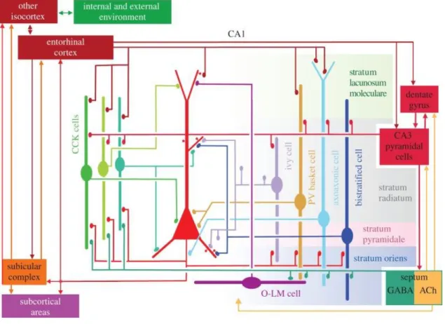
Figure 2. Spatial interaction between pyramidal neurons and several classes of interneurons in the hippocampal CA1 region, summerize the main synaptic connections of the cells. The same somatic and dendritic domains receive differentially timed input from several types of GABAergic interneuron (Somogyi et al., 2014).
The hippocampal CA1 region represent three types of CCK-containing cells (basket cell, perforant path-associated cell and Schaffer collateral-associated cell), ivy cells and PV-positive basket, axo-axonic, bistratified and O-LM interneurons. Figure 2.
shows the local projections, afferents and efferents of these neurons in the hippocamapal CA1 region.
1.4.2. Activity patterns of the different interneurons during hippocampal oscillations
Several in vivo and in vitro studies paid attention to understanding the functional properties of the pyramidal cells and different interneurons during SPW-Rs, theta and gamma oscillations and define specific firing characteristics of identified neurons (summarizes: (Somogyi et al., 2014). Different domains of the pyramidal neurons are innervated by different interneuronal subtypes (Figure 2). During theta and ripple activities cells fire with distinct patterns which are coupled to field oscillations. The different temporal dynamics could come from interneurons expressing CCK or PV that innervate parallel the soma and dendrite of principal cells.
During theta activity recorded in CA1 pyramidal layer, the firing rate of pyramidal neurons is the lowest at the peak of the theta cycle, while axo-axonic cells, CCK- and PV-positive basket cells fire the most. Basket, ivy and O-LM cells show theta phase coupling and reach the maximal probability of their firing at the trough of a theta cycle. During gamma oscillations bistratified and ivy cells show the highest firing frequencies and their spike showed strong phase coupling in CA3 area. The firing probability of PV-positive cells and bistratified interneurons is the maximal during ripple oscillations such as pyramidal cell discharge, whereas the axo-axonic cells fire only the first half of the ripple activities (Figure 3.). O-LM cells could be inhibited or activated during ripples (Varga et al., 2012).
Figure 3. Main types of interneurons and their activities during hippocampal network oscillations. Their spike timing is coupled to field gamma, ripples and theta oscillations at different degrees (Somogyi et al., 2014).
1.5.
Role of fast spiking PV-positive interneurons in SPW-R activities
As I have already mentioned, the role of FS-PV INs in SPW-Rs generation is extremely crucial. Early studies showed that the firing activity of the hippocampal FS- PV INs is strongly phase-locked to the peak of SPW-R oscillations (Klausberger et al., 2003, Bahner et al., 2011). To understand how these interneurons can integrate and convey the information even at such a high frequency range as ripple oscillations, I have to concern the basic properties of these types of neurons.
FS-PV INs are a subclass of interneurons which could be well-identified by distinct electrophysiological properties and molecular markers. These interneurons selectively express the Ca2+ binding protein, called parvalbumin (PV) which can be found in every compartment of the cell (Meyer et al., 2002). The extended thin, aspiny dendritic and axonal arbour (Figure 4A), the number of synapses and boutons, the ion channel distribution and types help to facilitate the generation of fast excitatory
postsynaptic potentials (EPSPs) on PV INs (Geiger et al., 1997). These features of FS- PV INs assist the better and faster information flow from the input of the cell to its output, to axonal boutons, which are equipped with machinery for fast transmitter release. These parameters support the fast signalling and signal transmission. These details are well-summarized in a recently published review by Peter Jonas and his colleagues (Hu et al., 2014).
1.5.1. Basic properties of hippocampal FS-PV INs
In the stratum pyramidale of CA1three types of PV-containing interneurons exist: PV basket cells, axo-axonic cells and bistratified cells. The somata of the O-LM cells are located in the stratum oriens, thus these neurons can be selectively separated often from the other three neuron types. PV as a neurochemical ‘marker’ is suitable for the post hoc identification of these cells (Celio, 1986, Eggermann and Jonas, 2012).
Furtheremore the PV gene can be used for selective genetical targeting of these cells i.e.
by enhanced green fluorescent protein (GFP) (Meyer et al., 2002) or viral vectors (Stark et al., 2014). The population of PV-containing neurons is about 2.6% of the total neuron population (24% within the interneuronal population) (Bezaire and Soltesz, 2013). PV interneurons innervate the postsynaptic cells mainly in their perisomatic region, i.e. at their somata and proximal dendrites (basket cells) or axons, axon initial segments (axo- axonic cells or chandelier cells) (Freund and Katona, 2007). The bistratified neurons innervate the proximal dendritic compartments of the pyramidal neurons. This means that the PV basket and axo-axonic cells regulate the site of the neurons where the action potentials (APs) are generated, thus adding a great impact directly to the output of the principal cells, whereas bistratified interneurons mostly have an effect on synaptic plasticity, LTP and LTD. The synaptic domain of the FS-PV INs contains P/Q type Ca2+
channels in order to shorten the synaptic delay and increase the temporal precision of transmitter release (Hefft and Jonas, 2005, Zaitsev et al., 2007).
In the stratum pyramidale of the CA regions these PV interneurons have similar electrophysilogical parameters (Figure 4B). According to Pettilla terminology (Ascoli et al., 2008), they are all fast spiking interneurons. The passive intrinsic membrane properties measured in a whole-cell current-clamp configuration show that the resting membrane potential is between -65.1 and -69.2 mV. Their input resistance is
between 31 to 73 MΩ, while their membrane time constant is between 7.7 to 18.6 ms.
The action potential (AP) half width is 364 to 527 µs; somatically injected current can evoke a maximal firing rate at 120Hz to 300 Hz, showing a low accommodation in firing (Buhl et al., 1994, Buhl et al., 1996, Lamsa et al., 2007, Ascoli et al., 2008).
These parameters defines their fast spiking phenotype.
Figure 4. Morphological and functional properties of the FS-PV INs. A: Reconstruction of a PV-containing basket cell in the CA1 hippocampal region. Soma and dendrites are shown in black whereas axon in red. SR: stratum radiatum; SP: stratum pyramidale; SO: stratum oriens.
(Soruce: (Lapray et al., 2012). B: Fast spiking AP phenotype. Long somatic current pulse evoked a high-frequency train of AP in the recorded neuron (Hu et al., 2010).
1.5.2. Relevant functions of FS-PV INs
PV-expressing interneurons play a role in feed-forward and feed-back inhibition. Feed-forward inhibition of PV neurons is triggered by excitatory inputs which arrive from the surrounding areas. Feed-forward inhibition narrows the temporal summation of excitatory postsynaptic potentials (EPSPs) and AP initiation in principal neurons (Pouille and Scanziani, 2001). GABAergic inputs originating from PV interneurons generate fast inhibitory conductance right before the AP initiation in principal neurons (Hu et al., 2014), thus regulating the activity in a great number of principal cells. Their role in feed-back inhibitions is also crucial. In the winner-takes-it- all mechanism the pyramidal cell which gets the strongest input fires earlier than the
A A A B B B
others, thus the remaining cells get inhibited (de Almeida et al., 2009). This mechanism is important for the understanding of, for instance, how grid cells respond in a well- determined place in the EC and the grid-place code conversion (Hafting et al., 2005, Leutgeb et al., 2007, de Almeida et al., 2009). The feed-back inhibition is compound of two types: the early inhibition is mediated by perisomatic interneurons while the late one is mediated by dendrite targeting interneuons (Pouille and Scanziani, 2001). Feed- forward and feed-back inhibition of perisomatic interneurons are also crucial in the local microcircuits which generate high frequency oscillations during SPWs as discussed above.
As I already mentioned, perisomatic interneurons have a crucial role in the generation, organization and synchronization of the SPW and ripple activities as well.
High frequency ripple oscillations recorded in pyramidal neurons in whole cell mode shows oscillating postsynaptic potentials IPSPs, which indicate the important role of the perisomatic inhibitory inputs in the generation of the high frequency oscillations. The excitatory current temporally preceeds the inhibitory current in the LFP recording during SPW-R acitivities (Maier et al., 2011, Schlingloff et al., 2014) indicating that the surrounding pyramidal neuronal population can stimulate the local subnetwork of perisomatic interneurons, mostly including the basket cell population (Schlingloff et al., 2014, Stark et al., 2014). The activity is not uniform in the different hippocampal layers.
Basket cells mediated inhibition to the pyramidal cells in the deep layers is stronger than those located in the superficial layers (Lee et al., 2014). Moreover, an in vivo study showed that interneurons receive excitatory inputs from diverse CA3 and CA1 pyramidal cell assemblies (Patel et al., 2013). Even tonic excitatory drive can entrain the activity of the reciprocally connected PV-positive basket cells, which then start ripple frequency range spiking activity. This activity is phase locked through reciprocal inhibition (Schlingloff et al., 2014).
1.6. Dendritic integration and role in SPW-R oscillation of fast spiking PV interneuron
1.6.1. Dendritic signal integration and dendritic Ca2+ spike
The theory of passive dendritic properties has assumed that dendritic arbour works as an antenna. In this case the signal propagation depends only on the membrane conductance, membrane capacitance, membrane resistance and the dendritic morphology (branch number, dendritic diameter) of the cells. These parameters indicate linear summation of the signal (Rall et al., 1966, Abrahamsson et al., 2012, Vervaeke et al., 2012). In this model no active ion channel contributes to the generation of signal transmission. The presence of active conductance on the dendrites, in contrast to this theory, predicts that these voltage-gated ion channels amplify the signals and thus results in sub-and supra-linear summations of the signals. The signal integration, the localization and density (Lorincz and Nusser, 2008) of the different ion channels in the dendrite effect synaptic events which may even lead in some cases to the firing of the neurons. The simultaneously arrived inputs to the dendrites can increase the local membrane potentials up to the threshold of voltage-gated ion channels. These responses are similar to action potential but emerge locally in dendrites. These local regenerative events are called dendritic spikes (Llinas et al., 1968, Golding et al., 1999, Gasparini et al., 2004). The types of dendritic spikes depend on what kind of voltage sensitive ion channel plays a role in its generation. Thus we can distinguish calcium (Llinas and Sugimori, 1980, Larkum et al., 1999, Waters et al., 2003, Larkum et al., 2009), sodium (Ariav et al., 2003, Magee and Johnston, 2005, Losonczy and Magee, 2006) and NMDA (Schiller et al., 1997, Schiller et al., 2000, Larkum et al., 2009) dependent spikes.
Dendritic Ca2+ spikes can induce LTP on pyramidal cells (Golding et al., 2002) and can affect the network activity through somatic action potential modulation (Golding et al., 1999). In vivo study demonstrated that Ca2+ spikes could modify the oscillative SPW-R network activity by local processing in hippocampal pyramidal neurons (Kamondi et al., 1998). The Ca2+ spikes can travel below the lowpass filtering threshold of the dendrites due to their slower rise and decay times. They emerge from multiple events as fast, burst-like activity which followed by a sort of synaptic events that increase the membrane potential to a threshold of the VGCC activation (Schiller et
al., 1997, Kamondi et al., 1998). In addition there is another type of Ca2+ spikes with the characteristic of longer depolarization at the dendrites which leads to Ca2+ plateau potentials that start with a short, burst like event and then keeping the dendrite at a hyperpolarized state for a longer period of time (Golding et al., 1999).
Single neuronal computation in the neuronal networks can be achieved by synaptic integration. In pyramidal cells, around 6 synchronously activated spines are needed for supralinear summation of signals (Losonczy and Magee, 2006). Similarly to the pyramidal cells’ spines, the interneurons have a well-defined functional compartmentalization of responses (Goldberg et al., 2003a, Goldberg et al., 2003b, Rozsa et al., 2004) which makes for similar integration properties possible.
1.6.2. Dendritic properties of FS-PV INs
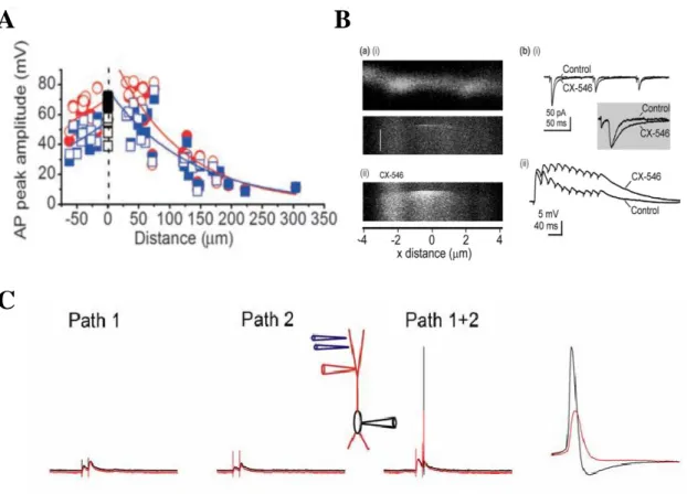
In contrast to the dendritic backpropagation AP calcium response in pyramidal cells, calcium signals in PV INs (basket and axo-axonic cells) show a high restricted spatial extent (Goldberg et al., 2003b, Aponte et al., 2008, Hu et al., 2010, Camire and Topolnik, 2014). This is in agreement with the literature, namely the dendrites of PV containing neurons are passive (Figure 5). Low densitiy of voltage gated Na+ channels (Hu et al., 2010) and high densitiy of K+ channels (Goldberg et al., 2003b) indicate high dendritic ratio of K+ to Na+ channels, which basically distinguishes their dendritic properties from other type of neurons (Stuart and Sakmann, 1994, Golding and Spruston, 1998, Martina et al., 2000, Vervaeke et al., 2012). With this ion channel content, it is understandable that dendritic spikes cannot be evoked neither by dendritic current injection nor by synaptic stimulation, at least in PV INs located in DG (Hu et al., 2010). In thin dendrites of PV interneurons AMPA receptor-mediated conductances generate EPSPs with large peak amplitude (Norenberg et al., 2010) which can reach the high activation threshold of Kv3 type of voltage-sensitive K+ channels (Rudy and McBain, 2001). These channels show fast activation and fast deactivation kinetics (Rudy and McBain, 2001). Activation of these K+ channels help in shortening the decay time of the EPSPs, shortening the time period of temporal summation and promoting AP initiation with high speed and temporal prescision (Fricker and Miles, 2000, Hu et al., 2010). K+ channels activation supports sublinear integration and makes PV cells sensitive to distributed excitatory inputs but not clustered ones (Hu et al., 2010).
Figure 5. Passive properties of FS-PV INs. A: Action potentials in basket cell dendrites show robust amplitude’ attenuation as a function of the distance, indicating passive action potential back-propagation. Positive distance, apical dendrite; negative distance, basal dendrite; both measured from the center of the soma. (Source: (Hu et al., 2010)). B: Local Ca2+ signaling and fast sublinear integration. Left: Thin, aspiny dendrite of a perisomatic interneuron with the representation of Ca2+ microdomain mediated by Ca2+-permeable AMPA receptors. CX-546 inhibits AMPA receptor deactivation, resulting prolonged Ca2+ influx and summation of EPSPs (Right). (Source: Goldberg et al. 2005). C: Stimulation of two inputs fails to evoke dendritic spikes indicating the lack of dendritic spikes in basket cells (Source: (Hu et al., 2010)).
1.6.3. Activity of FS-PV INs during physiologically relevant SPW-R oscillations
During hippocampal SPW-Rs reactivation of previously established cell assemblies correspond to synchronized population dischargies and plays a crucial role in establishing long-term memory traces in the neocortex (Girardeau et al., 2009, Buzsaki, 2010, Buzsaki and Silva, 2012, Lorenz et al., 2012). A stochastic transient increase in pyramidal cell firing is generated autonomously in CA3 evoking depolarization in pyramidal cells and interneurons in CA1, leading to the generation of
A B
C
A B
C
A B
C
network ripple oscillations (Traub and Bibbig, 2000, Buzsaki and Silva, 2012). The precisely timed input activity of CA3 neurons is nonlinearly transformed to neuronal output by the somatic and dendritic compartments of downstream neurons (Losonczy and Magee, 2006, Larkum et al., 2009, Katona et al., 2011). Voltage-gated ion channels contribute to the nonlinear dendritic processing which interacts through locally propagating and attenuating membrane potential fluctuations. Dendritic signal integration can be clustered in small (~10 µm) dendritic computational subunits (’hot- spots’) (Polsky et al., 2004, Katona et al., 2011). In addition, voltage-gated ion channels activation can induce more global signals, thus regenerative dendritic spikes are engendered when more synaptic inputs are activated in synchrony (Stuart, 1999, Schiller et al., 2000, Larkum et al., 2009).
Several facts suggest that SPW-R-associated cell assemblies can activate dendritic hot-spots, but the relationship between SPWs, field ripple oscillations and dendritic hot-spots have not yet been studied. Synchronized cell assemblies have been shown to activate dendritic hot-spots (Kleindienst et al., 2011, Makino and Malinow, 2011, Takahashi et al., 2012) during a SPW-R event up to 10% of the total neuronal population discharges in the hippocampus, making SPW-Rs the most synchronized cell assembly pattern in the entire cortex (Buzsaki and Chrobak, 1995, Buzsaki, 2010).
Moreover, synaptic inputs in dendritic hot-spots have been reported to be locally synchronized for an interval of around 60 ms (Takahashi et al., 2012), which matches the average length of individual SPW-R events (Buzsaki and Silva, 2012).
However, how and why these SPW-R-associated cell assemblies activate dendritic hot-spots and if this activation changes the dendritic computation and AP output of individual neurons, have not been investigated yet. Hippocampal FS-PV INs show higher activity rate than other types of neurons during SPW-Rs and their firing is strongly phase-locked to ripple oscillations (Klausberger et al., 2003, Bahner et al., 2011). It was shown that FS-PV INs play a crucial role in the generation of synchronized cell assembly activities, even in the SPW-R generation (Sohal et al., 2009, Buzsaki and Silva, 2012, Lapray et al., 2012, Taxidis et al., 2012, Tukker et al., 2013, Schlingloff et al., 2014, Stark et al., 2014). According to the generally accepted view, FS-PV INs act in cortical circuits as fast and, essentially, passive integrators of synaptic
inputs (Buhl et al., 1996, Pouille and Scanziani, 2004, Glickfeld and Scanziani, 2006, Hu et al., 2010).
Several papers support the passive properties of FS-PV INs: accelerated kinetics of excitatory postsynaptic potentials (EPSPs), a reduced, sub-millisecond temporal window for dendritic integration, and precise and fast coupling between EPSPs and AP outputs (Fricker and Miles, 2000, Goldberg et al., 2003b, Pouille and Scanziani, 2004, Goldberg and Yuste, 2005, Hu et al., 2010). These parameters were measured under the conditions of low network activites, when incoming synaptic activity is low. Ca2+ dynamics have been found to be fast in the aspiny dendrites of FS- PV INs and are strongly related to approximately 1 µm long, dendritic microdomains (Goldberg et al., 2003a, Goldberg and Yuste, 2005, Topolnik, 2012). According to the literature, regenerative dendritic spikes cannot be evoked in these cells and back- propagating APs are severely attenuated (Goldberg et al., 2003b, Hu et al., 2010).
However, dendritic integration and EPSP-AP coupling can be different under high- activity conditions such as SPW-Rs when neurons receive precisely timed dendritic inputs (Katona et al., 2011).
2. Aims
I. One of my main goals was to reveal active, regenerative Ca2+ events in dendrites of FS-PV INs during spontaneous SPW-R activities.
II. Second, I addressed the connection between dendritic spontaneous Ca2+ events and the associated membrane potential signals to examine the input-output transformation of FS-PV INs.
III. My third aim was to define how many inputs require to evoke regenerative Ca2+
events in the distal dendrites of FS-PV INs and to specify the nonlinear dendritic integration mechanisms in the generation of Ca2+ events and associated membrane potential signals in FS-PV INs.
IV. Finally, I clarified the types of ion channels which play a role in the generation and propagation of dendritic Ca2+ events and membrane potential signals.
3. Methods
3.1. Mouse line and slice preparation
All experiments were carried out in accordance with the Hungarian Act of Animal Care and Experimentation (1998; XXVIII, section 243/1998.). Acute brain slices were prepared from transgenic mice line where the PV-containing neurons express enhanced green fluorescens protein (eGFP) (Meyer et al., 2002).
Acute hippocampal slices were prepared from 15-30-day old animals. The animals were deeply anesthetised with isoflurane before decapitation. The brain was removed into the ice cold cutting solution, containing (in mM): 2.8 KCl, 1 MgCl2, 2 MgSO4, 1.25 NaH2PO4, 1 CaCl2, 10 D-glucose, 26 NaHCO3 and 206 sucrose. Two types of slices were prepared: 450 µm thick slices for the spontaneous SPW-R measurements and 300 µm thick for the uncaging and pharmacological experiments. Horizontal slices were cut with a Vibratome 3000 (Vibratome, Bannockburn, IL, USA). After the cutting procedures the slices were stored at an interface-type chambers at 35°C in normal artificial cerebrospinal fluid (ACSF) solution containg (in mM): 126 NaCl, 2.5 KCl, 2 CaCl2, 2 MgCl2, 1.25 NaH2PO4, 26 NaHCO3 and 10 glucose (Rozsa et al., 2004). After the preparation procedures the slices were incubated for at least 1 hour while the normal ACSF solution cooled down to room temperature.
3.2. Recording chambers
For the electrophysiological experiments two types of submerged recording chamber were used. For the recording of the spontaneous hippocampal SPW-R activities we developed a modified version of the dual superfusion recording chamber (Hajos et al., 2009) to improve two-photon imaging (Figure 6.A-C). In this type of chamber the 450 µm thick slices were laid on a nylon mesh. Both sides of the slices were oxygenated by simultaneously perfused ACSF at a higher perfusion rate (>10ml/min). The neuronal operation under this conditions might better approximate the physiological conditions, since the spontaneous activities such as SPW-R could be detected. The optical recording was optimized by changing the metal mesh into a nylon slice supporting grid (Warner Instruments; thickness: 100 µm; mesh size: 1.0 mm x 1.0
mm). The Pair of bubble traps was developed to eliminate bubbles from the perfusion tube system and to prevent to reach the recording chamber (Figure 6.D-E).
Figure 6. Schematic drawings of the optimized dual superfusion chamber and the bubble trap. A-B: Double perfusion system improved oxygenation of the 450 µm thick acute hippocampal sclices. The slices lay on a polypropylene mesh, the perfusion fluid flows on both sides of the slices. The polypropylene mesh and the glass coverslip on the bottom helped to optimize the optical recordings. C: Picture of experimental setup, during recordings. D-E:
Polycarbonate bubble trap. The upper inlet and lower outlet eliminate the bubbles and reduce the vibrations of the perfusion system.
For the pharmacological experiments a commercially available chamber was used (Luigs&Neumann, Ratingen, Germany) that was equipped with single perfusion tubing. In this system 300 µm thick slices were used where spontaneous activities were
outlet Two inlets
Upper and lower parts of the perfusion flow Cover glass glued
by silicone sealant
A
B C
Inlet
Outlet Bubble trap controller
D E
low. In this case the neuronal operation could be limited, which resulted a smaller neuronal activity.
3.3. Electrophysiology
Whole cell patch-clamp recordings were performed from FS-PV INs in the stratum pyramidale of CA1 region of the hippocampus. The LFP was recorded from the CA1 (close to the patched FS-PV IN) or the stratum pyramidale of the CA3. Both recordings were acquired by a MultiClamp 700B amplifier and digitized with a Digidata 1440 Data Acquisition System (Molecular Devices, Sunnyvale, CA, USA) and MES (Femtonics Ltd., Budapest, Hungary) software. Both patch clamp and LFP recordings were performed with 6-9 MΩ-resistance borosilicate glass electrodes. For LFP recordings, the intrapipette solution was normal ACSF. For whole cell recordings the pipette contained (in mM): 125 K-gluconate, 20 KCl, 10 HEPES, 10 Di-Tris-salt phosphocreatine, 0.3 Na-GTP, 4 Mg-ATP, 10 NaCl, and 0.008 biocytin. Patch pipettes also contained 100 µM Fluo-4 (Invitrogen, Budapest, Hungary) or 60 µM Oregon Green BAPTA-1 (OGB-1, Invitrogen), both in combination with 100 µM Alexa 594 (Invitrogen). The LFP signals were band-pass filtered offline (1Hz – 3 kHz and 100 – 300 Hz) using a built-in filtering function of the MES software package. Juxtacellular signals were recorded using glass electrodes (6-9 MΩ) filled with normal ACSF solution comparable to that obtained during LFP recordings. The electrophysiological recordings were performed at 32-34°C (in-line heater: Supertech; chamber heater: Luigs
& Neumann).
FS-PV INs were visualized using a 880 nm infrared oblique illumination and two-photon imaging (830-900 nm) (Figure 7). For the physiological and optical recordings, cells were accepted with a resting membrane potential more negative than – 50 mV. The recorded interneurons represented the typical electrophysiological properties of fast-spiking interneurons (Lamsa et al., 2007) (maximum firing frequency
= 206.66 ± 43.33 Hz; firing adaptation = 7.8 ± 1.7 %; AP amplitude = 52.6 ± 12.4 mV;
resting membrane potential = -63.9 ± 7.6 mV; n=47 cells). Backpropagating action potentials (bAPs) were induced by somatic current injections (700 pA for 5 ms; five bAPs were evoked at 40 Hz). Step depolarization was also induced by somatic current injections (1500-1700 pA for 100 ms).
Figure 7. Identification of hippocamapal FS-PV INs in the CA1 region. A: Schematic drawing of the hippocampus, black box indicates the scanned CA1 region. B: Maximum intensity two-photon image stack projection of PV-containing interneurons in the strata pyramidale and oriens. The PV cells were visualized by 900 nm two photon imaging. C:
Transmission infrared image of the stratum pyramidale. Red arrow indicates the localization of a FS-PV IN. D: Two-photon TiR and PMTgreen channel image are overlapped to show the PV content of the targeted cell. E: Maximum intensity two-photon image stack projection of the patched interneuron (PMTred). For whole cell recording, glass electrode was filled with intrapipette solution, containing Fluo-4 calcium sensitive dye (100µM) and ALEXA 594 (10µM). Inset: Enlarge image of the distant dendrites of the neuron (merged of the PMTgreen and PMTred images). F: Somatic voltage trace from the cell upon step depolarization. The recorded interneurons had the typical electrophysiological properties of fast spiking interneurons.
For the classification of the FS-PV INs, passive membrane properties (Table 1) were measured in a whole-cell current-clamp configuration (Buhl et al., 1996, Gloveli et al., 2005). No holding current was introduced during the estimation for resting membrane potential. Input resistance (Rin) and the membrane time constant (τ) were calculated from voltage responses to current injections (500 ms, 50 pA). The AP properties (e.g. half width) were measured for the first AP, evoked by an increased depolarizing current injection. To determine the AP accommodation, we increased the
CA1
100µm
B
B:
50µm 50µm
C D
100µm
20µm
E F
250ms 60mV
A
somatically-injected current until the cell reached its maximal firing frequency (~800 pA, 500 ms). The level of the accommodation was estimated by the rate of the first and last 100 ms interval firing frequencies. Five bAPs were induced with somatic current injections (500-700 pA for 5 ms, five current steps at 40 Hz). LFP signals were recorded in a ~ 50 µm distance from the measured cell’s soma of the hippocampal CA1 region. In a different set of experiments, LFP signals were recorded at the same area where the dendritic segments were activated and then the pipette was gradually moved away from the activated dendritic segment. Electrophysiological data were filtered at 3-10 kHz and sampled at 20 kHz. In some measurements, LFP signals were further filtered with a 3-5 kHz low-pass Bessel filter before baseline subtraction method (see later). Spectrograms are shown in the 0-600 Hz range (15-40 ms window side). In some cases, a sine wave was fitted to the low-pass filtered raw traces and was then subtracted in order to remove the 50 Hz noise.
3.4. Pharmacological experiments
All drugs were applied in the bath, except local TTX injection. Tetrodotoxin (TTX) (1 μM), nimodipine (20 μM), mibefradil (10 μM), ω-connotoxin MVIIC (0.5 μM), 6-cyano-2,3-dihydroxy-7-nitro-quinoxaline (CNQX) (10 μM) and D,L-2-amino-5- phosphonopentanoic acid ( DL-AP5) (60 μM) were purchased from Tocris Bioscience.
The cocktail of voltage-gated calcium channel (VGCC) blockers contained ω- connotoxin MVIIC, nimodipine, and mibefradil as well. During local puff of TTX we injected 10 µM TTX in combination with Alexa 594 using a patch pipete (6-9 MΩ).
3.5. Two-photon imaging
Two-dimensional two-photon imaging in the dendrites was performed during the glutamate-uncaging and pharmacological experiments where spontaneous neuronal network activity was low. 2D and 3D two-photon Ca2+ imaging was achieved when spontaneous SPW-R activities were occurred (Figure 8). Two-photon imaging started 15–20 min after obtaining the whole-cell configuration on a two-photon laser-scanning system (Femto2D, Femtonics Ltd., Budapest) equipped with a femtosecond laser tuned to 830 nm (Mai Tai HP, SpectraPhysics, Mountain View, CA, USA). The use of