Research Article
Maternal Smoking Highly Affects the Function, Membrane Integrity, and Rheological Properties in Fetal Red Blood Cells
Krisztina N. Dugmonits,1Payal Chakraborty,1Réka Hollandi,2Szabolcs Zahorán,1 Gabriella Pankotai-Bodó,3Péter Horváth,2Hajnalka Orvos,4and Edit Hermesz 1
1Department of Biochemistry and Molecular Biology, Faculty of Science and Informatics, University of Szeged, H-6726 Szeged, Közép fasor 52, Hungary
2Institute of Biochemistry, Biological Research Centre, Hungarian Academy of Sciences, Szeged, Hungary
3Department of Pathology, Faculty of Medicine, University of Szeged, Szeged, Hungary
4Department of Obstetrics and Gynecology, Faculty of Medicine, University of Szeged, Szeged, Hungary
Correspondence should be addressed to Edit Hermesz; hermesz@bio.u-szeged.hu
Received 6 June 2019; Revised 25 September 2019; Accepted 15 October 2019; Published 29 November 2019
Academic Editor: Juan Gambini
Copyright © 2019 Krisztina N. Dugmonits et al. This is an open access article distributed under the Creative Commons Attribution License, which permits unrestricted use, distribution, and reproduction in any medium, provided the original work is properly cited.
An understanding of the basic pathophysiological mechanisms of neonatal diseases necessitates detailed knowledge about the wide range of complications in the circulating fetal RBCs. Recent publications on adult red blood cells (RBCs) provide evidence that RBCs carry an active nitric oxide synthase (NOS3) enzyme and contribute to vascular functioning and integrity via their active nitric oxide synthesis. The aim of this study was to determine the effect of maternal smoking on the phenotypical appearance and functionality of fetal RBCs, based on morphological and molecular studies. We looked for possible links between vascular dysfunction and NOS3 expression and activation and its regulation by arginase (ARG1). Significant morphological and functional differences were found between fetal RBCs isolated from the arterial cord blood of neonates born to nonsmoking (RBC-NS, n= 62) and heavy-smoking (RBC-S, n= 51) mothers. Morphological variations were quantified by Advanced Cell Classifier, microscopy-based intelligent analysis software. To investigate the relevance of the newly suggested “erythrocrine”
function in fetal RBCs, we measured the levels of NOS3 and its phosphorylation in parallel with the level of ARG1, as one of the major influencers of NOS3 dimerization, by fluorescence-activated cell sorting. Fetal RBCs, even the “healthy-looking” biconcave-shaped type, exhibited impaired NOS3 activation in the RBC-S population, which was paralleled with elevated ARG1 level, thus suggesting an increased redox burden. Our molecular data indicate that maternal smoking can exert marked effects on the circulating fetal RBCs, which could have a consequence on the outcome of in utero development. We hypothesize that any endothelial dysfunction altering NO production/bioavailability can be sensed by circulating fetal RBCs. Hence, we are putting forward the idea that neonatal RBC could serve as a real-time sensor for not only monitoring RBC-linked anomalies but also predicting the overall status of the vascular microenvironment.
1. Introduction
There is increasing evidence that environmental agents can exert a marked impact on the outcome ofin uterodevelop- ment and even mediate long-lasting health consequences [1]. Many of the compounds present in cigarette smoke are regarded as harmful toxicants playing crucial roles in the pathogenesis of certain severe illnesses [2]. It has been hypothesized that many of the adverse effects may result
from oxidative damage to proteins, lipids, and nucleic acids.
Such damages could be traced back to direct attack of oxi- dants present in cigarette smoke and to the activation of the endogenous sources of reactive oxygen species (ROS) [3, 4].
Around 20-30% of women continue to smoke during full-term pregnancy [5]. The harmful pollutants in the vapor and tar phases might diffuse into the placenta and pass down to the fetal circulatory system through the umbilical cord.
With the exception of its most proximal segment, the human
Volume 2019, Article ID 1509798, 10 pages https://doi.org/10.1155/2019/1509798
umbilical cord lacks innervation and thereby the main regu- lator of their vascular tone and bloodflow is the nitric oxide (NO), a signaling molecule produced by the endothelial nitric oxide synthase (NOS3) from L-arginine [6, 7].
Chronic smoking jeopardizes proper endothelial func- tioning by decreasing the formation and increasing the deg- radation of NO, via generation of ROS or other strong oxidants like peroxynitrite anion (ONOO-) [8].
The crucial function of NOS3 is the bioactive NO pro- duction, which depends on its subcellular localization and on the active dimeric conformational state [6]. NOS3 is dynamically regulated by cofactors like BH4, modulating the coupling of electron to the dimeric form [9], the availabil- ity of the substrate L-arginine [10], diverse interacting pro- teins [11], and the phosphorylation at Ser1177 residue via the PI3-kinase/Akt kinase pathway [12].
RBCs play an important role in tissue oxygenation and regulating blood pressure by metabolizing large quantities of NO. Our understanding on RBCs’ biological function has been expanded by new groundbreaking data. Recently published experimental evidences make it highly supposable that adult RBCs not only are passive regulators of the endothelium-derived NO level but also actively control sys- temic NO bioavailability by synthesizing, transporting, and releasing it [13, 14]. This new“erythrocrine function”helps to maintain the vascular tone and blood flow by releasing bioavailable NO synthetized by the posttranslationally mod- ified RBC-NOS3.
The RBCs’ high nanomechanical plasticity or deform- ability allows them to be dynamically adapted to the contin- uous changing flow conditions along the vascular system.
The nanomechanical properties of RBCs are highly compro- mised by any imbalance in the redox homeostasis; thus, con- tinuous formation of oxidants is one of the important indicators of various pathological processes [15].
Fetuses are more sensitive to the toxicity of many sub- stances, simply because of their lower detoxification and anti- oxidant capacities, lower immunologic competence, and higher proliferation rate. However, the physiological and the underlying molecular effects of tobacco smoke in par- ticular on embryonic/fetal development still remain poorly understood.
We studied the consequences of an inappropriate mater- nal lifestyle, viz., smoking during pregnancy, on RBCs col- lected from umbilical cord arteries at the moment of birth.
We looked for a potential rescue mechanism/compensatory role of fetal RBCs, based on the newly described“erythro- crine function”in case of any endothelial dysfunction.
2. Materials and Methods
2.1. Human Samples.Conforming to the principles outlined in the Declaration of Helsinki and with a pregnant mother’s informed consent, blood was taken from the umbilical cord artery of neonates born to nonsmoking mothers (NSn = 62) and mothers who continued smoking habit during preg- nancy (at least 10 cigarettes per day) (Sn = 51), in the Depart- ment of Obstetrics and Gynecology at the University of Szeged, Hungary. The Ethics Committee of the Department
of Obstetrics and Gynecology approved the study protocol (16/2014).
The blood samples were centrifuged at 200gfor 10 min at 4°C, and the lower two-thirds of the RBC phase was collected [13]. The RBCs were washed twice with 2 volumes of isotonic saline solution atpH = 7:0. The purity of the RBC prepara- tion was checked by immunostaining with an RBC-specific mouse anti-glycophorin A antibody. Purity of the samples was>95% for RBCs.
2.2. Immunofluorescence Staining, Fluorescence-Activated Cell Sorting (FACS), and Microscopic Analysis. RBCs were fixed and stained as described by Chakraborty et al. [16].
Briefly, cells were fixed in 4% (w/v) paraformaldehyde in 0.05 M phosphate buffer (PB, pH = 7:4) at 4°C for 60 min.
After extensive washing with PB, they were permeabilized with 0.1% Triton X-100 for 20 min and incubated for 1 h in PB containing 1% bovine serum albumin and 10% normal goat serum to block nonspecific antibody binding. RBCs were immunolabelled with primary antibodies (single or double staining) at 4°C overnight. Incubations with the pri- mary antibodies was followed by washing and incubation with goat anti-mouse Alexa®647- and/or goat anti-rabbit Alexa®488-conjugated secondary antibodies for 1 h at room temperature (for the antibody list, see supplementary mate- rial online, Table S1).
After washing, RBCs were either processed for quantita- tive analysis (FACS, BD FACSCalibur™, BD Biosciences) [17] or were mounted in ImmunoHistoMount (Sigma- Aldrich, Saint Louis, Missouri, USA) and examined under a confocal laser-scanning microscope (Zeiss LSM 880, Axio- cam 503 mono, 40x oil immersion objective, numeric aper- ture: 1.4; Carl Zeiss Microscopy GmbH, Germany). Eight- bit pictures were taken by using ZEN 2.1 (black) software (Carl Zeiss Microscopy GmbH 1997-2015) and analyzed by using ImageJ 1.51n and ZEN 2.1.
2.3. Ex Vivo Experiments
2.3.1. Heavy Metal Treatment.Total blood of four indepen- dent individuals (NS = 4,S = 4) was subjected to Cd2+treat- ment in anex vivoexperiment. Samples were incubated with and without cadmium acetate (CdðCH3COOÞ2× 2H2O in 0.9% NaCl) solution at thefinal concentration of 0.5 ng/μL, 5 ng/μL, 10 ng/μL, 20 ng/μL, and 50 ng/μL for Cd2+[18].The incubation time was 30 minutes at 37°C. After treatment, mor- phological changes of parallel samples with and without Cd2+
were compared on eosin-stained blood smears.
2.3.2. Candida Infection. Total blood of NS = 19 and S = 9 individuals was incubated for 20 hours at 37°C in a CO2ther- mostat (5%) with and withoutCandida parapsilosis(ATCC 22019).Candida parapsilosiswas maintained on YPD plates (1% yeast extract, 2% bactopeptone, 2% glucose, and 2.5%
agar) at 4°C. Prior to the experiments, it was grown overnight in liquid YPD medium (1% yeast extract, 2% bactopeptone, and 2% glucose) at 37°C in a shaker incubator. Cells were harvested by centrifugation, washed twice with PBS (pH 7.4), and counted in a Bürker chamber, and the cell number was adjusted to 109/mL. The final concentration was 107
fungi/mL total blood. After treatment, morphological changes of the parallel samples were compared on eosin- stained blood smears.
After treatment, morphological changes of the parallel samples were compared on eosin-stained blood smears.
Eosin-stained smears’image processing was done in the scientific calculation software MATLAB. All the images went through quality control as a preprocessing step in the initial phase of the analysis, which consisted of illumination correc- tion using the MATLAB tool CIDRE [19] and exclusion of poor-quality ones (e.g., too noisy or out-of-focus images).
The intelligent analysis software Advanced Cell Classifier [20] (ACC, available on http://cellclassifier.org) was utilized to (automatically) identify and populate distinct phenotypes present in the data, then refine decision planes between them with advanced machine learning methods such as active learning and neural networks, with minimal user interaction requirement. Based on such a model trained, all cells were predicted as a given phenotype. In data analysis, the reports created in ACC provided insight into the distribution of each phenotype of interest compared to the control one, while their mere frequency was also given.
2.4. Determination of ONOO- Level. To determine the ONOO- levels, we followed the method of Chakraborty et al. [16]. Spectrophotometric measurements were per- formed at 302 nm, using a GENESYS 10S UV-Vis spectro- photometer (Thermo Fisher Scientific, Madison, WI, USA).
The total RBC fraction was hemolysed by the addition of dis- tilled water at a ratio of 1 : 9. The hemolysate of each samples was diluted in 1 M NaOH solution in a ratio of 1 : 250. The increase in absorbance was measured until it reached a stable equilibrium; then, samples were added into 100 mM PB (pH = 7:4) in the same ratio as the reference. On this neutral pH, the ONOO- decomposed, and the decrease in absor- bance was observed till the equilibrium point [21]. The ONOO- concentration was calculated as the difference in the absorbances at the two distinct pH values, according to the Lambert-Beer law (ƐONOO−= 1670 M−1cm−1). Thefinal results were normalized with protein concentration (μmol/mg protein) [22].
2.5. Statistical Analysis. All statistical analyses were calcu- lated with one-way analysis of variance (ANOVA) (Graph- Pad Statistical Software version 4.0) using the Newman- Keuls multiple comparison test. Significant differences were accepted at ∗p< 0:05, ∗∗p< 0:001, ∗∗∗p< 0:001, and ∗∗∗∗p
< 0:0001.
3. Results
3.1. Characteristic Parameters of the Neonatal Study Populations.Mother age below 18 years, gestational age less than 37 weeks, complications during delivery, previous infec- tion, inflammatory conditions or disorders such as cardio- vascular diseases, diabetes mellitus, and malformations and evidence of genetic disorders were considered exclusion fac- tors during sample collection. Significant differences were found between the NS and S study groups in the major inde- pendent characteristic parameters, such as the head and chest circumference, APGAR score at 1 min, and birthweight. The head and chest circumference values in the S group were 6- 6.5% lower than those of NS origin. The overall birthweight difference was about 16% between the two examined groups (Table 1). Major difference was found in the birthweight- based distribution at weight under 2500 g and above 3500 g.
55% of neonates born to nonsmoking mothers had birth- weight above 3500 g, while this value was only 13% in the case of neonates of smoking mothers (Suppl. Figure S1).
3.2. Increased Level of Morphological Abnormalities in RBC-S Populations.Pathological conditions affecting RBCs can lead to significant alteration in their characteristic, biconcave disc shape, and deformability. Altogether, we screened over 25000 cells of RBC-NS (n= 7956cells from 20 independent individ- uals) and RBC-S (n= 15745cells from 17 independent indi- viduals) origins for phenotypic variants, utilizing the Advanced Cell Classifier intelligent analysis software. In the RBC-S population, the echinocyte phenotype was about 15% of the total cell count, whereas in the RBC-NS group, the frequency of this phenotype was less than 2%. The fre- quency of teardrop-like RBCs showed an elevated tendency in the RBC-S population, but the increase was not significant (Figure 1(a)). To demonstrate that active cigarette smoking Table1: Parameters of the study groups.
Neonates born to nonsmoking mothers Neonates born to smoking mothers
n 62 51
Birthweight (kg) 3:4883 ± 0:4465 2:9469±0:5850∗∗∗∗
Gestational age at delivery (week) 39:295 ± 1:252 38:492 ± 2:481
Blood sample pH 7:277 ± 0:068 7:283 ± 0:080
Chest circumference (cm) 33:083 ± 1:763 31:101±2:802∗∗∗∗
Head circumference (cm) 34:65 ± 1:400 32:575±1:873∗∗∗∗
APGAR score at 1 min 9:33 ± 0:90 8:969 ± 1:189∗
Maternal age (years) 32:453 ± 5:637 28:015 ± 5:729
Total blood samples were collected from the umbilical cord arteries of neonates born to nonsmoking (NS,n= 62) and smoking (S,n= 51) mothers and analyzed according to the experimental protocols. The data are expressed asmeans ± standard deviation. Statistical significances were accepted at∗p< 0:05and∗∗∗∗p
< 0:0001by one-way ANOVA using the Newman-Keuls multiple comparison test. APGAR: appearance, pulse, grimace, activity, and respiration.
could be one of the etiological factors for the adverse out- comes in morphological changes, we induced controlled oxi- dative damage by incubating the total blood with and without Cd2+ in 0.5, 5, 10, 20, and 50 ng/μL concentration for 30 minutes at 37°C in anex vivoexperiment. In the NS popula- tion, Cd2+, at a concentration as low as 0.5 ng/μL, trans- formed 16-18% of RBCs to echinocytes. On the contrary, in the RBC-S population, we detected a modest increase (~5%) only at 20 ng/μL Cd2+concentration and above, com- pared to the appropriate control group without Cd2+
(Figure 1(b)). In parallel, we tested the effect on morpholog- ical appearance by an additional stressor, mimicking Can- dida parapsilosis infection, which was also reported to increase free radical production.Candidatreatment induced morphological changes in both RBC populations, regardless of their origin, though cells in the NS population exhibited higher sensitivity (Figure 1(c)).
3.3. Decreased NOS3 Activation Indicates Impairment in the Functionality of Cells in the RBC-S Population.To investigate the relevance of the newly suggested“erythrocrine function” in fetal RBCs, we measured the NOS3 level and its phosphor- ylation status by FACS analysis. During the evaluation of the histograms, we set an intensity value of 102as a borderline between basal and high NOS3-expressing populations.
Though the NOS3 expression in the RBC-S population was somewhat lower than that in the RBC-NS group, the differ- ence was not statistically significant. Even the frequency of the basal and high NOS3-expressing cell was very similar between the two groups (2/3 and 1/3) (Figures 2(a) and 2(b) and Suppl. Figure S2A-B). In parallel with NOS3 detection, we investigated the phosphorylation level at
Ser1177. In general, NOS3 proteins were highly phosphorylated in the RBC-NS population, while in the RBC-S group, the phosphorylation lagged behind.
Phosphorylation in the basal and high NOS3-expressing cells was about 25% and 50% of the levels detected in the matching RBC-NS populations, respectively (Figures 2(c) and 2(d) and Suppl. Figure S2A and C).
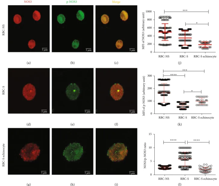
The shape of RBCs helps us to critically predict their bio- logical function. Here, we looked for possible correlation between morphological variation and functional behavior of RBCs. In parallel with FACS analysis, anti-NOS3/p-NOS3 double-labelled RBCs were subjected to ImageJ evaluation of Z-stack confocal images (Figures 3(a)–3(i)). The pheno- typic variants in the RBC-S population are affected at differ- ent levels causing impairment in NOS3 regulation. In the biconcave disc-shaped cells of RBC-S origin, there was no significant alteration detected in the NOS3 level, while the phosphorylation was about 50% of the morphologically matched RBC-NS cells (Figures 3(j) and 3(k)). The echino- cytes of S origin exhibited low NOS3 expression, about 55%
of the biconcave disc-shaped cells from either of origins (Figure 3(j)). This lowered NOS3 level was paralleled with lower phosphorylation (Figure 3(k)), and as a consequence, the NOS3/p-NOS3 ratio was comparable to that found in the biconcave disc-shaped cells with NS origin (Figure 3(l)).
3.4. Increased ARG1 Level Indicates an Altered NOS3 Activation. Adult human RBCs carry a functional ARG1 enzyme, which competes with NOS3 for their common sub- strate, L-arginine. We also could detect ARG1 in fetal RBCs.
To assess the alterations that may underlie the difference in NOS3 activation between groups RBC-NS and RBC-S, we
Frequency of morphological changes (%) RBC-NS RBC-S RBC-NS RBC-S
0 5 10 15 20 25
Echinocyte phenotype
Teardrop phenotype
⁎⁎⁎⁎
(a)
Frequency of echinocyte phenotype (%) RBC-NS RBC-NS-0.5 ng/𝜇l Cd2+ RBC-NS-20 ng/𝜇l Cd2+ RBC-S RBC-S-0.5 ng/𝜇l Cd2+ RBC-S-20 ng/𝜇l Cd2+
0 10 20
30 ⁎⁎⁎⁎
⁎⁎⁎⁎
⁎⁎⁎⁎ ⁎⁎⁎
⁎⁎⁎
(b)
Frequency of echinocyte phenotype (%) RBC-NS RBC-NS- infected RBC-S RBC-S-infected
0 20 40 60 80
⁎⁎⁎⁎⁎⁎⁎ ⁎⁎
(c)
Figure1: Distribution of phenotypic variations in RBC-NS and RBC-S populations. Frequencies of echinocyte and teardrop phenotypes in RBC-NS (n= 7956cells from 19 individual samples) and RBC-S (n= 15745cells from 45 individual samples) populations (a). Frequency of echinocytes (b), after incubation of total blood with Cd2+at 0.5 and 20 ng/μL concentration for 30 minutes at 37°C, RBC-NS (n= 7956cells) and RBC-S (n= 15745cells) investigated from 4 independent individuals in each group.Candida parapsilosisinduced morphological changes in RBC-NS and RBC-S populations (c). Total blood samples were incubated for 20 hours at 37°C at 5% CO2with and withoutCandida parapsilosis(ATCC 22019) (107fungi/1 mL). After theex vivotreatment, morphological changes were counted in RBC-NS (n= 3548cells from 19 independent samples) and RBC-S (n= 4173 cells from 9 independent samples) on eosin-stained blood smears. Phenotypic variants were analyzed by the Advanced Cell Classifier software. Statistical significances were accepted at∗p< 0:05,∗∗p< 0:01,∗∗∗p<
0:001, and∗∗∗∗p< 0:0001by one-way ANOVA using the Newman-Keuls multiple comparison test.
followed ARG1 and NOS3 coexpression, measuring the fre- quencies and intensities of ARG1- and NOS3-positive RBCs by FACS analysis. The ARG1 expression was generally low in the RBC-NS group; 98% of the cells can be characterized with low ARG1 level (Figures 4(a) and 4(b)). RBCs with S origin demonstrated a significantly higher ARG1 expression. In about 75% of the cells, we detected high ARG1 level (Figure 4(b)), and around 90% of these high ARG1- expressing cells expressed the basal level of NOS3 (Figure 4(c) and Suppl. Figure S3A-C).
3.5. Elevated ONOO- Level Indicates an Increase in Free Radical Production.In case of impaired activation, NOS3 is unable to produce bioavailable NO but increases the produc- tion of superoxide radicals leading to the imbalance of the redox homeostasis. Cells of RBC-S origin had a significantly increased level of ONOO-(~1.5-fold) (Figure 5).
4. Discussion
Sustained maternal smoking had been escalated to a world- wide health issue, with insufficient oxygen supply that can induce marked effects on thein utero development [5, 23,
24]. Smoking-induced vascular endothelial dysfunction highlights the circulating RBCs as vital sensor entities, in the purview to operate any compensatory mechanism [14, 25, 26]. This project was based on the expectation that the status of the circulating fetal RBCs will be reflecting the con- sequences on a long-term exposure to the harmful substances that originated from an improper maternal lifestyle and remained unfiltered by the placental barrier.
Our results indicated that RBCs of the developing fetus born to a smoking mother undergo morphological and molecular alterations/aberration. We demonstrated that fetal RBCs carry functional NOS3, and during long-termin vivo exposure to harmful stimuli, the NOS3 level and its activa- tion pathways are altered in a morphology-dependent man- ner. Moreover, ARG1 level is elevated in fetal RBCs of smoking origin and high ARG1-expressing cells show low NOS3 level. We also demonstrated that cells in the RBC-S population become a source of oxidizing agents, carrying the possibility of further inactivation of the NOS3 pathway by induced ARG1 level.
The phenotypic variations in the RBC-S population, even the characteristic biconcave disc-shaped cells, can be charac- terized with impaired NOS3 activation. One of the critical
Frequency of basal NOS3-expressing cell populations RBC-NS
RBC-S
100 80 60 40 20 0
(a)
Frequency of high NOS3-expressing cell populations RBC-NS
RBC-S
50 40 30 20 10 0
(b)
RBC-NS RBC-S
150 100
50 0
MFI of p-NOS3 within basal NOS3 expressing cell populations
(arbitrary unit)
⁎⁎⁎⁎
(c)
MFI of p-NOS3 within high NOS3- expressing cell populations
(arbitrary unit) RBC-NS
RBC-S
250 200 150 100 50
0
⁎⁎
(d)
Figure2: Quantification of the immunolabelled NOS3 from RBC-NS and RBC-S populations. Graphical representation of the frequency of basal (a) and high (b) NOS3-expressing cell populations in RBC-NS (n= 62independent clinical subjects) and RBC-S (n= 51independent clinical subjects), quantified by FACs analysis. Summary of NOS3 phosphorylation level (pNOS3) within basal (c) and high (d) NOS3- expressing cells. Statistical significances were accepted at∗∗p< 0:01and∗∗∗∗p< 0:0001by one-way ANOVA using the Newman-Keuls multiple comparison test. MFI: meanfluorescence intensity.
influential steps in NOS3 activation is the phosphorylation of the Ser1177 residue via phosphatidylinositol-3 kinase and the downstream serine/threonine protein kinase Akt pathway [12]. In the RBC-S populations, the bulk of the healthy- looking biconcave disc-shaped cells showed a significantly lower level of phosphorylation, compared to their morpho- logical match in the control population. Contrarily, in the increased echinocyte population, probably the phosphoryla- tion pathway was unaltered but the NOS3 level was signifi- cantly lowered. This kind of alteration could be explained
by either an alteredde novobiosynthesis or an enhanced rate of NOS3 monomerization, accompanied by NOS3 degrada- tion [27].
In clinical practice, phenotypic variants of RBCs serve as a preliminary platform for many clinical diagnoses [28–32].
However, recognizing and understanding the molecular alter- ations in RBCs preceding the detectable phenotypic changes would allow us to recognize and intervene into the possible complications at earlier stages. In this study, we provided evi- dence that RBCs could be functionally impaired before their
5 𝜇m NOS3
RBC-NS
5 𝜇m
p-NOS3 Merge
5 𝜇m
5 𝜇m
RBC-S
5 𝜇m 5 𝜇m
5 𝜇m
RBC-S echinocyte
5 𝜇m 5 𝜇m
(a) (b) (c) (j)
0 200 400 600 800 1000
RBC-S echinocyte RBC-S
RBC-NS
MFI of NOS3 (arbitrary unit)
(d) (e) (f) (k)
(g) (h) (i) (l)
0 100 200 300
RBC-S echinocyte RBC-S
RBC-NS
MFI of p-NOS3 (arbitrary unit)
⁎⁎⁎
⁎⁎⁎⁎
⁎
0 5 10 15
NOS3/p-NOS3 ratio
RBC-S echinocyte RBC-S
RBC-NS
⁎⁎⁎⁎ ⁎⁎⁎⁎
⁎⁎⁎
⁎
Figure3: Visualization of the confocal images with varying morphology in relation to the phosphorylation of NOS3 in RBC-NS and RBC-S populations. The representative confocalZ-stack images show RBC-NS (a–c) and RBC-S (d–f) with regular biconcave and RBC-S echinocyte (g–i) with the echinocyte phenotypes. The panels (a, d, and g) show the RBCs immunolabelled with a mouse primary anti-NOS3 antibody followed by an Alexa Fluor® 647 secondary antibody. The panels (b, e, and h) represent the RBCs immunolabelled with a rabbit anti-p- NOS3 (p-NOS3) primary antibody, followed by an Alexa Fluor® 488 secondary antibody. The microscopic parameter, i.e., the thickness of z-axis, was 3.845μm/slice, and the scale bar of 5μm was kept constant at all instances. The RBCs were randomly selected from both RBC- NS (n= 40-45 regular biconcave-shaped cells from each of 3 independent samples) and RBC-S (n= 30-35 regular biconcave cells from each of 5 independent samples andn= 10echinocytes from each of 4 independent samples) groups. In the highlighted zone of interest, zooming ratios were 4.486, 6.865, and 10.019 in (a)–(c), (d)–(f), and (g)–(i), respectively. The ImageJ 1.51n software was used to quantify the mean intensity levels from three middle slices in the RBC-NS, RBC-S, and RBC-S echinocyte for NOS3 (j) and p-NOS3 (k). (l)The ratio between NOS3 and p-NOS3 intensity levels. All statistical analyses were accepted by one-way ANOVA using the Newman-Keuls multiple comparison test at∗p< 0:05,∗∗∗p< 0:001, and∗∗∗∗p< 0:0001. MFI: meanfluorescence intensity.
detectable morphological alterations. Phenotypical variants are connected to changes in the cytoskeletal network. We have no supporting data about the timing and extent of these changes, which marks as the limitation to our conclusion.
However, the changes in NOS3/p-NOS3 could serve as an early marker alerting for possible risk in later development.
Additional evidence also supports our conclusion that before the appearance of obvious phenotypic variants, RBCs are already influenced in their functional capacity by mater- nal smoking. Theex vivotreatment of RBCs with the oxi- dizing agent cadmium indicated that cells in the RBC-S population developed resistance against metal or metal- induced oxidative insults, regardless of the phenotypic appearance. There were almost two orders of magnitude dif- ference in Cd2+concentration necessary to induce the echi- nocyte phenotype between the NS and S populations. On the other hand, we did not observe significant protection against the fungal infection, with known harmful outcome on RBC’s membrane [33]; it led to an increased number of echinocytes in both the NS and S populations.
Another important regulator molecule in the NOS3- NO production pathway is the ARG1 enzyme. It meta-
bolizes L-arginine to urea and L-ornithine. Enhanced ARG level or activity has been implicated in many diseases including cardiovascular dysfunction [34–36], partly because the excessive production of ornithine may be involved in the formation of vascular structural problems [37]. On the other hand, the competition between NOS3 and ARG1 for their common substrate L-arginine could lead to the impairment of NO production [38], because the maximal catalytic rate of ARG1 is at least 1000-fold of NOS3 [39]. Recent studies provided novel insights into oxidative stress increasing ARG1 expression in endothelial cells [40–42], and the upregulation of ARG impairs endothelium-dependent NO-mediated dilatation [40]. Con- tinuously growing data indicate that RBCs themselves are able to contribute to vascular functioning and integrity; a NOS3- derived NO export from RBCs mediates protection against vascular injury. This protective effect of RBC-NOS3 can be tightly regulated by ARG1 in an acute oxidative stress con- dition with excessive overproduction of ROS [25, 43]. Under a pathological condition, an intimate crosstalk between dysfunctional endothelial cells and the circulating RBCs was recently reported where RBCs originated from patients with type 2 diabetes-induced vascular endothelial dysfunction in healthy rat aorta via upregulated vascular ARG1. This induced dysfunctionality can be prevented by inhibition of ARG activity and/or increased ROS production [25].
We could detect ARG1 in fetal RBCs and demonstrated that ARG1 is upregulated in the RBC-S populations. It is well known that pregnancy itself is associated with an enhanced metabolism and demand for O2, which may lead to the over- production of ROS and the development of oxidative stress.
Maternal smoking during pregnancy leads to an extra source of ROS because the unfiltered prooxidants/oxidants could be a direct inducer of ARG1 in the RBC-S population. Another possibility for ARG upregulation is an elevated intrinsic ROS source from reduction of BH4 cofactor to BH2. Under this condition, NOS3 uncoupling may occur, resulting in the for- mation of superoxide anion (O2⋅-) instead of NO [10]. The O2⋅-itself and the highly cytotoxic oxidant ONOO-, formed in a spontaneous reaction of O2⋅-with NO [8] are the poten- tial inducers of ARG1 [40].
Mean of ARG1 intensity
0 20 40 60
80 ⁎⁎⁎⁎
RBC-NS RBC-S
(a)
Frequency of low and high ARG1 (%) 0 20 40 60 80 100
Low High
⁎⁎⁎⁎
⁎⁎⁎⁎
RBC-NS RBC-S RBC-NS RBC-S (b)
Frequency of NOS3 within high ARG1-expressing cells (%) 0 20 40 60 80 100
Basal NOS3
High NOS3
⁎⁎⁎⁎
RBC-S RBC-S
(c)
Figure 4: Quantification of the immunolabelled ARG1 cells by FACS analysis from RBC-NS- and RBC-S-derived samples. Graphical representation of the intensity (a) and frequency (b) of ARG1 expression level based on FACS analysis of cell populations, originated from RBC-NS (n= 10independent clinical subjects) and RBC-S (n= 15 independent clinical subjects) samples. (c) The NOS3 intensity level within the high ARG1-expressing RBC-S population. Statistical significances were accepted at∗∗∗∗p< 0:0001by one-way ANOVA using the Newman-Keuls multiple comparison test.
ONOO− concentration (𝜇mol/mg protein) 0.0 0.2 0.4 0.6 0.8 1.0
RBC-S RBC-NS
⁎⁎⁎
Figure5: Spectrophotometric measurements of the strong oxidant in RBC-NS and RBC-S populations. Peroxynitrite (ONOO-) levels were measured in RBCs from the umbilical cord arteries of independent clinical subjects in each group with NS and S origin (n= 10for RBC-NS andn= 15for RBC-S). Statistical significance was accepted at ∗∗∗p< 0:001 by one-way ANOVA using the Newman-Keuls multiple comparison test.
Recent publications already demonstrated in the adult system that ARG plays an essential role in the regulation of the complex mechanism of NOS3 uncoupling under various pathological conditions [42] and that NOS3 uncoupling forms a key step in the progression of major vascular dysfunc- tion [44]. Cortese-Krott and Kelm published decreased RBC- NOS3 expression and activity in correlation with endothelial dysfunction in case of coronary artery disease [14]. Based on our data, it is most likely that fetal RBCs also lose their NOS3 activity under certain pathological circumstances.
5. Conclusion
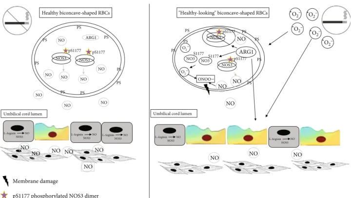
In this work, we demonstrated that RBCs from the smoker origin become a source of reactive oxygen and nitrogen spe- cies and lose their characteristic structural and functional features. Assuming an intimate crosstalk between the vascu- lar endothelium and the circulating RBCs, any endothelial dysfunction altering NO production/bioavailability can be sensed by fetal RBCs acting as a biosensor; however, in the RBC-S population, the NOS3-NO production is most likely unavailable as a compensatory mechanism to improve the blood flow to the fetus. Moreover, the changed protein expression profile might even augment and synergizes the prognosis of vascular dysfunction/comorbidities (for a graphical summary, see Figure 6).
Data Availability
The data used to support thefindings of this study are avail- able from the corresponding author upon request.
Conflicts of Interest
The authors declare that they have no conflicts of interest.
Acknowledgments
The authors would like to thank Zoltán Villányi, Atanu Chakraborty, and Miltu Kumar Ghosh for their useful com- ments on the manuscript. This work was supported by the European Union and the Hungarian Government in the framework of the GINOP-2.3.2-15-2016-00035 project.
P.H. and R.H. acknowledge the support from the Lendület BIOMAG grant (2018-342).
Supplementary Materials
Table 1. List of the sources and dilutions of primary and sec- ondary antibodies. Figure S1. Birthweight-based distribution of neonates with nonsmoking and smoking origins. Figure S2. Quantification of the immunolabelled NOS3 and p- NOS3 by FACS analysis from RBC-NS and RBC-S popula- tions. Figure S3. Quantification of the immunolabelled ARG1 by FACS analysis from RBC-NS- and RBC-S-derived samples.(Supplementary Materials)
References
[1] I. Berlin and C. Oncken,“Maternal smoking during pregnancy and negative health outcomes in the offspring,” Nicotine &
Tobacco Research, vol. 20, no. 6, pp. 663-664, 2018.
[2] C. J. Smith and C. Hansch, “The relative toxicity of com- pounds in mainstream cigarette smoke condensate,” Food and Chemical Toxicology, vol. 38, no. 7, pp. 637–646, 2000.
NO
NO NO
NO
.
Umbilical cord lumen
NO NO NO
NO NO NO NO
NO
NO
L-Arginin NO
NOS3 L-Arginin NO
NOS3
Umbilical cord lumen NO NO
NO
PS PS
PS
PS
PS PS
PS
PS
ARG1
NOS3 pS1177
NOS3 pS1177
NO NO NO
NO
Membrane damage
pS1177 phosphorylated NOS3 dimer
PS
PS
PS PS PS
PS
PS
ARG1
NO3 S1177 NO3
S1177
NOS3 pS1177 .O2–
O2– .
ONOO–
NOS3 pS1177
.
. .
.
L-Arginin NO NOS3
NO
L-Arginin NO
NOS3 L-Arginin NO
NOS3
Healthy biconcave-shaped RBCs "Healthy-looking" biconcave-shaped RBCs
O2– O2– O2– O2– O2–
Figure6: Graphical summary.
[3] K. H. Al-Gubory, P. A. Fowler, and C. Garrel,“The roles of cellular reactive oxygen species, oxidative stress and antiox- idants in pregnancy outcomes,”The International Journal of Biochemistry & Cell Biology, vol. 42, no. 10, pp. 1634–1650, 2010.
[4] M. Chelchowska, J. Ambroszkiewicz, K. Jablonka-Salach et al.,“Tobacco smoke exposure during pregnancy increases maternal blood lead levels affecting neonate birth weight,”
Biological Trace Element Research, vol. 155, no. 2, pp. 169–175, 2013.
[5] A. Hackshaw, C. Rodeck, and S. Boniface,“Maternal smoking in pregnancy and birth defects: a systematic review based on 173 687 malformed cases and 11.7 million controls,”Human Reproduction Update, vol. 17, no. 5, pp. 589–604, 2011.
[6] G. H. Oliveira-Paula, R. Lacchini, and J. E. Tanus-Santos,
“Endothelial nitric oxide synthase: from biochemistry and gene structure to clinical implications of NOS3 polymor- phisms,”Gene, vol. 575, 2 Part 3, pp. 584–599, 2016.
[7] A. Mauro, M. Buscemi, S. Provenzano, and A. Gerbino,
“Human umbilical cord expresses several vasoactive peptides involved in the local regulation of vascular tone: protein and gene expression of orphanin, oxytocin, ANP, eNOS and iNOS,” Folia Histochemica et Cytobiologica, vol. 49, no. 2, pp. 211–218, 2011.
[8] R. Radi, G. Peluffo, M. N. Alvarez, M. Naviliat, and A. Cayota,
“Unraveling peroxynitrite formation in biological systems,” Free Radical Biology & Medicine, vol. 30, no. 5, pp. 463–488, 2001.
[9] J. K. Bendall, N. J. Alp, N. Warrick et al.,“Stoichiometric rela- tionships between endothelial tetrahydrobiopterin, endothelial NO Synthase (eNOS) activity, and eNOS coupling in Vivo,” Circulation Research, vol. 97, no. 9, pp. 864–871, 2005.
[10] U. Förstermann and W. C. Sessa,“Nitric oxide synthases: reg- ulation and function,”European Heart Journal, vol. 33, no. 7, pp. 829–837, 2012.
[11] B. C. Kone, “Protein-protein interactions controlling nitric oxide synthases,” Acta Physiologica Scandinavica, vol. 168, no. 1, pp. 27–31, 2000.
[12] S. Dimmeler, I. Fleming, B. Fisslthaler, C. Hermann, R. Busse, and A. M. Zeiher,“Activation of nitric oxide synthase in endo- thelial cells by Akt-dependent phosphorylation,” Nature, vol. 399, no. 6736, pp. 601–605, 1999.
[13] M. M. Cortese-Krott, A. Rodriguez-Mateos, R. Sansone et al.,
“Human red blood cells at work: identification and visualiza- tion of erythrocytic eNOS activity in health and disease,” Blood, vol. 120, no. 20, pp. 4229–4237, 2012.
[14] M. M. Cortese-Krott and M. Kelm,“Endothelial nitric oxide synthase in red blood cells: key to a new erythrocrine func- tion?,”Redox Biology, vol. 2, pp. 251–258, 2014.
[15] M. Girasole, S. Dinarelli, and G. Boumis,“Structure and func- tion in native and pathological erythrocytes: a quantitative view from the nanoscale,”Micron, vol. 43, no. 12, pp. 1273– 1286, 2012.
[16] P. Chakraborty, K. N. Dugmonits, A. G. Végh et al.,“Failure in the compensatory mechanism in red blood cells due to sus- tained smoking during pregnancy,”Chemico-Biological Inter- actions, vol. 313, article 108821, 2019.
[17] O. Mizrahi, E. Ish Shalom, M. Baniyash, and Y. Klieger,
“Quantitativeflow cytometry: concerns and recommendations in clinic and research,”Cytometry Part B: Clinical Cytometry, vol. 94, no. 2, pp. 211–218, 2018.
[18] M. Witeska, E. Kondera, and K. Szczygielska,“The effects of cadmium on common carp erythrocyte morphology,”Polish Journal of Environmental Studies, vol. 20, pp. 783–788, 2011.
[19] K. Smith, Y. Li, F. Piccinini et al.,“CIDRE: an illumination- correction method for optical microscopy,”Nature Methods, vol. 12, no. 5, pp. 404–406, 2015.
[20] P. Horvath, T. Wild, U. Kutay, and G. Csucs,“Machine learn- ing improves the precision and robustness of high-content screens: using nonlinear multiparametric methods to analyze screening results,”Journal of Biomolecular Screening, vol. 16, no. 9, pp. 1059–1067, 2011.
[21] R. E. Huie and S. Padmaja,“The reaction of no with superox- ide,”Free Radical Research Communications, vol. 18, no. 4, pp. 195–199, 1993.
[22] O. H. Lowry, N. Rosebrough, A. Farr, and R. Randall,“Protein measurement with the Folin phenol reagent,”Journal of Bio- logical Chemistry, vol. 193, no. 1, pp. 265–275, 1951.
[23] M. Chełchowska, J. Ambroszkiewicz, J. Gajewska et al.,“Influ- ence of active exposure to tobacco smoke on nitric oxide status of pregnant women,”International Journal of Environmental Research and Public Health, vol. 15, no. 12, p. 2719, 2018.
[24] J. A. Courtney, J. F. Cnota, and H. N. Jones,“The role of abnor- mal placentation in congenital heart disease; cause, correlate, or consequence?,”Frontiers in Physiology, vol. 9, 2018.
[25] Z. Zhou, A. Mahdi, Y. Tratsiakovich et al.,“Erythrocytes from patients with type 2 diabetes induce endothelial dysfunction via arginase I,”Journal of the American College of Cardiology, vol. 72, no. 7, pp. 769–780, 2018.
[26] J. Pernow, A. Mahdi, J. Yang, and Z. Zhou,“Red blood cell dys- function: a new player in cardiovascular disease,”Cardiovascu- lar Research, vol. 115, no. 11, pp. 1596–1605, 2019.
[27] W. Chen, H. Xiao, A. N. Rizzo, W. Zhang, Y. Mai, and M. Ye,
“Endothelial nitric oxide synthase dimerization is regulated by heat shock protein 90 rather than by phosphorylation,”PLoS One, vol. 9, no. 8, article e105479, 2014.
[28] V. Kuhn, L. Diederich, T. C. S. Keller IV et al.,“Red blood cell function and dysfunction: redox regulation, nitric oxide metabolism, anemia,” Antioxidants & Redox Signaling, vol. 26, no. 13, pp. 718–742, 2017.
[29] E. M. Strohm, E. S. L. Berndl, and M. C. Kolios,“Probing red blood cell morphology using high-frequency photoacoustics,” Biophysical Journal, vol. 105, no. 1, pp. 59–67, 2013.
[30] J. Jung, L. E. Matemba, K. Lee et al.,“Optical characterization of red blood cells from individuals with sickle cell trait and dis- ease in Tanzania using quantitative phase imaging,”Scientific Reports, vol. 6, no. 1, article 31698, 2016.
[31] R. L. Harris, G. L. Cottam, J. M. Johnston, and C. R. Baxter,
“The pathogenesis of abnormal erythrocyte morphology in burns,”The Journal of Trauma: Injury, Infection, and Critical Care, vol. 21, no. 1, pp. 13–21, 1981.
[32] A. Sinha, T. T. T. Chu, M. Dao, and R. Chandramohanadas,
“Single-cell evaluation of red blood cell bio-mechanical and nano-structural alterations upon chemically induced oxidative stress,”Scientific Reports, vol. 5, article 9768, 2015.
[33] M. C. Furlaneto, D. Favero, E. J. G. França, and L. Furlaneto- Maia, “Effects of human blood red cells on the haemolytic capability of clinical isolates of Candida tropicalis,” Journal of Biomedical Science, vol. 22, no. 1, pp. 1–8, 2015.
[34] T. Hein, C. Zhang, W. Wang, C. Chang, N. Thengchaisri, and L. Kuo,“Ischemia-reperfusion selectively impairs nitric oxide- mediated dilation in coronary arterioles: counteracting role of
arginase,”The FASEB Journal, vol. 17, no. 15, pp. 2328–2330, 2003.
[35] D. E. Berkowitz, R. White, D. Li et al.,“Arginase reciprocally regulates nitric oxide synthase activity and contributes to endothelial dysfunction in aging blood vessels,”Circulation, vol. 108, no. 16, pp. 2000–2006, 2003.
[36] R. B. Caldwell, H. A. Toque, S. P. Narayanan, and R. W. Cald- well, “Arginase: an old enzyme with new tricks,” Trends in Pharmacological Sciences, vol. 36, no. 6, pp. 395–405, 2015.
[37] W. Durante, “Role of arginase in vessel wall remodeling,” Frontiers in Immunology, vol. 4, article 111, 2013.
[38] C. Moali, J. L. Boucher, M. A. Sari, D. J. Stuehr, and D. Mansuy,“Substrate specificity of NO synthases: detailed comparison of L-arginine, homo-L-arginine, their Nω- hydroxy derivatives, and Nω-hydroxynor-L-arginine,” Bio- chemistry, vol. 37, no. 29, pp. 10453–10460, 1998.
[39] R. W. Caldwell, P. C. Rodriguez, H. A. Toque, S. P. Narayanan, and R. B. Caldwell,“Arginase: a multifaceted enzyme impor- tant in health and disease,” Physiological Reviews, vol. 98, no. 2, pp. 641–665, 2018.
[40] N. Thengchaisri, T. W. Hein, W. Wang et al.,“Upregulation of arginase by H2O2 impairs endothelium-dependent nitric oxide-mediated dilation of coronary arterioles,”Arteriosclero- sis, Thrombosis, and Vascular Biology, vol. 26, no. 9, pp. 2035–2042, 2006.
[41] C. Zhang, T. W. Hein, W. Wang et al.,“Upregulation of vascu- lar arginase in hypertension decreases nitric oxide-mediated dilation of coronary arterioles,”Hypertension, vol. 44, no. 6, pp. 935–943, 2004.
[42] Z. Yang and X. F. Ming,“Arginase: the emerging therapeutic target for vascular oxidative stress and inflammation,”Fron- tiers in Immunology, vol. 4, p. 149, 2013.
[43] J. Pernow and C. Jung,“Arginase as a potential target in the treatment of cardiovascular disease: reversal of arginine steal?,” Cardiovascular Research, vol. 98, no. 3, pp. 334–343, 2013.
[44] R. Kietadisorn, R. P. Juni, and A. L. Moens,“Tackling endothe- lial dysfunction by modulating NOS uncoupling: new insights into its pathogenesis and therapeutic possibilities,”American Journal of Physiology-Endocrinology and Metabolism, vol. 302, no. 5, pp. E481–E495, 2012.