V. 2. THE ROLE OF SH GROUPS IN THE INTERACTION OF MYOSIN WITH PHOSPHATE COMPOUNDS AND WITH ACTIN *
J . Gergelyt/ A. Martonosi, and M. A. Gouvea
Cardiac Biochemistry Research Laboratory, Departments of Medicine, Massachusetts General Hospital and Harvard Medical School, Boston, Massachusetts
I. Introduction 297 II. Experimental 298
1. Materials 298 a. Proteins 298 6. Commercially Obtained Substances 299
c. Isotopic P P 299 2. Methods 299
a. Pyrophosphate Equilibrium Dialysis 299 b. Counting of P P82 and Calculation of Bound PP 300
c. Light Scattering Measurements 300 d. Miscellaneous Determinations 300
3. Results 301 a. P P Binding to Various Proteins 301
b. Light Scattering Studies on Actomyosin 307
III. Discussion 311 IV. Summary 313
I. Introduction^
It is generally accepted that SH groups play an important role in the ATPase activity of myosin, in the interaction between actin and myosin, and in the interaction of nucleotides and other compounds with actomyosin
(1). It has been postulated by Bailey and Perry (2) that the binding of
* Supported by grants from the National Heart Institute, U. S. Public Health Service (H-1066-C6), the Muscular Dystrophy Associations of America, and the Life Insurance Medical Research Fund.
t This work was carried out during the tenure of an Established Investigatorship of the American Heart Association.
t The following abbreviations will be used in this paper : ATP, adenosinetriphos- phate; ADP, adenosinediphosphate ; AMP, adenosinemonophosphate ; I D P , inosine- diphosphate; PP, inorganic pyrophosphate; PCMB, p-chloromercuribenzoate ; N E M , iV-ethylmaleimide ; EDTA, ethylenediaminetetraacetate ; D N P , 2,4-dinitrophenol ; CSH, cysteine; AM, actomyosin; O.D., optical density; P i8 2, inorganic orthophos- phate labeled with P8 2; PP8 2, inorganic pyrophosphate labeled with P3 2; Pt, inorganic orthophosphate; c.p.m., counts per minute.
297
298 J . GERGELY, A. MART0N0SI, M. A. GOUVEA
A T P to myosin involves the direct participation of an SH group. N o direct evidence, however, has been forthcoming for this point of view. Bradley and Kielley (3) have recently shown that the blocking of some SH groups in myosin leads to an activation of the ATPase in the presence of C a+ +. Studies with ATP itself are complicated by the fact that it is both bound to and hydrolyzed by myosin. In view of the fact that P P affects the AM system in many respects similarly to A T P and is also known to influence the ATPase activity of myosin (4-6) it seemed of interest to study its binding to myosin, in the presence and absence of SH reagents, and to correlate these findings with the effect of P P and nucleotides on the physi- cal state of AM. Studies of this type may open up further possibilities for obtaining information on the nature of the active center in myosin.
II. Experimental
1. MATERIALS
a. Proteins
Myosin (crystalline) was prepared essentially according to Szent- Györgyi (7) as described earlier (8). Actin was extracted according to Feuer et al. (9) and F-actin purified by ultracentrifugation according to Mommaerts (10). F-actin was depolymerized by dialysis against an as- corbic acid-ATP solution (40 μg. per ml. of each) and repolymerized in 0.1 M KCl and 0.001 M MgCl2, after removal of insoluble material by centrifugation. It was found important to insure thorough internal mixing during the dialysis in order to achieve rapid depolymerization and avoid denaturation in a drawn out process. The procedure was repeated once more.
Dog heart myosin was extracted for 30 minutes with a solution con- taining 0.3 M KCl, 0.2 M potassium phosphate buffer, pH 6, and .02 M potassium PP, with the pH adjusted to 6.2. The rest of the procedure is essentially identical with the one used for the preparation of skeletal myosin. Tryptic and chymotryptic meromyosins (8, 11) were prepared as described earlier with the modification of using 3-minute digestion times for the isolation of the heavy fractions (12). Natural AM (myosin-B) was prepared by extraction with the Weber-Edsall solution for 16 hours, with occasional stirring, at 2°. Two precipitation steps followed; the pre- cipitates were in each case dissolved in 0.6 M KCl, pH 7, and the solutions filtered through eight layers of cheese cloth. The redissolved second pre- cipitate was centrifuged for one hour at 35,000 X g in the presence of 1 0 ~3M PP. The addition of P P reduces the viscosity and facilitates the removal of particulate impurities still present. The supernatant is limpid
INTERACTION OF MYOSIN WITH PHOSPHATE COMPOUNDS 299
and shows opalescence. In order to remove the P P two precipitations fol- low at 0.1 and 0.3 M KCl, respectively.
b. Commercially Obtained Substances
Crystalline N a - A T P , the N a salt of A D P , and A M P (free acid) were obtained from Pabst Laboratories. The A D P preparation contained less than 0.1% ATP. Adenosine was obtained from Nutritional Biochemicals Corp., I D P and crystalline P C M B from Sigma Chem. Corp. and Salyrgan from Winthrop-Stearns, Inc.
c. Isotonic PP
P P3 2 was prepared by heating 100 /mioles of K2H P 04 in the presence of essentially carrier free P3 2, total activity 2-5 m c , obtained from the Union Carbide Nuclear Co., in a platinum crucible, for five minutes in an 02-flame. The red hot crucible was placed immediately afterward into a desiccator containing CaCU. The dry material was taken up in 5 ml. of water and chromatographed on Dowex 1-formate (13). Two radioactive peaks were obtained, the first containing P i3 2, the second containing the P P3 2 (about 1% of the total activity appeared as Pi). Pi determinations were carried out on the effluent fractions both directly and after seven minute hydrolysis in 1 Ν HCl. The P P3 2 containing fraction was lyophi- lized and the ammonium formate removed by heating with an infrared lamp as described by Hurlbert et al. (13).
2. METHODS
a. PP Equilibrium Dialysis
2.5 ml. of the protein solution were placed in an %2 in- dialysis bag, previously thoroughly washed with deionized, glass-distilled water. Pre- liminary experiments showed that there was no binding of P P to the tubing, and it was thus found unnecessary to use specially treated (boiled in hot water) tubing. In a few pilot experiments it was also found that the use of boiled tubing did in no way affect the experimental results. The filled dialysis bags containing the protein were placed in test tubes con- taining P P and P P3 2. The tubes were closed with rubber stoppers, and placed in a test tube rack, which was attached to a rocking apparatus in the cold room at 2°. The results of preliminary experiments showed that after 24 hours of dialysis no further concentration changes occurred. In most of the experiments here reported the dialysis time was 24 hours. The volume of the outer liquid was 5 ml. At the end of the dialysis period ali- quote were taken both from the liquid inside and the liquid outside the dialysis bag, and the radioactivity was determined.
300 J . g e r g e l y , a. m a r t o n o s i , m . a. g o u v e a
b. Counting of P P3 2 and Calculation of Bound PP
The amounts of bound P P were determined on the basis of differences between the counts in the inside and outside fluids.* Samples taken from both were placed in stainless steel cups, dried, and counted with an end window Geiger tube.
Let: V0 be the volume of the liquid outside the dialysis bag in liters, Vi be the volume of the liquid inside the dialysis bag in liters, N* be the specific activity of P P3 2, c.p.m. per mole,
C0 be the outside concentration of PP, ikf/liter, C i be the inside concentration of PP, M/liter,
N0 be the number of counts due to P P3 2 outside, in c.p.m., N i be the number of counts due to P P3 2 inside, in c.p.m., r be the number of moles of P P bound to one mole of protein, Ρ be the total number of moles of protein.
At equilibrium
Co r Co d r
c. Light Scattering Measurements
Light scattering measurements were carried out in a Brice-Speiser type apparatus (Phoenix Instrument Co.). The samples were placed in a cylin- drical cell of circular cross-section and the measurements were taken at 90° with a Brown Recorder attached to the photomultiplier circuit.
d. Miscellaneous Determinations
Protein concentrations were determined with a standard micro Kjeldahl procedure and the factor 6.2 was used for converting Ν values into protein.
pH was determined in a Beckman model G-instrument with a glass elec-
* I n view of the high concentration of KCl present, and considering the net charge on myosin under the experimental conditions, the Donnan effect would have been less than 0.1%.
d - r(P), and Ci - a
(P) No VoN*'
N i
• Substituting
and
one obtains ViN*
1 ( N i _ N o \ ( P ) N * \ V i V j
INTERACTION OF MYOSIN WITH PHOSPHATE COMPOUNDS 301
trode. The spectrophotometric measurements were carried out on a Beck- man model D U instrument and on a Cary recording spectrophotometer.
3. RESULTS
a. PP Binding to Various Proteins
(1) Myosin. In order to obtain information on the combining weight of the protein and on the affinity constant of the combination, experi- ments were carried out on several myosin preparations employing a fairly wide range of P P concentrations. The results could be best presented by the following equation (14) :
j^=Kn-Kr,
where r is the number of moles of P P bound per mole of protein, the mo- lecular weight having provisionally been assumed to be 5 Χ 105 ; Κ is the binding constant defined as
(protein site—PP)
~~~ (protein site) (PP)'
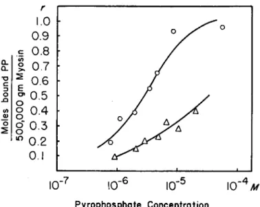
η the maximum number of moles of P P bound per the assumed molecular weight, and (A) is the free P P concentration in moles per liter. It is as- sumed in the above equation that, if η does not equal 1, there is no inter- action between sites. The intercept on the abscissa equals n, that on the ordinate, nK. A series of such experiments is shown in Fig. 1. The affinity constant for the myosin P P binding is 1.01 X 10e, η is 0.88. From this ex- periment, assuming the true value of η to be 1, the minimum molecular weight would be 5.69 Χ 105. It was found, however, that the value of n, and thus the apparent molecular weight, varies somewhat with the age of the myosin preparation. With preparations only a few days old as low a value for the apparent molecular weight as 4.26 Χ 105 was obtained, in good agreement with recent determinations by other methods (4, 15-17).
The highest combining weight found by the P P binding method was 6.7 X 10⁵ gm., on a preparation 3 weeks old.
We have also carried out some experiments with cardiac myosin and the molecular weight suggested by the maximum binding is of the order of 4.5 Χ 105. The affinity constant seemed to be somewhat lower than that for skeletal myosin.
(2) Meromyosins. The meromyosins, that is the two fractions obtain- able from the tryptic and chymotryptic digest of myosin were also investi- gated for P P binding. The light fraction, known to be devoid of ATPase activity, was found to bind no measurable amount of P P . P P binding to
302 J . GERGEL Υ, Α. MARTONOSI, Μ. Α. GOUVEA
FIG. 1. Binding of P P by skeletal myosin and tryptic and chymotryptic heavy frac- tions. Ordinate: Moles of bound PP per assumed molecular weight of protein per free PP concentration at the end of the experiment. Abscissa: moles of bound PP per assumed molecular weight of protein. The assumed molecular weight of myosin is 500,000; that of the heavy fractions, 300,000. Composition of solution: 0.6 M KCl, 0.015 M phosphate buffer, pH 7, 0.001 M Mgcl2. Amount of protein in dialysis bag : myosin, 17.5 mg.; trypsin heavy fraction, 15.5 mg.; chymotrypsin heavy fraction, 24.2 mg. KEY: Ο — Ο » trypsin heavy fraction; Δ — Δ , chymotrypsin heavy fraction;
H—B> skeletal myosin. For details see text.
the heavy fractions from the two digests is shown in Fig. 1. The affinity constants appear to be of the same order as the one for myosin, and mo- lecular weights calculated from such experiments yield values around 3 X 10⁵, in fair agreement with recent determinations by other physico-chemi- cal methods (12) * There is some variation among the preparations and a careful correlation of the apparent molecular weight and the time of diges- tion will be necessary before we can draw definite conclusions from this type of experiment.
F-actin, both skeletal and cardiac, was found to be completely devoid of measurable P P binding.
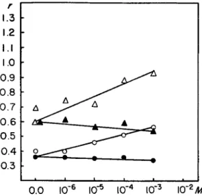
(3) The Actomyosin System. Figure 2 shows a series of experiments with myosin and reconstituted AM (cardiac). It will be seen that actin
* S. Lowey, personal communication.
INTERACTION OF MYOSIN WITH PHOSPHATE COMPOUNDS 303
r
Ο-υ.
1.0 • 0.9 - .s 0.8 -
in
%. 0.7 ·
Ο
T 5 C Ο
§
0.4 - g0.3 ·
(Λ Q)
Ο
0.2 • 0.1 ·
10"
r 710"
Γ610"
Γ5 Ι Ο "4 Λ/Pyrophosphate Concentration
FIG. 2 . The effect of dog heart F-actin on PP binding to dog heart myosin. Equi- librium dialysis for 3 6 hours. In the myosin experiment the dialysis bag contained 14.70 mg. of protein. In the experiment with actin the amount of myosin used was 11.6 mg., the amount of actin 2 . 5 4 mg. Both the inner and the outer solution con- tained 0.6 M KCl, 0.015 M PO*, pH 7, 0.001 M MgCla. The initial PP concentration of the outside solution varied from 7.95 X 10~* M to 9.23 Χ lO^Af. Abscissa: con- centration of free PP ; Ordinate : moles of P P bound per 5 Χ 1 05 g of myosin. KEY : Ο — Ο » heart myosin; Δ — Δ , heart myosin and heart actin. For details see text.
has a significant effect on the binding of P P and leads to a considerable reduction of the amount bound. It has not been possible to utilize con- centrations high enough to reach the amount bound in the case of myosin alone because at higher concentrations the difference between the inner and outer solutions becomes rather small.
(4) Ion Effects. In view of the known effects of the various ions on the ATPase activity of myosin and on the interaction between myosin and F-actin, it appeared of interest to undertake a limited investigation of such effects on the P P binding by myosin (Fig. 3 ) . C a + + has only very slight inhibitory effects, if at all significant, and Mg+ + increases the P P binding.
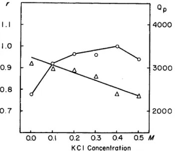
Potassium ions are known to influence the calcium activated ATPase of myosin and of the heavy fractions (8). A comparison of the K + effect on the P P binding to the trypsin heavy fraction with the effect on ATPase shows no correlation (Fig. 4 ) . Thus, the likelihood arises that the potas- sium effect is due to an influence on the splitting rather than on the bind- ing process.
304 J . GERGEL Υ , Α . M A R T O N O S I , Μ . Α . G O U V E A
0.0 I0"6 I0"5 I0"4 I0'3 \0'2M
FIG. 3 . Ca++ and M g++ effect on PP binding by myosin. Equilibrium dialysis for 2 4 hours. Amount of protein used in experiments with lower PP concentration 17.5 mg., in experiments with higher PP concentration 4 7 mg. Composition of solution, 0.6 M KCl, 0.015 M PO* buffer, pH 7. Ca++ and M g++ concentration as indicated in the figure. Ordinate: number of moles of PP bound per 5 χ 1 05 gm. of myosin.
Abscissa: C a++ or M g++ concentration, M. KEY: O—O, initial (PP) = 2 . 6 6 X lO^M, M g++ as indicated on abscissa; Δ — Δ , initial (PP) = 2 . 6 6 X lO^Af, M g++ as indi- cated ôn abcissa; # — φ , initial (PP) = 2 . 6 6 X 1 0 ~eM , Ca++ as indicated on abcissa;
A—A, initial (PP) = 2.66 Χ 10"5 M, Ca++ as indicated on abcissa. Initial PP con- centration is calculated on the basis of the total volume.
tion occurs are 0.1 to 0.2 /mioles per mg. These values are of the same order of magnitude as those found by Kielley and Bradley (3) for inhibi- tion of myosin ATPase activity. Changes in the P P concentration had no apparent effect on the inhibition by mercurials. This point was further checked by spectrophotometric experiments. 1 0 ~2 M P P did not affect the reaction of P C M B with the SH groups of cysteine or myosin, detectable at 250 τημ (18). This experiment appears to exclude an effective displace-
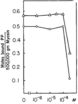
(5) Effect of Sulfhydryl Reagents. As shown in Figs. 5 and 6, both P C M B and salyrgan exert an inhibitory effect on the binding of P P both to myosin and to the proteolytic heavy fractions. This inhibitory effect begins at a mercurial concentration of about 5 X l O ~4M and reaches a virtual abolition of the binding in a saturated P C M B solution. If one ex- presses the amount of mercurial present in the system as /mioles per mg.
of protein, one finds that the concentrations at which substantial inhibi-
r 1.3 1.2 I.I 1.0 0.9 0.8 0.7 0.6 0.5 0.4 0.3
I N T E R A C T I O N O F M Y O S I N W I T H P H O S P H A T E C O M P O U N D S 305 ment of the mercurial by P P from the S H sites and also makes it rather unlikely that the inhibitory effect of mercurials on P P binding is based on a complex formation between P P and the inhibitor.* K3[ F e ( C N )6] , iodo- acetic acid, and N E M , all at a concentration of 1 0 ~8 Μ, were without effect on the P P binding. Similarly without effect was 0.1 M glycine, a
0.0 O.I 0.2 0.3 0.4 0.5 M K C l Concentration
FIG. 4 . Effect of KCl on P P binding and Ca++ activated ATPase activity of trypsin heavy fraction. Left hand side ordinate : number of moles of bound P P per 3 Χ 1 05 gm.
protein. Binding experiments : equilibrium dialysis for 3 0 hours. Composition of solu- tion: 0.015 Ai PO* buffer, pH 7, 10"8Af MgCl2, KCl concentration as stated in figure.
Dialysis bag contained 15.5 mg. trypsin heavy fraction. Initial PP concentration in outer solution, 1.02 χ 10"5. ATPase experiments: Pt liberation was measured at 3 8 ° during a 5 minute period. Total volume 2 ml., containing 0.01 M CaCla, 0.1 M glycine buffer pH 9 , 10"3Af ATP, 1 5 mg. protein, KCl concentration as stated in figure.
Right hand side ordinate: activities expressed as Qp. KEY: Ο—0> P P binding;
Δ — Δ , ATPase. For details see text.
reagent which has been shown to interfere with the determination of S H groups in proteins {19).
(6) Nucleotide and Nucleoside Effects. In view of the similarities be- tween the effect of P P and of A T P on the A M system (4, 5), it appeared of interest to carry out a limited investigation of the effect of some adenine
•There is no change in the spectrum of PCMB or salyrgan on addition of PP.
In view of the studies to be described below the effect of ATP, ADP, and adenosine, on the spectra of the two mercurials and on the reaction of PCMB with cysteine and myosin was also checked. All these experiments were negative.
306 J. GERGELY, A. MARTONOSI, M. A. GOUVEA
0.6
I0"
5I0~
4 MFIG. 5. Effect of mercurials on PP binding by myosin. Equilibrium dialysis. Ordi- nate: number of moles of bound P P per 5 Χ 105 gm. of myosin. 0.6 M KCl, 0.015 M PO* buffer, pH 7, 10"8 M MgCl2. In the case of the PCMB experiments the amount of myosin in the dialysis bag was 47.0 mg., initial PP concentration outside 1.15 χ 10"5 Μ, in the salyrgan experiment amount of myosin 17.5 mg., initial PP concentra- tion outside 5.75 X 10"EM. KEY: Q—O, salyrgan; Δ — Δ , PCMB. For details see text.
nucleotides. It was not feasible to use substantially higher P P concentra- tions, since the percentage binding would have decreased to the point where the experimental errors became greater than the expected results.
In some experiments with the heavy fraction isolated from a tryptic digest of myosin, 1 0 ~3 M A T P or A D P reduced the P P binding, at a P P concen- tration of 2.4 Χ 1 0 -5 M, by about 60%.
(7) Miscellaneous Reagents Affecting PP Binding. E D T A and D N P also inhibit P P binding at the concentrations shown in Fig. 8.
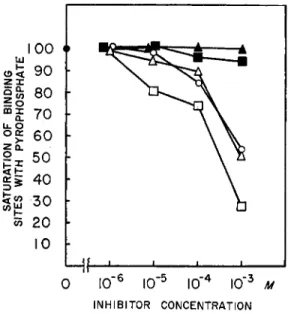
and hypoxanthine derivatives on the P P binding. Figure 7 shows that, of the compounds studied, ATP, A D P , and I D P exert a considerable inhibi- tion while A M P and adenosine are without effect. The effects of ATP and A D P appear to be identical ; this is to be expected in view of the fact that A T P would be broken down by myosin to A D P . Within the range of P P concentrations studied, from 7.6 X 1 0_ G M to 2.4 X 1 0 ~5 M, no increase in the binding was observed in the presence of the effectively inhibiting
I N T E R A C T I O N O F M Y O S I N W I T H P H O S P H A T E C O M P O U N D S 307
c i.o
I
Μ Η ' 1
0 ΙΟ"
6Ι Ο
5I0"
4Concentration PCMB
FIG. 6. Effect of PCMB on the PP binding to heavy fraction from tryptic and chymotryptic digests. Equilibrium dialysis for 24 hours. Composition of solution:
0.6 M KCl, 0.015 M PO* buffer, pH 7, 10~3 M MgCl2. Initial PP concentration in outer solution 1.02 χ 10~6M. Amount of trypsin heavy fraction in dialysis bag was 15.5 mg., that of chymotrypsin heavy fraction was 24.2 mg. KEY: Ο—O, trypsin heavy fraction; Δ — Δ , chymotrypsin heavy fraction. For details see text.
b. Light Scattering Studies
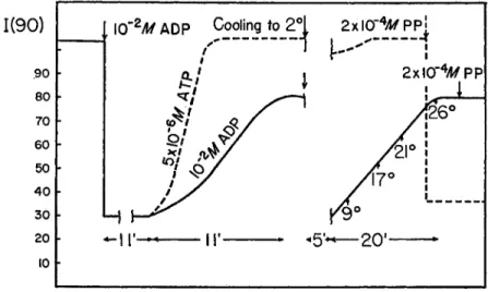
(1) PP and Adenine Derivatives. The decrease of the intensity of light scattered by actomyosin solutions on addition of ATP has been shown to be consistent with the dissociation of A M into actin and myosin {20). In view of the similarities between the P P and A T P effect (4, 5) it will be assumed in the following that reagents that cause a change in the intensity of the scattered light at 90° are causing dissociation of AM, and the change of the intensity of the scattered light is related, although not in a linear form (4)j to the degree of dissociation. The interrelations between the ATP, A D P , and P P effects are illustrated in Fig. 9. A D P in concentra- tions higher than 10 ~3 M has a small but significant dissociating effect on AM at room temperature. Owing to a very small amount of A T P present in the A D P preparations at higher A D P concentrations there is a transient A T P effect, leading to a complete dissociation followed, as the A T P is re- moved by the ATPase activity of myosin, by a partial reversal in the tur-
3 0 8 J . GERGELY, A. MARTONOSI, M. A. GOUVEA
INHIBITOR CONCENTRATION
FIG. 7. The effect of adenine derivatives and IDP on PP binding by myosin. Equi- librium dialysis. Ordinate : binding expressed in per cent of that with PP only. Amount of myosin in dialysis bag 4 7 . 0 mg., in the experiments with IDP 23.5 mg., initial PP concentration in outside liquid 1.15 X 10_ 5Af, 0.6 M KCl, 0.015 M PO* buffer, pH 7, 10"*Λί M g C k Other additions as indicated. K E Y : O, ATP; Δ , ADP; • , I D P ; • , A M P ; A, adenosine. For details see text.
bility that the A D P effect is due to a small amount of myokinase present in the myosin preparation that could have produced a low steady state concentration of ATP. We think that this is ruled out, or made extremely unlikely, by the following facts: addition of 1 0 ~2 M A M P has no effect on the turbidity in the presence of A D P ; nor has addition of hexokinase and glucose although they quickly reverse light scattering changes caused by 2 X 1 0 -5 M ATP. Addition of P P in the presence of 1 0 ~2 M A D P is in- effective, as indicated by Fig. 9. This fact is consistent with the inhibition bidity change. A D P seems to have a pronounced effect upon the rate of this reversal as a comparison between the reversal after addition of 5 X 1 0 ~E
M ATP, and the reversal after the effect of the presumably small amount of ATP in the presence of A D P shows. In the presence of A D P , cooling has a very marked effect, and the amount of A D P which causes a rather small change at room temperature will produce complete dissociation at 2 ° . Two points need be mentioned here: we wanted to exclude the possi-
I N T E R A C T I O N O F M Y O S I N W I T H P H O S P H A T E C O M P O U N D S 309
of P P binding by A D P . It is somewhat difficult to compare the two effects in the same concentration range, since the concentrations used in the P P - binding experiments are too low for light scattering studies.
(2) Salyrgan and PCMB Effects. It has been known from the work of various investigators that salyrgan and other mercurials cause a viscosity drop in an actomyosin solution. Similar changes can be observed in the light scattering system which we interpret as the dissociation into actin
I 0 0 h
INHIBITOR CONCENTRATION
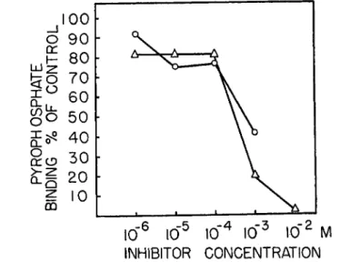
FIG. 8 . The effect of D N P and EDTA on the PP binding to myosin. Equilibrium dialysis, 24 hours. Abscissa: inhibitor concentration; ordinate: PP binding expressed as percentage of binding in the absence of inhibitors. In the D N P experiment the amount of myosin in the dialysis bag was 47.0 mg., the initial PP concentration in the outside liquid was 1.02 χ 10"5ikf. Both the inside and outside solution contained 0.6 M KCl, 0.015 M PO* buffer, pH 7, 10"8 M MgCl2. In the EDTA experiments the dialysis bag contained 47.0 mg. of myosin, the initial PP concentration of the outer liquid was 1.02 χ 10~6 M, other components as in the previous experiment, except that no M g++ was present. KEY: O—O, D N P ; Δ — Δ , EDTA. For details see text.
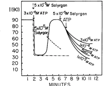
and myosin (Fig. 10). In the absence of ATP, salyrgan and P C M B cause a fairly slow dissociation of actomyosin, in comparison with the A T P effect. If A T P and salyrgan are added simultaneosuly, the A T P effect de- velops as though salyrgan had not been present, but the lower turbidity level persists indefinitely, presumably owing to the known inhibition of myosin ATPase by mercury compounds. Addition of salyrgan during the rising phase of the light scattering curve, when the A T P concentration in
3 1 0 J . G E R G E L Y , A . M A R T O N O S I , M . A . GOTJVEA
10 h
FIG. 9. Effect of ADP on the actomyosin PP system. Light scattering experiments.
Ordinate: scattering intensity at 90°, in arbitrary units. Volume of solution 10 ml., containing 3.82 mg. of myosin and 1.65 mg. of actin, 0.6 M KCl, 0.015 M PO* buffer, pH 7, 10"3 Μ M g C k Other additions as indicated. The first arrow shows the addition of 10~2 M ADP. As discussed in the text the ADP solution contains a small amount of ATP. The broken line shows the effect of 5 χ 1 0e M ATP. At the second arrow the solution was taken to 0°, and kept at that temperature for 5 minutes. It was then allowed to come to room temperature in the apparatus, while the temperature and light scattering were being measured. The solution having reached the original tem- perature 2 X 10"* M PP was added.
The rate of decrease is, however, very slow even on addition of A T P at 1 0 ~3 ikf, a concentration 1 , 0 0 0 times higher than the one leading to an instantaneous drop. P C M B in concentrations of the order of 1 0 ~5 M has the same effect as salyrgan.
In contrast to the inhibition of the A T P effect by salyrgan, the effect of P P and A D P (the latter to be observed only in the cold) on the turbid- ity of A M remains practically unchanged in concentrations of 2 X 1 0 ~4 M and 1 0 ~3 ikf, respectively.
the system is extremely low, stops the further rise of the scattered light intensity and causes a slow dissociation, taking place at about the same rate as would prevail if salyrgan alone were added to AM. If A T P is added about one minute or more after the addition of salyrgan, then, depending on the concentration of the A T P added, either no change occurs in the rate of decrease or rates intermediate between that observed in the presence of salyrgan alone and that found with ATP alone are seen.
I N T E R A C T I O N O F M Y O S I N W I T H P H O S P H A T E C O M P O U N D S 311
1(90) 90 80 70 60 50 40 30 20 10
I 2 3 4 5 6 7 8 9 10 II 12 MINUTES
FIG. 1 0 . The effect of salyrgan in the ATP-AM system. Light scattering experi- ments. The total volume was 1 0 ml. and the solution contained 4.5 mg. of natural AM, 0.6 M KCl, 0.015 M PO* buffer, pH 7, 10"8 M MgCl2. Other additions as indicated in the figure. Ordinate: scattering intensity at 9 0 ° , in arbitrary units. The first solid arrow indicates the addition of ATP, 3 χ 10~αΜ. K E Y : —·—·—, indicates simul- taneous addition of salyrgan with ATP ; , indicates addition of salyrgan in the ascending phase of the ATP effect. The family of curves following the solid arrow indicates the addition of ATP in the amounts shown for each curve about one minute after the addition of salyrgan.
III. Discussion
The study of the binding of P P to myosin can be a source of informa- tion on the mechanism of the binding of adenosine nucleotides to myosin only if the binding of both types of compounds involves the same sites or site. Our experiments suggest that this, in fact, is the case, for higher con- centrations of A D P are able to depress the binding of P P and do also in- hibit the action of P P on the light scattering of A M solutions. Furthermore, the fact that A D P inhibits the ATPase activity of myosin {21), renders it more probable that a common center is involved in the binding of these compounds and in the enzymatic activity. If this view is correct then the discrepancy between the effect of K + , D N P , and E D T A on the binding of P P and their effect on the ATPase activity of myosin {8, 22-24) > should be attributed to their being not so much involved in the binding process, as to their being modifiers of the specific rate of hydrolysis of the phosphate group. An obvious experiment to clinch the identity or at least the ex-
312 J . G E R G E L Y , A. M A R T O N O S I , M . A . GOTJVEA
tremely close relationship of the binding sites for these various compounds would be to study the effects of P P on the binding of labeled A D P to myosin. The inhibition of P P binding occurs at the same mercurial to myosin ratio as found by Kielley and Bradley (S) to inhibit ATPase ac- tivity. Assuming that the mechanism is the same in the case of P P and A T P it would appear that the ATPase inhibition by mercurials is based on the inhibition of the combination of the substrate with the enzyme. P P binding appears to be restricted to that part of the myosin molecule which after proteolytic digestion appears as the so-called heavy meromyosin fraction. The remainder of the molecule seems to be devoid of any binding ability.
Bailey and Perry have suggested that the combination of myosin and actin depends on the participation of SH groups (2), and more recently Perry has proposed (25) that the link between myosin and actin is medi- ated through an A D P - M g - S H bridge. The experiments here reported lend further support to such a concept and appear to furnish some evidence for the identity of the binding site for P P and other substances with the one for actin, since actin effectively inhibited the binding of P P to myosin.
The results of the light scattering experiments allow further tentative conclusions to be drawn about the more detailed properties of the active site. Since the dissociating action of the mercurials on actomyosin is rather slow, it is possible to investigate the availability of the dissociating site to other substances while the turbidity is still near the initial value. It is thus found that under these conditions A T P is rendered largely ineffective, while P P even in a lower concentration is able to produce rapid dissocia- tion. This suggests that the mercurial is bound to the actomyosin even be- fore a change in turbidity occurs and, secondly, that there exists another site which, in the presence of the mercurial bound to the first site, is ac- cessible only to a smaller molecule, such as P P , but not to ATP. This pic- ture would further suggest that the slow dissociation caused by the mer- curials is due to a rate-limiting step whereby the mercurial reaches the second postulated site. We would say on the basis of the actin effect on P P binding that actin is bound to myosin at this second site. If P C M B or salyrgan are added to myosin first, subsequent addition of actin causes no increase in turbidity, indicating that in the absence of actin both types of sites are immediately available for the mercurials. This view should not be taken to imply that actin is, of necessity, bound to an SH group; we simply suggest that the SH group is in the domain in which the contact between myosin and actin is established.
INTERACTION OF MYOSIN WITH PHOSPHATE COMPOUNDS 313
IV. Summary
(1) P P combines with skeletal and cardiac myosin and with the tryp- tic and chymotryptic "heavy-meromyosins" in a ratio of about one mole of P P per mole of protein. Binding constants have been evaluated. Actin and the "light-meromyosins" do not combine with P P .
(2) Mercurials and F-actin interfere with the combination of P P with the above proteins. The effect of some other compounds, including A D P , on the binding has been investigated.
(3) Light scattering studies have been carried out to study the inter- relation of P P , A D P , and mercurials on the actin-myosin binding.
(4) The results are discussed from the point of view of the structure of the active center of myosin.
REFERENCES
1. K. Bailey, in "The Proteins" (H. Neurath and K. Bailey, eds.), p. 951. Academic Press, New York, 1954.
2. K. Bailey and S. V. Perry, Biochim. et Biophys. Acta 1, 506 (1947).
3. W. W. Kielley and L. B. Bradley, J . Biol. Chem. 218, 653 (1956).
4. J. Gergely and H. Kohler, "Conference on the Chemistry of Muscular Contrac- tion," p. 14. Igaku Shoin, Tokyo, 1957.
5. Y. Tonomura, F. Morita, and K. Yagi, J . Phys. Chem. 61, 605 (1957).
6. P. Gallop, C. Franzblau, and E. Meilman, Biochim. et Biophys. Acta 24, 644 (1957).
7. A. Szent-Györgyi, "Chemistry of Muscular Contraction." Academic Press, New York, 1951.
8. J. Gergely, M. Gouvea, and D . Karibian, J . Biol. Chem. 212, 165 (1955).
9. G. Feuer, F. Molnâr, Ε. Pettko, and F. Β. Straub, Acta Physiol. Acad. Sei. Hung.
1, 150 (1948).
10. W. F. H. M. Mommaerts, J . Biol. Chem. 188, 559 (1951).
11. A. G. Szent-Györgyi, Arch. Biochem. Biophys. 42, 305 (1953).
12. J. Gergely, H. Kohler, W. Ritschard, and L. Varga, Abstr. Biophys. Soc. p. 46 (1958).
13. R. B. Hurlbert, H. Schmitz, A. Brumm, and V. R. Potter, J . Biol. Chem. 209, 23 (1954).
14. I. M. Klotz, in "The Proteins" (H. Neurath and K. Bailey, eds.), p. 727. Academic Press, New York, 1953.
16. K. Laki, and W. R. Carroll, Nature 175, 389 (1955).
16. A. Holtzer and S. Lowey, / . Am. Chem. Soc. 78, 5955 (1956).
17. W. F. H. M. Mommaerts and B. B. Aldrich, Science 126, 1294 (1957).
18. P. D . Boyer, / . Am. Chem. Soc. 76, 4331 (1954).
19. J. P. Greenstein and J. T. Edsall, J . Biol. Chem. 133, 401 (1940).
20. J. Gergely, J . Biol. Chem. 220, 917 (1956).
21. H. M. Kalckar, J . Biol. Chem. 153, 355 (1944).
22. G. D . Greville and D . M. Needham, Biochim. et Biophys. Acta. 16, 284 (1955).
23. J. B. Chappell and S. V. Perry, Biochim. et Biophys. Acta. 16, 285 (1955).
314 J . GERGELY, A. M A R T O N O S I , M . A . G O U V E A U. W. J. Bowen and T. D. Kerwin, J. Biol. Chem. 211, 237 (1954).
26. S. V. Perry, Physiol Revs. 36, 1 (1956).
Discussion
MORALES: I think that Dr. Gergely has given us several demonstrations that pyrophosphate is similar to ATP which is very gratifying. This had been thought for a long time because pyrophosphate has been well known to be a competitive inhibitor of ATPase. We differ on some things he said but I think those differences are beyond the scope of this meeting. However, there is one little item that I think fits into this puzzle, and perhaps somebody has a good idea about this. A couple of years ago Dr.
Kielley wrote a very interesting paper in which he showed that as you add PCMB to myosin in 0.6 M KCl, that the first half of the PCMB that goes on actually stimu- lates the ATPase activity. Recently we have been experimenting along lines which now seem very related. We have been studying substances like cysteine ethyl ester and AET* on ATPase activity, and we now think that these compounds form mixed disulfides with the enzyme, so that probably they are doing the same thing as Dr.
Kielley 5s PCMB. But the curious thing is that they do not of themselves increase the ATPase activity. What they do is to make the ATPase enormously sensitive to cal- cium. If you always test in calcium, then you observe simply an acceleration of the splitting rate. But it, can be shown that the effect is that when you tie up the SH group with either PCMB or by forming a mixed disulfide, you then make the system very much more sensitive to calcium. So something is going on long before you get an interference with pyrophosphate bond splitting.
Very recently Dr. Blum has communicated to me some work which appears to point to the fact that what is going on is some interference with the binding of the ring end of the ATP. I think this is very interesting. I think it is maybe also har- monious with the fact that somehow the first PCMB's that go on or the first AET's that go on, interfere with the ring end and the later ones interfere with the splitting of the pyrophosphate structure.
RACKER : I would like to draw your attention to a procedure for measuring ATPase activity which we find more accurate and reproducible than the conventional method.
We use catalytic amounts of ATP together with an ATP regenerating system, e.g., excess phosphoenolpyruvate and pyruvatekinase, and measure liberation of ortho- phosphate. While in the conventional system an excess of ATP is used and ADP is allowed to accumulate, by the above procedure ADP is continuously removed. Several ATPases have been reported to be inhibited by ADP. With these enzymes, much higher activity values can be obtained in the ATP regenerating system than by the conventional assay. For example, in the case of mitochondrial ATPase, which Kielley found to be inhibited by ADP, and which is known to be difficult to analyze, good proportionality between concentration and activity can be obtained. Moreover, the activity values are 4-5 times higher than without the regenerating system. I would like to ask Dr. Gergely whether some of his findings may not be explained by ADP inhibition which may be more pronounced under certain experimental conditions.
GERGELY: In reply to Dr. Racker's suggestion, I think this is a very good suggestion and very often useful. In the case of myosin—and I think Dr. Morales agrees with me—the amount of ADP needed for inhibition is much larger than the amount that
* Aminoethylthiouronium salts.
I N T E R A C T I O N O F M Y O S I N W I T H P H O S P H A T E C O M P O U N D S 315 is liberated in the course of the enzymatic reaction. Furthermore, the phosphate liberated is proportional to time, showing no falling off as ADP is formed.
MORALES: I was going to say that it can be shown that the velocity is strictly linear with enzyme concentration.