The Milky Diseases
S. R. DUTKY
Entomology Research Division, Agricultural Research Service, United States Department of Agriculture, Beltsville, Maryland
I. Introduction 75 II. Development of Disease in the Field 76
III. D e v e l o p m e n t w i t h i n the Insect Host 80 A. Effect of Temperature 81 B. Effect of Food 85 C. Effect of Dosage 89 D. Lethality of Disease 96
IV. Host Range 97 A. Virulence of Milky-Disease Strains 102
V. Artificial Culture Studies 103 A. Effect of p H 104 B. Relation to Oxygen 105 C. Carbohydrate Requirement 107 D . Nitrogen Requirement 108 E. Mineral Requirements 110 F. Growth Factors 110 G. Primary Isolation of Cultures 112
VI. Concluding Remarks 113
References 114
I. INTRODUCTION
Extensive studies over nearly thirty years have shown that milky diseases are important control agents of the Japanese beetle, Popillia japonica Newman, and the large-scale use of type A milky disease for the control of this destructive pest is one of the impressive successes of biological control by man's use of an insect pathogen. Special methods for the propagation and application of the causative organism in amounts sufficient for this use were developed and patented to protect
75
the public interest (Dutky, 1940, 1941a, 1942a, b; Dutky and Fest, 1942;
W h i t e and Dutky, 1940).
While most of this effort was directed toward the milky diseases of the Japanese beetle and their use in the control of this insect, almost from the first it was recognized that these organisms also attacked a n u m b e r of related insect species in m u c h the same way, and some of these species could be controlled by the same organisms or by closely related ones (Dutky, 1941a, b).
In a few instances other scarabaeids were found in field surveys to have a milky disease. I n most of these, the organisms were very similar morphologically to the causative organism of type A milky disease, Bacillus popilliae Dutky. Significant numbers of Cyclocephala larvae infected by a similar organism were also found. T h i s organism has been designated as Bacillus popilliae, Cyclocephala strain (White, 1947;
Harris, 1959). I n a smaller n u m b e r of other scarabaeids, organisms resembling that of the type Β milky disease, Bacillus lentimorbus Dutky, occurred.
As most of the recoveries were m a d e in areas where milky disease of the Japanese beetle was well established, or the diseased larvae were encountered d u r i n g the course of laboratory tests, it could not be certain
£hat the diseased insects h a d been infected independently of the presence of milky disease of the Japanese beetle larvae. I n point of fact, the recent discoveries of milky diseased scarabaeids in Switzerland (Wille, 1956), France (Hurpin, 1955), and Australia (Beard, 1956) have been m a d e years after milky disease material from Japanese beetle sources was introduced and studied in these countries.
A n u m b e r of reports have been m a d e on the Federal-State co
operative distribution of milky disease for the control of the Japanese beetle (White and Dutky, 1942; Adams and Wheeler, 1946; Easter, 1947; Cory and Langford, 1950; W h i t e and McCabe, 1951; Polivka, 1956), and the reader is referred to these for the control aspects of the subject.
II. DEVELOPMENT OF DISEASE IN THE FIELD
I n the field, after disease has become generally distributed by natural spread or artificial introduction, spores of the causative organism are present in the soil in enormous numbers. T h e u p p e r levels of the soil are highly infective, and healthy larvae held in such soils rapidly be
come infected. Since these soils can be diluted manyfold (as m u c h as 1:250) without materially reducing their infectivity, it is estimated that they may contain the equivalent of nearly 100 billion spores per kilo
gram in the u p p e r inch of the soil.
T h e spores are not uniformly distributed in the soil b u t are present in spots of high concentration. T h e spores released from decomposed dead diseased larvae become tightly b o u n d by the soil particles and do not move far from the point of release. W h e n high host population levels are present, this localized distribution is broken u p since larvae in their feeding and movement churn u p the soil, loosen it, and re
distribute it to a depth of several inches.
Most of the spores are present in the u p p e r inch or so of the soil, since d u r i n g the period of greatest larval activity, the larvae feed close to the surface. Most of the diseased larvae die without descending to lower levels to overwinter or p u p a t e , consequently these lower levels remain fairly free of spores. In certain soil types and in sod of certain deeply rooted grasses, larvae feed on roots at lower depths. In these cases, larvae dying of disease eventually cause accumulation of spores at these depths, even where spore dust has been applied as a surface application. Factors that shift the depth at which larvae feed will alter the relative exposure of larvae to residual spores. T h i s may explain why in some years many first and second stage larvae are infected and killed by milky disease whereas in others the infection rate of young larvae is much lower.
T h e seasonal life cycle of the Japanese beetle in the latitude and elevation of the Philadelphia vicinity of Pennsylvania (USA) as it per
tains to disease development is as follows:
Eggs are laid normally from July to September in the soil at a depth of 2 to 4 inches below the surface. T h e eggs hatch after 14 to 21 days and give rise to first-instar larvae which occur in fair abundance in August. T h e newly hatched larvae feed for a few days at the depth at which the eggs were laid and gradually work their way nearer to the surface. T h e y feed and grow in size to about one-third of an inch (10.5 mm) in length, molting after about 3 weeks. T h e second-instar larvae feed for about the same period of time and attain a length of about three-fourths of an inch (18.5 mm). T h e third instar is generally reached by the latter part of September, when the larvae are about an inch in length (32 mm) (Boving, 1939). T h e y continue to feed actively until the latter part of October when downward movement of the larvae begins. T h e larvae now weigh about 200 mg. T h i s downward movement continues through November. W h e n the soil temperature drops to 50°F (10°C) in late November, all activity ceases. T h e larvae form cells in which they pass the winter, usually not more than 6 inches below the surface and never at depths greater than 1 foot. I n April after over
wintering the larvae again rise toward the surface and resume active
feeding on the grass roots. T h e larvae continue to feed until the latter part of May when most of them empty the hindgut, stop feeding, and enter the p r e p u p a l stage, forming a cell about 2 to 4 inches below the surface. P u p a t i o n follows about 10 days after the cessation of feeding and the p u p a l stage lasts from 8 to 20 days, depending on temperature.
T h e threshold for p u p a t i o n is about 60°F (15.5°C). T h e beetles start emerging from the p u p a l skin about the second week in J u n e . T h e adults live for about 30 to 45 days. D u r i n g this time a female may lay as many as 40 to 60 eggs, a few at a time. T h e sex ratio is very nearly 1:1; the females are larger and somewhat longer-lived than the males.
It is evident from this description that d u r i n g the period that tem
peratures are o p t i m u m for disease development, the larval soil popula
tion is at a m i n i m u m . I n more n o r t h e r n latitudes and at higher eleva
tions, there occurs a complete or partial 2-year cycle, and in these localities there is a rather high larval population throughout July and August which would favor the continuous spread and development of disease. T h i s may more than compensate for the lower m a x i m u m soil temperature and shorter season for disease development.
It should be noted that the success of milky disease as a control factor is quite dependent on the range of soil temperatures encountered and more particularly on the threshold temperatures of the various stages of the insect. W i t h the Japanese beetle, the threshold for p u p a t i o n is only a degree or so below that for disease development. Hence, in areas where the threshold for p u p a t i o n is quickly exceeded, there is little chance in most seasons for many larvae to escape infection. I n an unusual situation it might be possible for the soil temperature to dwell in the narrow range between the two critical temperatures, and u n d e r this condition the explosive type A milky disease development usually observed in the spring in this species could not occur. W i t h other insect species—Hylamorpha elegans (Burmeister) is a good example—the threshold for p u p a t i o n appears to be m u c h lower and there is a good possibility that disease development in this species would be m u c h reduced (Dutky, 1957). Careful studies must be m a d e to evaluate the merit of milky disease u n d e r these conditions.
W i t h type Β milky disease where infection is largely initiated in the early instars and diseased larvae frequently molt several times before further development is arrested, a very high rate of infection of third- instar larvae may occur in the fall. Infection rates as high as 85 percent have been observed. T h e overall effect on the population, however, is n o greater than that of the lower rate observed of type A milky disease infection in this instar at the same time (15 to 30 percent), since larvae infected with type A in one instar generally die in that instar without
molting, and the population of third instars has therefore already been reduced by previous mortality.
Since larvae cease to feed several days before molting and do not resume feeding for a day or so after, it follows that type A milky- diseased second-instar larvae are not found until about a week after the first occurrence of second-instar larvae in the population. Similarly, the first milky diseased third-instar larvae show u p in the field 2 weeks after the first occurrence of third-instar larvae in the population. T h i s delay in disease development, the failure to molt, and the longevity of diseased larvae change the stage distribution of type A milky-diseased larvae so that when healthy larvae are predominantly third instars, diseased larvae are mostly second instars with a few surviving first and newly infected thirds. I n contrast, the stage distribution of type Β milky-diseased larvae does not differ markedly from that of healthy larvae. T h i s is consistent with the observations mentioned above that infection with type Β is largely restricted to the first instar and diseased insects can molt.
Diseased larvae may overwinter b u t usually die soon after tempera
tures reach the threshold for resumed activity in early spring. T h e n the type Β diseased larvae take on a chocolate-brown coloration some time before death.
T h e threshold for larval activity is about 12°C whereas the threshold for disease development is nearly 16°C, so that most of the larvae will ingest quantities of spores before this latter temperature is reached.
T h e larvae at this time are very resistant to type Β infection and despite the large n u m b e r of spores ingested, few develop the disease. O n the other hand, these larvae are highly susceptible to type A infection, and consequently the disease develops explosively, numerous insects show
ing external symptoms at the same time. Nearly all the third instar larvae infected die without pupating, and the few that might p u p a t e could hardly emerge as adult, since the length of time between cessation of feeding prior to p u p a t i o n and the p u p a l period is long enough to permit disease development to lethality. Microscopic examination of large numbers of beetles collected in field surveys and trapped by Japanese Beetle Laboratory personnel in areas where the disease rate among larvae was very high failed to produce a single instance of natural infection in the adult, a result indicating that, as would be expected, this incidence must be very low. Experimentally infected adults could be produced regularly by injecting p u p a e late in the p u p a l stage when coloration of the adult cuticle was well advanced. T h e susceptibility of adults to infection by injection of spores is about the same as for larvae, and adults can be used in inoculation studies and for mass propagation
of the disease. T h i s last has been pointed out correctly (Langford et al., 1942), but the data presented on the natural or induced occurrence of the disease among adults are misleading.
Eggs from adults collected from areas where disease incidence was high were hatched and larvae reared from them were free from disease.
W h e n tested for susceptibility by exposing them to inoculated soil, it was found that these reared larvae were as susceptible as those collected from newly infested areas free from disease. Beetles emerging from treated areas are readily susceptible to infection by injection, and there is no indication of development of resistance to milky disease.
T h e effect of low temperatures on the fate of the rods of the caus
ative organism in the blood deserves some mention. About 2 weeks after soil temperatures h a d fallen to 10°C in the field, microscopic examination of larvae collected at this time showed that the rods were not refringent, and it was assumed that they were dead. Laboratory studies to test this hypothesis were made; it was found that when larvae injected with spores and held for 2 and 3 days at 30°C were placed at 10°C for 2 weeks, u p o n second incubation at 30°C these larvae failed to develop infection, whereas larvae injected with spores and then placed directly at 10°C for a like period developed disease normally u p o n incubation at the elevated temperature. Larvae held for longer periods before cooling died as a consequence of disease u p o n reincubation with
out any large increase in the n u m b e r of spores produced.
III. DEVELOPMENT WITHIN THE INSECT H O S T
After infection by any method, the course of the several milky dis
eases is similar. T h e organisms in some way reach the blood and there multiply and sporulate. I n the typical case, the infected insect lives for a fairly long period and the organism develops in sufficient numbers to produce marked turbidity of the normally clear blood. I n most cases, this turbidity results from the accumulation of spores; in third-instar Popillia larvae infected with B. popilliae, turbidity is observed when the insect contains about 60 million spores. W i t h B. lentimorbus, which does not produce a refringent paraspore, about 100 million spores are required to produce recognizable turbidity. I n the case of the organism we have designated as "Maryland type B," morphologically similar to Bacillus euloomarahae Beard (Beard, 1956), the vegetative forms develop in such numbers that in spite of their low refringency they produce observable turbidity before sporulation occurs. As the disease progresses, the increased turbidity due to accumulating spores obscures the internal organs; the milkiness of the blood at this stage is the basis for the name
"milky disease" (Dutky, 1940).
T h e processes of vegetative development followed by sporulation continue to occur in sequence as long as the infected insect serves as an adequate substrate for this development. D u r i n g this period, vegetative multiplication a n d sporulation are occurring simultaneously. O u r studies of development with larvae injected with spores and vegetative rods from artificial cultures indicate that the cells undergo a limited n u m b e r of vegetative doublings (less than 8), and proceed to sporulate.
T h i s process is repeated in turn by daughter cells of the first divisions.
I n the species producing paraspores, these are produced after the process of endospore formation is completed.
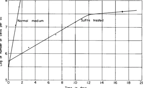
T h e n a t u r e of the paraspore and its pathological significance has not been fully established. Vago and Delahaye (1961) report electron microscope studies on the paraspores. W e have studied the germination of spores in media, using sulfite-treated brain-heart infusion to restrict subsequent vegetative development, and find that with B. popilliae the vegetative cell produced from the spore leaves the sporangium from the end away from the paraspore, and the paraspore remains in the spo
rangium morphologically intact after the process is completed.
A. Effect of Temperature
All the milky-disease organisms thus far studied have a narrow range of temperature in which development can occur. T h e lower limit is about the same for all, 15.5°C (60°F). T h e u p p e r limit for B. popilliae a n d its related strains is nearly 36 °C (96°F). At temperatures between these limits, except for the increased time required as the temperature is reduced, the general sequence is similar. T h e organism develops in the blood a n d sporulates there. T h e larvae assume a milky appearance and finally succumb to the disease. Both the time required for the on
set of gross symptoms a n d the longevity of diseased larvae after symp
toms first appear increase with reduction of temperature. T h e r e is some indication that at the u p p e r extreme of temperature (and possibly also at the lower) sporulation is reduced, b u t throughout the midrange of temperature there is little change in the numbers of spores produced.
T h e slower development of the organism at lower temperatures is compensated by the greater longevity of the host larvae. Extensive tests at the Japanese Beetle Laboratory showed only a very slight reduction in spore yields (about 1 percent) attributable to increased temperature between 20°C and 30°C.
T h e exact relationship between the time required for onset of symp
toms and temperature has not been fully established. Several studies have been reported (Dutky, 1940; Beard, 1945; T a s h i r o and White, 1954;
H u r p i n , 1959) that give data for analysis, b u t these data are not com-
parable because of differences in dosage, host insect, and possibly other factors, including the method of diagnosis and equipment used. Beard takes exception to describing the relationship as rectilinear and sug
gests that it is more nearly exponential. My analysis of his data does not confirm this statement, however. T h e curves he presents indicate a perfect hyperbolic relationship. I would be inclined to agree that such a relationship should exist between the temperature and the time of onset of symptoms, time of death, or any other definable period that is a function of the doubling time of the organism (Dutky, 1959).
Unfortunately, repeated tests at the Japanese Beetle Laboratory have failed to establish this simple relationship with any of three milky- disease organisms studied extensively in Popillia japonica.
T h e expected relationship would be Eq. (1)
T i m e (t — 60) = Κ (1) where Κ is a constant dependent on the phase defined, the dosage of
inoculation, and the nutritional level of the host, and t — 60 is the n u m b e r of degrees Fahrenheit above the lower limit of development.
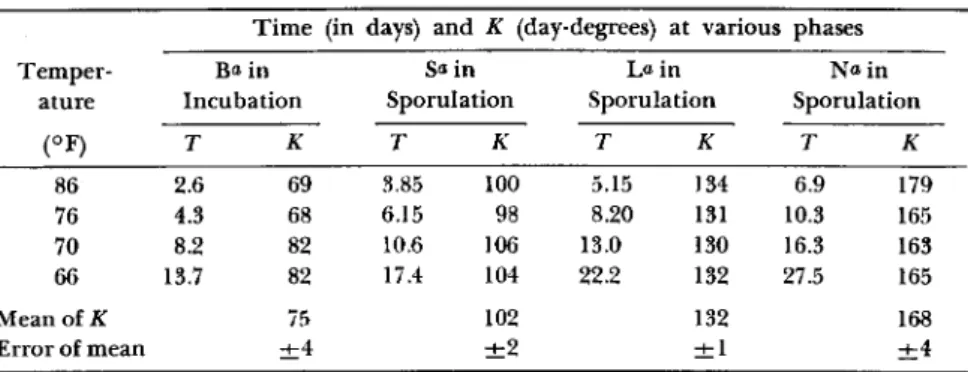
T A B L E I
EFFECT OF TEMPERATURE ON DISEASE DEVELOPMENT T i m e (in days) and Κ (day-degrees) at various ί phases
Temper Ba in S a i n L« in Να in
ature Incubation Sporulation Sporulation Sporulation
( oF) Τ Κ Τ Κ Τ Κ Τ Κ
86 2.6 69 3.85 100 5.15 134 6.9 179
76 4.3 68 6.15 98 8.20 131 10.3 165
70 8.2 82 10.6 106 13.0 130 16.3 163
66 13.7 82 17.4 104 22.2 132 27.5 165
Mean of Κ 75 102 132 168
Error of mean ± 4 ± 2 ± 1 ± 4
α Beard (1945) in his graph Fig. 12 has labeled the ordinate "Invasion, Incubation, Sporulation" to designate "phases" in development. I have used letters of the above words to indicate the ordinate values I have chosen in extracting data from his curves (cf. Fig. 1).
For centigrade, Κ would be five-ninths of this value and 15.5 would be used as the lower limit.
T h e data I have extracted from Beard's curves (Beard, 1945; Fig. 12, p. 525) are presented in T a b l e I. T h e data are plotted in the accom
panying graph, Fig. 1. T h e i r intercepts are in remarkable agreement with the estimated threshold and they point out a convenient tool for establishing the validity of a determined temperature threshold. W h e n
the data are plotted, the reciprocal of time against temperature, all four series are straight lines. T h e intercepts on the temperature axis indicate a common threshold (60.5 °F) a n d the slope ( t e m p e r a t u r e / reciprocal time) is Κ in day-degrees for each phase.
0.45 0.40
0.35
& 0.30
TD ο
c
I
0.25ö
0.20Ο ο
v . Q .
I
0.150.10
0.05
w" " 5 5 60 70 80 9 0
Temperature Fahrenheit
FIG. 1. Effect of temperature on development of Bacillus popilliae. (Data from Beard, 1945; Fig. 12, p. 525.)
T o be complete, a study of the effect of temperature on disease de
velopment should include larvae injected parenterally with two spore dosages. A dosage of 1 million spores and one of 100,000 spores per larva are recommended. These will give nearly 100 percent infection, a n d the difference in the m e d i a n times of infection at the two dosages at the various temperatures divided by 3.322, the n u m b e r of doublings equivalent to a tenfold increase in the dosage, will be the doubling
time of the milky disease organism at these temperatures. At 30°C, B. popilliae has a doubling time in Japanese-beetle larvae of about 6 hours determined by this method. I n the best artificial media used to date, doubling times are from 4.8 to 6.2 hours.
Enteral injections are not recommended since the results reported indicate that the method is not very reliable and, furthermore, the dosage required to produce infection is many times that known to produce consistent infection by exposing larvae to soils inoculated with spore dust. Exposure of larvae to inoculated soil at different tempera
tures should yield valuable information. Here again, dosages at two levels should be used. T w o million and one million spores per gram are recommended. Results of hundreds of tests at 30°C indicate that the median time of infection is 10.2 days at the higher level of inocu
lation, whereas a smaller n u m b e r of tests at 22°C show the median time of infection to be about 21 days at this same level of inoculation.
All series should include uninoculated insects as controls at each temperature u n d e r investigation.
T h e effect of temperature on disease development is not simply a relationship of the development of the causal organism in respect to temperature. T h e host insect's response to temperature is also involved.
Its nutritional state at the time of inoculation and the quantity a n d type of food also have a bearing. If sprouted seed is used as food, especially if the nutritional state of the host initially is poor, then the growth rate of the sprouts at the different temperatures also will enter into the relationship. It is interesting to note that tests with Japanese- beetle larvae collected in the fall and in good nutritional state (mean weight 200 mg) showed that the larvae m a d e the same weight gain (30 mg in 3 days, 55 mg in 7 days) at 30°C as at 22°C when held in soil-seed mixtures. T h e feeding rate, as determined by collecting and weighing frass pellets, and the rate of m a t u r a t i o n are b o t h m u c h greater at the higher temperature, however. T h e same gain at the two
temperatures indicates that the greater basal metabolic rate at the higher temperature offsets the larger quantity of food ingested.
These factors can and should be evaluated in a complete study.
Larvae of different nutritional states can be produced by prefeeding one group b u t not the other. Tests can then be made with both groups with and without food at various temperatures. Consideration should also be given to the type of equipment used. Most constant-temperature cham
bers have an appreciable variation, even at the thermostat, and under
shoot a n d overshoot the set temperature by a larger a m o u n t than is indicated by the monitoring thermometer. T h i s variation is not sig-
nificant at mid-ranges b u t becomes highly so when one is attempting to establish the points of discontinuity. Also, the individual boxes con
taining moist soil or other substrate must permit some evaporative loss of water to sweep out carbon dioxide accumulated from the metabolic activities of the insect, the seed sprouts, and the soil organisms. T h i s evaporative loss may cause a considerable reduction of temperature in the microenvironment as compared with the thermostat. T h i s last should be checked by inserting thermocouples in representative boxes.
T h e frequency of observation should be great enough to obtain a reliable median time of development, b u t it must be remembered that observation usually entails a change of conditions. I n view of the many factors involved, it is not particularly surprising that different investi
gators have arrived at somewhat different interpretations from the results of their tests.
A purely empirical relationship derived from data of reasonable accuracy, such as that given in my original description of B. popilliae (Dutky, 1940), can be very useful.
T i m e in days for onset of symptoms = 24 — 0.6 (temperature °C) (2) T h i s equation was used with good results to forecast the probable date when infection would occur in the field by determining the time re
quired for samples of field-collected larvae to develop symptoms at 30°C and adjusting this value to the expected soil temperature range for the area u n d e r consideration. T h e accuracy of the formula u n d e r field con
ditions was confirmed in b o t h 1935 and 1936 when data from thermo
graph records were analyzed. I n 1935 a total of 313 hours of tempera
tures of 60°F and above h a d occurred u p to the time when the first newly infected larvae showing visible symptoms of disease were re
covered on J u n e 3. T h e computed value was 312 hours. Similarly, in 1936, 331 hours of 60°F and above were recorded u p to the time the first infected larvae were found on May 18. T h e computed value from the formula was 324 hours. Both determinations agree with the com
p u t e d values within 2 percent.
B. Effect of Food
T h e nutritional state of the larvae at the time of infection and the a m o u n t of food available d u r i n g the course of the disease has a pro
found effect on its development. I n general, the better the initial con
dition of the larvae, and the better its n u t r i t i o n thereafter, the longer will the larvae survive after infection, and the greater will be the num
ber of spores produced. T h e r e is a very close correlation between the longevity of diseased larvae and spore yields. Increasing the a m o u n t of
food available does not prevent mortality due to disease, b u t rather postpones it for a time.
W h e n larvae are infected by injection or by feeding in contaminated soil, the a m o u n t of vegetative development and sporulation that the milky-disease organisms undergo is limited by the nutritional level of the blood of the host insect. As we have determined from artificial culture studies, these organisms have a very limited spectrum of sub
stances that they can attack and utilize for growth. T h e complex materials to which the insect converts its food for growth and storage are not attacked, and the pathogen must intercept the intermediates before conversion or else it must depend on the host to reconvert the complex materials to forms that are available to it, usually a fairly slow process. T h e growth of the pathogen depending on this source of nutrients will therefore be slow.
T h e nutritional state of the insect in terms of the requirements for rapid growth of the pathogen is not directly related to the whole organic content of the host, b u t rather to its blood volume a n d to the level of certain substances in it. Insects with large amounts of fat and other reserve materials have a lower blood volume in relation to their weight than do insects that are growing rapidly and have accumulated little stores. T h e blood volume in cubic millimeters of the latter is just about one-half their weight in milligrams whereas in insects that have well- developed fat bodies the blood volume may be as low as one-third the weight in milligrams. W e have found this relationship to hold for a n u m b e r of scarabaeid species. A very simple method for estimating the blood volume of milky-diseased larvae (Dutky, 1957) is to determine the n u m b e r of spores per cubic millimeter of blood and then, by triturat
ing the insect in water, to determine the total n u m b e r of spores. T h i s total divided by the n u m b e r per cubic millimeter is equal to the blood volume in cubic millimeters. These counts are m a d e at proper dilutions with a hemacytometer. T h e blood volume of uninfected insects can also be estimated by the same process. O n e has only to inject a known n u m b e r of spores, wait a few minutes for them to be distributed, and then fill the counting chamber with an undiluted blood sample and determine the n u m b e r of spores per cubic millimeter. T h e same formula again gives the volume of blood in cubic millimeters.
Insects with large amounts of reserves therefore do not necessarily produce as high yields or live so long as do insects with lesser amounts that are ingesting quantities of food sufficient to supply their own re
quirements and to maintain their blood levels in spite of the removal of materials therefrom by the pathogen. Unfortunately for the infected
insect, the pathogen increases in numbers in response to the nutritional level and consequently demands an ever increasing share of the nutrients that the insect derives from its food. T h i s deprivation of essential food elements a n d the accumulation of toxic products finally takes its toll;
the ability of the insect to feed is reduced, and the injured insect dies.
Soil insects must have a certain nutritional level to be able to m a i n t a i n their water balance against the higher water potential of moist soil. Insects of good nutritional state m a i n t a i n their weight for some time in moist soil even u n d e r starvation conditions, b u t eventually they begin to imbibe water and inflate. Larvae of poor nutritional state quickly become hydrated and will die rapidly as a result. T h e y may show a weight gain that is m u c h larger u n d e r starvation conditions than when adequate amounts of food are present. These larvae are capable of supporting very small numbers of the pathogen, and spores are produced in such low numbers that visible symptoms of disease are not evident. I n spite of the lack of external symptoms, mortality due to disease is very high.
T h e nutritional state of larvae can be estimated by injecting larvae and holding them without feeding (each individual in a clean tightly stoppered vial) for 10 days at 30°C for B. popilliae and related organisms or 12 days at 28°C for B. lentimorbus and related organisms, and then determining the spore yield per larva. T h e n u m b e r of spores produced u n d e r these conditions is a reliable index of the nutritional state. If larvae are prefed before testing, a large increase in spore yield will occur if the initial nutritional state was low. Larvae held in soil with sprouted seed as food after inoculation will show a marked increase in spore yield in all cases if the larvae feed.
Popillia larvae obtained from the field in the early fall show wide variations in their nutritional state. From some areas in some years, a high proportion of the larvae are so well-developed that most of them can transform to adults without further feeding, while in other years few or none are capable of m a t u r a t i o n without considerable feeding.
D u r i n g storage at 7° to 10°C in the laboratory (and probably also in the field), there is a gradual loss of nutritional state; after a few months of storage, larvae held without food after injection will produce few spores.
Overwintering larvae collected early in the spring behave m u c h like fall-collected larvae after storage. T h e y are in poor nutritional state and, unless fed well, do not produce many spores; without feeding they are incapable of maturation. Larvae collected late in spring near pupa
tion do not yield as many spores as do freshly collected larvae in early
fall. These factors are of great importance in mass inoculation of larvae for spore production, and many tests have been made to develop o u r understanding of the rather complex relationship. A typical test with fall-collected stored larvae is presented in T a b l e II.
A careful study of the composition of larval blood should be made.
These studies should reveal the type and extent of damage caused by the disease, and also should disclose the requirements for sporulation and perhaps lead us to success in the development of a satisfactory arti
ficial culture procedure for mass producing spores of these pathogens.
T A B L E I I
EFFECT OF FOOD ON SPORE YIELD OF INJECTED LARVAE AFTER 1 0 D A Y S ' INCUBATION AT 3 0 ° C
Layer 1, Layer 2, Layer 3, Layer 4, Layer 5, 10 gm none 2.5 gm 5.0 gm 10 g m Parameter seed/kg added seed/kg seed/kg seed/kg N u m b e r surviving** (of 64) 46 45 42 44 44
Percent surviving 72 70 66 69 69
Mean weight (mg/larva) *> 222 195 173 202 228 Spore yield (109/larva)& 1.57 0.39 0.98 1.32 1.61 Mean weight (mg/larva)c 210 178 190 196 220 Spore yield (10»/larva)c 1.55 0.28 1.16 1.54 1.58 Mean of mean weight (mg) 216 ± 6 186 ± 9 181 ± 8 199 ± 3 224 ± 4 Mean spore yield (10®) 1.56 ± .01 0.33 ± .06 1.07 ± .09 1.43 ± .11 1.60 ± .0i Carbon (nig/larva)** 13.9 ± .3 5.22 ± .52 8.19 ± .54 10.4 ± .2 14.3 ± .3
α All surviving larvae were milky. Larvae were injected with 106 spores o n 11/26/48 and examined on 12/6/48.
ö Mean weight of 5 larvae weighed and triturated in water to determine spore yields. Counts in duplicate on each suspension.
c Second group of 5 larvae as above.
Λ Carbon was determined by wet combustion. Mean of two groups of 5 larvae.
Determinations in duplicate for each group.
Shotwell et al. (1962) report preliminary studies in this direction.
These studies should be made on larvae of different nutritional states so that a comparison of the composition of the hemolymph can be correlated with its ability to support growth and sporulation. If the nutritional state h a d not been determined, such blood analyses might be very misleading.
Larvae can be brought (as outlined above) to a very low nutritional state by storing them for several months at 7° to 10°C; b u t by pre- feeding them for a short time before testing, their ability to support growth and sporulation is increased markedly. It would be possible also with stored larvae to test the effect of injection of various materials (carbohydrates, vitamins, amino acids, metallic elements) on growth
and sporulation in these deficient insects in m u c h the same m a n n e r as one uses deficient culture media to determine such requirements.
C. Effect of Dosage 1. By Injection
W h e n milky-disease spores are injected parenterally, the n u m b e r of spores injected affects both the proportion of insects developing in
fection a n d the time of onset of disease symptoms. T h e results obtained will depend on the host insect species and the milky-disease organism involved. W i t h most species of host and pathogen, dosages of one mil
lion spores or more produce nearly 100 percent infection. At lower dosages, the percentage infected varies nearly exponentially with the dosage. A more precise interpretation is that the percentage of infec
tion varies directly with the square of the logarithm of the n u m b e r of spores injected, or the square root of the percentage of infection plotted against the logarithm of the dosage will be a straight line. In my thesis (Dutky, 1937), I expressed my findings with B. popilliae injected into Japanese beetle larvae by the following equation:
(Percent infection)1/* = 3.1 log1 02V — 9.2 (3) Subsequent modifications of the microinjector and improvements in
the technique gave a m u c h higher percentage infection at higher dosages, and the m i n i m u m dosage that would still infect a small percentage of larvae was likewise lowered. T h e general relationship was unchanged, however, and can be expressed by the equation
(Percent infection)1/* — 3.05 log1 0iV — 5.2 (4) Beard (1945) raises objection to this expression and claims that the
relationship is rectilinear when plotted on a logarithmic-probability grid, as he also reports to be true for his own data. His treatment of the data involved a tremendous a m o u n t of m a n i p u l a t i o n and still shows large variations. Furthermore, the data are limited to a small segment of the possible ranges of dosages that could be employed and do not test the applicability of the highly complex mathematical probit function. O u r studies (and those of other groups cooperating in the milky disease program) with h u n d r e d s of thousands of larvae injected in the mass production of spores indicate that at dosages of one million spores per larva, one can expect 20,000 of 20,000 larvae surviving injec
tion to be infected. T h i s would not be fulfilled from the probit relationship.
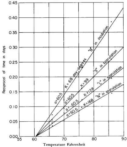
I n any event, a g r a p h of the data presented in his T a b l e 2 for test A, group A, Beard (1944) shows that these data also fit the general rela-
tionship given previously (Fig. 2). Of the 5 points plotted, 4 fall exactly on a line and the one off the line is not m u c h removed. T h i s point is also off the line by a similar a m o u n t when the data are plotted on log-probability paper (Fig. 3). T h e equation of the straight line that
ο ο
CT CO
Dutky et al (percent infection)1 3.05 log Ν - 5.20
Beard 1944 (percent infection)1 η 2.97 log Ν - 6.03—
/ Dutky 1937 (percent infection)' A-3.1 log N - 9.2
1/2
I
100 81
64 49
36
25 -
2 3 4 5 6 7 0
Log of number of spores injected
FIG. 2. Analysis of data from the literature: infection versus dosage.
fits his data so well, as determined by the intercepts 0 percent and 100 percent infection, is
(Percent infection)1/* = 2.97 log1 02V — 6.03 (5) T h i s equation is remarkably similar to Eq. (4) and differs only in the
values of the constants from Eq. (3).
Analysis of the data available from the literature shows a similar
relationship. Investigator and student alike are urged to treat these data similarly. T h e fine data presented by Beard (1944, 1945) are representative of his findings with milky disease in Popillia japonica.
Tashiro a n d W h i t e (1954) present data for Amphimallon majalis (Razou- mowsky), H u r p i n (1959) for Melolontha melolontha (Linnaeus).
T h e method outlined above requires no special graph paper, per
mits plotting both 0 and 100 percent values, and the conversion to logs
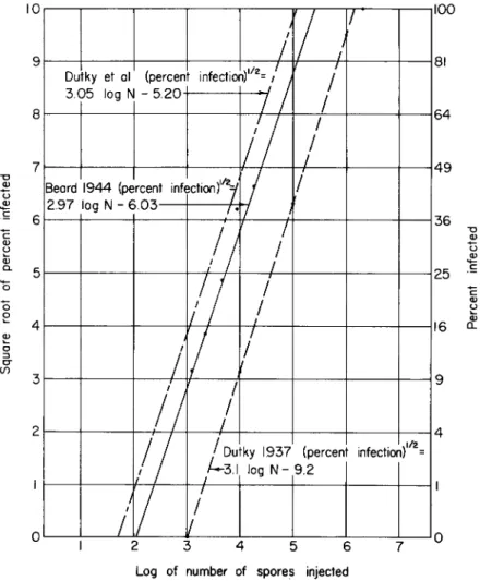
9 9 . 9 r - 1 1 I 1 1
9 9 . 8
W ' 1000 10,000 100,000 1,000,000
Number of spores injected
FIG. 3. Infection versus dosage probability-log plot of data from Fig. 2.
and square roots is easily done by means of slide rule or tables. For those not too familiar with mathematical treatment in the determination of empirical equations, the following discussion will be helpful.
T h e data are plotted as indicated. In most cases a straight line can be fitted to the data by inspection. T h e log (or exponent) of the dosage where the line crosses the 0 infection (x axis) is read from the scale.
In the center line of Fig. 2 (Beard's data) this value is 2.03 (the log of m a x i m u m dosage not producing infection), and similarly the log of the dosage where the line crosses the 10 level (100 percent infection) is also determined. Again from the line plotting Beard's data, this value is 5.38 (the log of the m i n i m u m dosage producing 100 percent infection).
T h e values are substituted into the general equation (Percent infection)^ = Κ log1 0]V -)- b
10 = 5.38 Κ + b 0 - 2.03 Κ + b
(6)
( 7 )
(8)
(10)
( 9 )
or
b = 6.03 (11)
Therefore the empirical equation for the line is Eq. 5:
(Percent infection)1^ — 2.97 log10AT — 6.03 (5) A critical review of the literature indicates that one should use cau
tion in interpreting results of injection tests. Some of the precautions required in microinjection have been outlined (Dutky, 1942a, b). T w o additional points must be added. First, our tests indicate that vegetative rods produce infection at m u c h lower dosages than spores; the median infective dose with rods is in the order of 10 rods per insect, or about 1/1000 that of spores. W h e n tests are made using fresh insect blood, one must make certain that rods are present in a very small ratio to spores or otherwise eliminate them. Second, fresh blood suspensions rapidly lose their infectivity. W i t h i n an h o u r after preparation, injec
tions at a level of 1 million spores per larva that initially produced 100 percent infection will drop to 90 to 95 percent. After several hours, infection rates as low as 25 to 50 percent will be obtained. T h i s d r o p in infectivity is paralleled by a darkening of the blood suspension, a n d may be associated with it. Spore suspensions m a d e from newly prepared dried blood films (less than 3 weeks old) behave similarly to fresh blood suspensions. Interpreting this loss of infectivity from the dosage-infec
tion pattern leads one to the conclusion that in the short holding time that caused infection to d r o p from 100 percent to 90 to 95 percent, the infectivity of 1,000,000 spores was reduced to that equivalent of 100,000 spores, or an apparent loss of 90 percent. Similarly, holding suspensions until the rate of infection dropped to 25 to 50 percent caused a reduc
tion of the infectivity of 1,000,000 spores to the level of 10,000 spores or an apparent loss of 99 percent. T h i s p h e n o m e n o n ought to be care
fully studied.
T h e interference of rods and also the rapid loss in infectivity are both avoided by use of dried-blood films for the preparation of stock
spore suspensions. Most of the rods are killed d u r i n g the drying process or die soon thereafter, and after about 6 months suspensions prepared from the dried films are very stable, do not darken, and show little or no loss in infectivity. T h e spores retain both virulence and ability to germinate indefinitely. T h e use of dried blood films also permits dupli
cation of injection tests and culture tests with the same spore stock months or years apart.
I n preparation of dried-blood films, treatment of larvae before bleed
ing to prevent clotting is most helpful. T h i s can be done by immersing them in hot water, 69.44°C (Beard, 1945). T h i s treatment is lethal to the larvae, however. W e have found that clotting may also be inhibited by immersing larvae in 95 percent alcohol for 5 minutes. T h i s treat
ment does not kill the larvae. Blood withdrawn from larvae immediately after treatment will still clot, b u t within 10 minutes after removal from the alcohol, the clotting power is lost. T h e clotting power is regained after 24 hours and the treated larvae are again normal. T h e late Τ . N . Dobbins worked out a combination of the two methods: larvae are exposed to 50 percent alcohol for 5 minutes and then immersed in water at 48 °C for 5 minutes. T h i s treatment works very well a n d pro
duces dried-blood films of superior quality and stability; it is the method of preference.
2. Soil Inoculation Studies
Soil inoculation studies using fresh blood suspensions or spore sus
pensions from dried-blood films as inocula have not given very uniform results. Some tests gave high rates of infection whereas in others little or no infection was obtained. Since tests with soils inoculated with spore dust give very uniform and reproducible results, it seems probable that the lack of uniformity obtained with spore suspensions is probably due to failure to get even distribution of spores in the soil.
I n tests with spore dust, the spore dust is added to air-dry soil and mixed by passing the m i x t u r e through a medium-mesh screen several times. Dry grass seed is then added to the inoculated soil (5 gm red top, Agrostis alba Linnaeus, and 5 gm white clover, Trifolium repens Lin
naeus, per kilogram of soil), and water is added to bring the moisture content to the desired level, 60 percent of ball point (Dutky, 1941c), and after mixing is distributed in tin cans. T h e seed used is the best grade obtainable in respect to purity and germination. Even so some lots of seed quickly d a m p off and thereby fail to provide an adequate a m o u n t of sprouts over a sufficiently long period. T h i s was prevented by using formaldehyde 40 percent USP solution diluted 1:1000 with distilled water to moisten the soil instead of water. At this concentration,
formaldehyde does not markedly affect germination of the seed nor does it interfere with milky-disease infection.
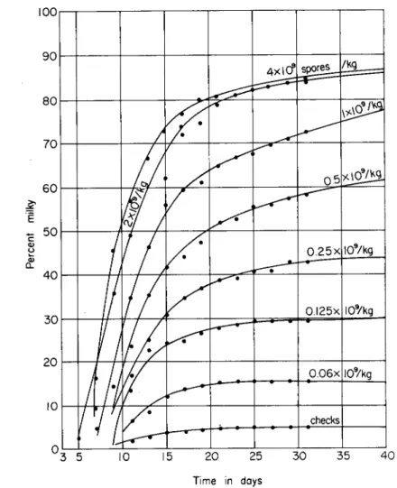
I n bioassay of type A milky-disease spore dust, duplicate samples of 20 gm are weighed out and added to kilogram portions of air-dry soil.
Ι Ο Ο γ — ι 1 1 1 1 1 1 1
π ι ι ι ι 1 1 1 1
U3 5 10 15 20 25 30 35 40
Time in days
FIG. 4. Infection of Popillia larvae in inoculated soil at 30°C. Soil was inoculated with Bacillus popilliae U S D A standard lot 7 / 5 / 3 9 .
After mixing, adding seed, moistening, and remixing, the inoculated soil is distributed into individual boxes, about 30 gm per box, and third-ins tar Popillia larvae reared in the laboratory or collected from disease free areas are added, one per box, twenty-five units per sample.
Soil inoculated with a tested laboratory lot serves as a standard. Un-
inoculated soil, to which has been added 2 gm of sterilized calcium carbonate precipitated USP a n d 18 gm of sterilized talcum USP and an equivalent a m o u n t of seed and water per kilogram, is used in the checks. T h e boxes containing larvae are then placed at 30°C and 88 percent relative humidity and examined every other day beginning on the third day after the start of the test. At each examination, the con
dition of each larva is recorded. T h e tests are m a d e at the equivalent concentration of 2 χ 109 spores per kilogram. Samples that deviate mark
edly from the standard in the n u m b e r infected or the median time of infection, and samples of commercially prepared material that have been submitted to confirm that they meet specification, are also tested at concentrations of 20, 10, 5, and 2.5 gm per kilogram and compared with soils inoculated with the standard spore dust at 2 χ 109, 1 χ 109, 0.5 χ W , 0.25 χ 109, 0.125 χ ΙΟ9, and 0.0625 χ 109 spores per kilogram. In all these tests, the total a m o u n t of calcium carbonate a n d talcum per kilogram of air-dry soil are maintained at levels equivalent to 2 g m a n d 18 gm, respectively. Data from a standard series are plotted in Fig. 4.
A n enormous a m o u n t of data has been accumulated from tests of this type starting with the first lots of experimental spore dust produced in 1937 and continuing at a m u c h reduced pace today. T h e records on indi
vidual larvae permit determination of the time of onset of infection and longevity of diseased larvae. I n some cases weight and spore yield at death were also determined. These data, as a whole, have not been sum
marized a n d treated statistically. T h e late Τ . N . Dobbins m a d e a sta
tistical treatment of 104 tests mostly of USD A lot 7 / 5 / 3 9 (used as the standard from 1940 to 1950) m a d e between 1940 and 1945 at levels of 2 χ 109, 1 χ 109, and 0.5 χ 109 spores per kilogram. T h e treatment was made to determine whether a standard curve could be used instead of the comparison standard included in each bioassay test. His general conclusion was that, in spite of a reasonably good general agreement, the variables introduced that might affect the results of specific tests, mostly in respect to the larvae used in the test, their size, nutritional state, maturation, and length of storage, were great enough to rule against elimination of the comparison standard.
Tests with other milky-disease organisms have not given as uniform results. Repeated tests with B. lentimorbus in which third-instar Popillia larvae were used confirm that this stage is resistant to infection, and although tests with first- and second-stage larvae gave somewhat better re
sults, these tests were quite variable, as were similar tests with B. popil
liae. T h i s variability is probably d u e to poor distribution. As the younger larvae ingest m u c h smaller quantities of soil, distribution has a greater effect.
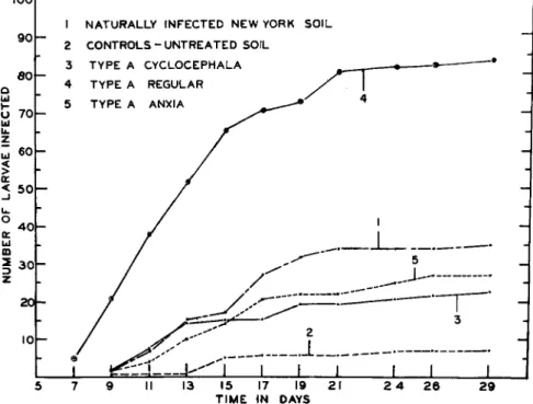
Results of feeding tests by Τ . N . Dobbins with Popillia larvae, for which experimental spore dusts were used, are presented in Fig. 5.
eoh
4 0 h
2 0 1 -
1 N A T U R A L L Y I N F E C T E D N E W YORK S O I L 2 C O N T R O L S - U N T R E A T E D SOIL
3 T Y P E A C Y C L O C E P H A L A 4 T Y P E A REGULAR 5 Τ Υ Ρ Ε A ANXIA
15 17 19 2 1 T I M E I N DAYS
2 9
FIG. 5. Infection of Popillia larvae in inoculated soil.
D . Lethality of Disease
A great deal of work remains to be done to determine the mode of action of this group of organisms. A large part of the effect of disease is certainly d u e to removal of essential nutrilites a n d nutrients from the insect blood a n d locking them u p in the vegetative cells a n d spores of the organisms. Studies on the growth requirements of the organisms on artificial media indicate that thiamine, nicotinic acid, tryptophan, a n d carbohydrate are utilized in vegetative growth a n d would be so removed as would the factors, n o t yet determined, that must also be required for sporulation. Culture tests show that diseased blood contains little or n o free thiamine a n d does n o t stimulate growth on thiamine-deficient media, whereas marked stimulation is obtained with healthy blood.
T o x i c products of the organisms are also involved in their lethality.
Cell-free filtrates of cultures of B. popilliae that show high virulence are lethal when injected in small amounts (0.003 ml of a 1:10 dilution).
These toxic substances are apparently heat labile since injection of these cultures heated to 50°C for 10 minutes did not injure the larvae. Tests on antibiotic interception of this organism also indicate that lethality is due to toxic products. T h e growth and development of B. popilliae in the blood of larvae can be intercepted by the injection of dihydrostrepto- mycin, 20 μg per larva. T h i s a m o u n t produces no injury to injected larvae. W h e n the antibiotic is administered within 48 hours after a challenging dose of one million spores, the disease is intercepted and the larvae survive as healthy insects. Administration of the antibiotic 72 hours or more after the challenging dose inhibits further development of the organism, b u t the insects receiving the antibiotic die at very nearly the same rate as those not receiving it. These tests infer that once the disease is well established the lethal process cannot be reversed by destroying the organism or inhibiting its further development. Peni
cillin G 300 units, Aureomycin hydrochloride 20 μg, and sulfadiazine sodium 200 μg did not intercept the disease development in larvae even though these materials inhibit the organism in artificial media.
T h e interception of disease development by cold has already been discussed near the end of Section II. Interception of the disease by this means, and also by antibiotics, should yield valuable information on its mode of action and on the development of immunity.
IV. H O S T RANGE
AS far as we know, the milky-disease organisms are able to infect only certain closely related beetles of the family Scarabaeidae. W h e n spores or cultures have been injected into other insects, n o disease development occurs.
I n larvae of Musca domestica Linnaeus injected with spores, blood samples showed spores in the hemolymph in numbers equivalent to the injection for several days. Thereafter, there was a gradual reduction in numbers of free spores in the hemolymph due to phagocytosis and e n t r a p m e n t by the fat body. After p u p a t i o n of the larvae and emergence of the adult, spores were again observed in the hemolymph equivalent to the numbers injected.
Larvae of white-fringed beetles, Pantomorus (Graphognathus), that I injected in Chile were observed to have a gradual increase of rods in the blood over a period of more than one m o n t h at 25 °C. These rods did not look like typical B. popilliae rods and did not sporulate in the blood, b u t were absent in control insects. I n repeated tests with larvae of Gal- leria mellonella (Linnaeus), using both spores and rods from cultures as inocula, no effect on the host was noted and no development of the organism occurred.
T A B L E III
SUSCEPTIBILITY OF W H I T E GRUBS TO Bacillus popilliae D U T K Y Insect species Literature citation^
Infected by direct injection
Adoretus sinicus Burmeister Carter (1945)
Amphimallon majalis (Razoumowsky) Tashiro and W h i t e (1954) Amphimallon solstitialis (Linnaeus) H u r p i n (1959)
Anomala innuba (Fabricius) Anomala lucicola (Fabricius) Anomala oblivia Horn (probably) Anomala orientalis Waterhouse
Aphodius howitti H o p e Beard (1956) Aphonus castaneus (Melsheimer)
Brachysternus sp. Dutky (1957) Cetonia aurata (Linnaeus) H u r p i n (1959) Cyclocephala borealis Arrow
Diplotaxis sp.
Heteronychus sanctae-helenae Blanchard Beard (1956) Hylamorpha elegans (Burmeister) Dutky (1957) Macrodactylus subspinosus (Fabricius)
Maladera (—Autoserica) castanea (Arrow)
Melolontha melolontha (Linnaeus) H u r p i n (1959) Melolontha vulgaris Fabricius—Melolontha melolontha Kern (1950)
Odontria zealandica W h i t e D u m b l e t o n (1945) Oryctes nasicornis (Linnaeus) H u r p i n (1959) Pelidnota punctata (Linnaeus)
Phyllophaga anxia (LeConte) Phyllophaga bipartita (Horn) Phyllophaga congrua (LeConte) Phyllophaga crassissima (Blanchard) Phyllophaga crenulata (Froelich) Phyllophaga drakei (Kirby) Phyllophaga ephilida (Say)
Phyllophaga forbesi Glasgow (or near) Phyllophaga forsten (Burmeister) (possibly) Phyllophaga fraterna Harris
Phyllophaga fusca (Froelich) Phyllophaga futilis (LeConte)
Phyllophaga glaberrima (Blanchard) (probably) Phyllophaga gracilis (Burmeister)
Phyllophaga hirticula (Knoch) Phyllophaga hornii (Smith) (probably) Phyllophaga implicita (Horn) Phyllophaga inversa (Horn) Phyllophaga micans (Knoch) Phyllophaga rugosa (Melsheimer) Phyllophaga quercus (Knoch) Phytalus georgianus H o r n Popillia japonica N e w m a n
« Where n o citation is given, tests were conducted at the Japanese Beetle Laboratory.
T A B L E III (Continued)
Insect species Literature citation**
Sericesthis pruinosa (Dalman) Beard (1956)
Strigoderma arboricola (Fabricius) Trichiotinus sp.
Infected by feeding in inoculated soil
Amphimallon majalis (Razoumowsky) Tashiro and W h i t e (1954) Anomala orientalis Waterhouse
Aphonus castaneus (Melsheimer) Cyclocephala sp.
Macrodactylus subspinosus (Fabricius)
Melolontha melolontha (Linnaeus) H u r p i n (1959) Odontria zealandica W h i t e D u m b l e t o n (1945) Phyllophaga anxia (LeConte)
Phyllophaga congrua (LeConte) Phyllophaga ephilida (Say)
Phyllophaga fraterna Harris (probably) Phyllophaga futilis (LeConte) (from Illinois) Popillia japonica N e w m a n
Not infected by injection Cotinis nitida (Linnaeus)
Cyclocephala immaculata (Olivier) Lichnanthe vulpina (Hentz)
Melolontha melolontha (Linnaeus) W i l l e (1956) Not infected by feeding in inoculated soil
Amphimallon solstitialis (Linnaeus) H u r p i n (1959)
Cetonia aurata (Linnaeus) H u r p i n (1959)
Lichnanthe vulpina (Hentz)
Maladera (—Autoserica) castanea (Arrow)
Oryctes nasicornis (Linnaeus) H u r p i n (1959)
Phyllophaga fusca (Froelich)
Phyllophaga futilis (LeConte) (from Wisconsin) Phyllophaga hirticula (Knoch)
Phyllophaga inversa (Horn) Phyllophaga rugosa (Melsheimer)
Found naturally infected in the field Anomala orientalis Waterhouse
Cyclocephala borealis Arrow
Maladera (—Autoserica) castanea (Arrow) Phyllophaga anxia (LeConte)
Phyllophaga fusca (Froelich) Phyllophaga futilis (LeConte) Phyllophaga fraterna Harris (probably) Phyllophaga hirticula (Knoch) Phyllophaga inversa (Horn) Popillia japonica N e w m a n Strigodermella pygmaea (Fabricius)
a Where no citation is given, tests were conducted at the Japanese Beetle Laboratory.
More tests o n different species have been made with B. popilliae than with any other milky-disease organism, a n d these were made to deter
mine mainly whether t h e strains used in preparation of standard spore dust would serve for t h e control of the species in question, or whether this species could serve as a host in advance of the spread of Popillia populations. I n some cases large numbers of a n insect species were col-
T A B L E I V
SUSCEPTIBILITY O F W H I T E GRUBS TO INFECTION BY Bacillus lentimorbus DUTKY Insect species Literature citation
By injection
Amphimallon majalis (Razoumowsky) Tashiro and W h i t e (1954) Anomala orientalis Waterhouse
Cyclocephala borealis Arrow
Hylamorpha elegans (Burmeister) Dutky (1957) Malader α (—Autoserica) castanea (Arrow)
Popillia japonica N e w m a n
Sericesthis pruinosa (Dalman) Beard (1956) Not infected by injection
Aphodius howitti H o p e Beard (1956)
Heteronychus sanctae-helenae Blanchard Beard (1956)
T A B L E V
SUSCEPTIBILITY OF W H I T E GRUBS TO Bacillus lentimorbus var. australis BEARD Insect species Literature citation
By injection
Anomala orientalis Waterhouse Beard (1956) Aphodius howitti H o p e Beard (1956) Heteronychus sanctae-helenae Blanchard Beard (1956) Popillia japonica N e w m a n Beard (1956) Sericesthis pruinosa (Dalman) Beard (1956)
Not susceptible
Amphimallon majalis (Razoumowsky) H u r p i n (1959) Cetonia aurata (Linnaeus) H u r p i n (1959) Malader α (—Autoserica) castanea (Arrow) Beard (1956) Melolontha melolontha (Linnaeus) H u r p i n (1959)
lected or made available, whereas i n others a few specimens only were available. Many of these species were injected with spores of B. popilliae incidental to mass inoculation of Popillia grubs for spore-dust produc
tion. Fewer tests were made with B. lentimorbus because of the limited n u m b e r of insect specimens available a n d because this organism was n o t employed in routine production.
A summary of early tests with B. popilliae has been published (Dutky, 1941a). A more comprehensive compilation of results with
Β. popilliae in t h e species tested at the Japanese Beetle Laboratory and of tests published by other investigators is given in T a b l e I I I . Re
sults with other milky-disease organisms have similarly been tabulated (Tables I V - I X ) .
T A B L E VI
SUSCEPTIBILITY OF W H I T E GRUBS TO Bacillus popilliae Cyclocephala STRAIN ( W H I T E , 1947) Insect species Literature citation
By injection Cyclocephala borealis Arrow
Cyclocephala immaculata (Olivier)
Hylamorpha elegans (Burmeister) Dutky (1957) Popillia japonica N e w m a n
By feeding Cyclocephala borealis Arrow
Popillia japonica N e w m a n
T A B L E VII
SUSCEPTIBILITY OF W H I T E GRUBS TO Bacillus popilliae Melolontha STRAIN H U R P I N « Insect species Literature citation
Infected by injection
Amphimallon majalis (Razoumowsky) H u r p i n (1959) Amphimallon solstitialis (Linnaeus) H u r p i n (1959) Melolontha melolontha (Linnaeus) H u r p i n (1959)
Not infected by injection
Cetonia aurata (Linnaeus) H u r p i n (1959) Oryctes nasicornis (Linnaeus) H u r p i n (1959)
Infected by feeding
Melolontha melolontha (Linnaeus) H u r p i n (1959) Not infected by feeding
Amphimallon majalis (Razoumowsky) H u r p i n (1959)
α H u r p i n (1955) reported the occurrence of milky disease i n Melolontha melo
lontha i n France. Wille (1956) reported disease i n this same species in Switzerland and n a m e d t h e organism B. fribourgensis, a n e w species. H u r p i n (1959) concludes that both are t h e same a n d d o not warrant species designation a n d considers that the milky-disease organism in this species should be designated as a strain of B.
popilliae; a n d I agree.
Milky-diseased specimens found in the field were bled to prepare dried-blood films so that the disease material could be preserved for future study; they were then injected with 95 percent ethyl alcohol in order to inflate a n d preserve them from decomposition a n d placed in vials containing 70 percent alcohol. T h i s was also done with representa
tive specimens from infection tests. These preserved specimens were checked against larval keys a n d tentatively identified; they were then