The Effects of Bilateral Theta-burst Stimulation on Executive Functions and Affective Symptoms in Major Depressive Disorder
Adrienn Holczer,aViola Luca Ne´meth,aTeodo´ra Ve´kony,a,bKrisztia´n Kocsis,aAndra´s Kira´ly,a,c Zsigmond Tama´s Kincses,a,dLa´szlo´ Ve´csei,a,ePe´ter Klive´nyiaand Anita Mustf*
aDepartment of Neurology, Faculty of Medicine, Albert Szent-Gyo¨rgyi Health Centre, University of Szeged, Szeged, Hungary
bLyon Neuroscience Research Center (CRNL), INSERM, CNRS, Universite´ Claude Bernard Lyon 1, Lyon, France
cCentral European Institute of Technology, Brno, Czech Republic
dDepartment of Radiology, Albert Szent-Gyo¨rgyi Health Centre, University of Szeged, Szeged, Hungary
eMTA-SZTE Neuroscience Research Group, Szeged, Hungary
fInstitute of Psychology, Faculty of Arts, University of Szeged, Szeged, Hungary
Abstract—Major depressive disorder (MDD) is characterized by severe affective as well as cognitive symptoms.
Moreover, cognitive impairment in MDD can persist after the remission of affective symptoms. Theta-burst stim- ulation (TBS) is a promising tool to manage the affective symptoms of major depressive disorder (MDD); however, its cognition-enhancing effects are sparsely investigated. Here, we aimed to examine whether the administration of bilateral TBS has pro-cognitive effects in MDD. Ten daily sessions of neuronavigated active or sham TBS were delivered bilaterally over the dorsolateral prefrontal cortex to patients with MDD. The n-back task and the attention network task were administered to assess working memory and attention, respectively. Affective symptoms were measured using the 21-item Hamilton Depression Rating Scale. We observed moderate evidence that the depres- sive symptoms of patients receiving active TBS improved compared to participants in the sham stimulation. No effects of TBS on attention and working memory were detected, supported by a moderate-to-strong level of evi- dence. The effects of TBS on psychomotor processing speed should be further investigated. Bilateral TBS has a substantial antidepressive effect with no immediate adverse effects on executive functions.Ó2021 The Author(s).
Published by Elsevier Ltd on behalf of IBRO. This is an open access article under the CC BY license (http://creativecommons.
org/licenses/by/4.0/).
Key words: major depressive disorder, theta-burst stimulation, working memory, attention, transcranial magnetic stimulation.
INTRODUCTION
Repetitive transcranial magnetic stimulation (rTMS) is now considered a therapeutic measure to reduce the affective symptoms of major depressive disorder (MDD) (seeLefaucheur et al., 2020for review). Over the dorso- lateral prefrontal cortex (DLPFC), both the left- hemispheric, facilitatory rTMS (5 Hz or above, high- frequency, HF-rTMS) (O’Reardon et al., 2007) and the right-hemispheric, inhibitory stimulation (1 Hz, low- fre- quency, LF-rTMS) are beneficial compared to sham stim- ulation (Fitzgerald et al., 2003, 2009; Isenberg et al.,
2005; Stern et al., 2007). A patterned version of rTMS, namely theta-burst stimulation (TBS), significantly reduces the duration and cost of the stimulation and seemingly exerts comparable effects to rTMS (Blumberger et al., 2012; Mendlowitz et al., 2019;
Nyffeler et al., 2007; Zafar et al., 2008). The inhibitory pat- tern of TBS is continuous TBS (cTBS), which applies an uninterrupted train of bursts, and the facilitatory is inter- mittent TBS (iTBS), which is fragmented by pauses among the trains of bursts (Huang et al., 2005). TBS over the DLPFC mitigates the clinical symptoms of MDD with an effect estimation similar to rTMS (Li et al., 2014;
Plewnia et al., 2014; Schwippel et al., 2019; Williams et al., 2018). In addition to unilateral stimulation, sequen- tially applied left facilitatory and right inhibitory (bilateral stimulation) by either rTMS or TBS appears to be similarly effective (Berlim et al., 2013a, 2013b; Chen et al., 2014;
Cheng et al., 2016; O’Reardon et al., 2007). Bilateral pro- tocols are based on the observations of interhemispheric imbalance in MDD (Grimm et al., 2008; Hecht, 2010), the resolution of which is suggested to improve affective
https://doi.org/10.1016/j.neuroscience.2021.03.001
0306-4522/Ó2021 The Author(s). Published by Elsevier Ltd on behalf of IBRO.
This is an open access article under the CC BY license (http://creativecommons.org/licenses/by/4.0/).
*Corresponding author. Address: Institute of Psychology, University of Szeged, Egyetem utca 2, Szeged, Hungary.
E-mail address:must.anita@med.u-szeged.hu(A. Must).
Abbreviations:ANCOVA, analysis of covariance; ANOVA, analysis of variance; ANT, Attention Network Task; BF, Bayes Factor; cTBS, continuous theta-burst stimulation; DLPFC, dorsolateral prefrontal cortex; HDRS, Hamilton Depression Rating Scale; iTBS, intermittent theta-burst stimulation; MDD, major depressive disorder; rMT, resting motor threshold; RT, reaction time; rTMS, repetitive transcranial magnetic stimulation; TBS, theta-burst stimulation.
N EUROSCIENCE
RESEARCH ARTICLE
A. Holczer et al. / Neuroscience 461 (2021) 130–139
130
symptoms. However, most studies have focused exclu- sively on affective changes and did not consider other characteristic symptoms of MDD, such as cognitive impairment. Here, we aimed at exploring the effective- ness of bilateral TBS on both the affective and cognitive symptoms of MDD.
Cognitive symptoms, especially deficits of executive functions including attention (Kaiser et al., 2015) and working memory (Ga¨rtner et al., 2018) as well as psy- chomotor retardation (Gorwood et al., 2014), are often present in MDD, further exacerbating the burden of dis- ease. Moreover, the impairment of all these cognitive domains may persist even after the remission of the affec- tive symptoms (Nebes et al., 2003; Rock et al., 2014). The effectiveness of pharmacotherapy appears to be limited to some cognitive subdomains (Pan et al., 2017), while the more promising results of rTMS are still preliminary and inconclusive (Demirtas-Tatlidede et al., 2013; Iimori et al., 2019; Martin et al., 2017) with reporting of no pro- cognitive effect (Wajdik et al., 2014). Concerning TBS, studies carried out on healthy participants revealed that it might modulate cognition at behavioral (Lowe et al., 2018; Ve´kony et al., 2018; Viejo-Sobera et al., 2017), electrophysiological (Chung et al., 2017), and neuro- chemical level (Suppa et al., 2016). Working memory and attention can be enhanced even after one session of TBS (He et al., 2013; Lowe et al., 2018; Xu et al., 2013). However, differences are present across cognitive domains, e.g., performance on tasks inquiring complex executive functions appears not to be affected (Lowe et al., 2018). Also, as the rationale of bilateral protocols derives from the clinical characteristics of MDD patients, the investigation of bilateral TBS in a preclinical setting is limited. To date, only a few studies have assessed whether TBS can mitigate cognitive impairment in MDD (Cheng et al., 2016; Scho et al., 2019) and only an even smaller proportion of these investigated bilateral TBS (Cheng et al., 2016). The present randomized, sham- controlled study aimed to examine the effects of 10 daily bilateral TBS sessions on the clinical symptoms and executive function in MDD. We assessed working mem- ory and attention using standardized neurocognitive tests:
the n-back and the Attention Network Task (ANT). Overall reaction times (RTs) for both tasks were also investigated to gather information on psychomotor processing speed.
Since TBS effects on the working memory domain seem to be the most reliable based on results of healthy partic- ipants (Lowe et al., 2018) and patients with neuropsychi-
atric disorders (Demirtas-Tatlidede et al., 2013), enhanced performance on the n-back task was expected.
As TBS is suggested to enhance attention (He et al., 2013), we also expected improvements on the ANT. To detect potential changes in clinical symptoms, the Hamil- ton Depression Rating Scale (HDRS) was administered.
Classical statistical analysis was supplemented by Baye- sian statistics to quantify the strength of the evidence.
EXPERIMENTAL PROCEDURES Participants
Patients diagnosed with unipolar MDD by experienced physicians were recruited from the Department of Psychiatry of the Albert Szent-Gyo¨rgyi Health Centre, University of Szeged. The diagnosis was established based on DSM-IV criteria using the Structured Clinical Interview for DSM-IV Axis I disorders. Patients with any confounding conditions such as comorbid major psychiatric disorders (e.g., substance abuse, psychosis) and individuals with a history of neurological disorders (e.g., stroke, epilepsy, head injury) were excluded.
Those who did not meet the safety restrictions of TBS (e.g., having metallic implants in the cephalic region or any implanted electronic devices) were excluded. Based on a meta-analysis, pharmacotherapy might support the development of more stable antidepressive effects (Kedzior et al., 2012). Therefore, TBS was applied as add-on therapy. Stable pharmacological status was required from at least two weeks before the commence- ment of the study and maintained throughout the TBS therapy. All participants signed informed consent. The experimental protocol was approved by the local Ethics Committee of the University of Szeged in accordance with the Declaration of Helsinki.
Overall, 25 participants have been recruited and randomly assigned to receive either active or sham stimulation. Three participants assigned to the sham group withdrew participation before the completion of all TBS sessions. Two additional participants were excluded: one participant from the active TBS group was excluded due to health concerns unrelated to TBS, and one from the sham group who requested changes in medication after reporting adverse effects. These drop-outs were deemed to be at random. Analysis of complete cases was carried out involving 20 participants (Table 1).
Table 1.Demographic and clinical characteristics of the total sample completing treatment and the subgroups (mean ± SD) Total sample Subgroups
Active group Sham group p
Sex (M/F) 5/15 1/9 4/6 0.303
Age (yr) 50.27 ± 13.24 51.86 ± 14.55 48.68 ± 12.35 0.605
Handedness (R/L) 19/1 9/1 10/0 0.352
Resting motor threshold (%) 60.6 ± 10.85 63.6 ± 10.59 57.6 ± 4.32 0.226
HDRS at baseline 17.2 ± 5.4 19.5 ± 5.7 15.0 ± 4.3 0.062
Benzodiazepine during treatment (number of patients) 3 1 2 1.000
Antidepressant during treatment (number of patients) 6 2 4 0.628
Antidepressant and benzodiazepine combined (number of patients) 11 7 4 0.370
Between group analyses were carried out using independent t-tests for continuous variables and Fisher’s exact tests for categorical variables.
Experimental design
Participants were assigned to active or sham group using computer-generated allocation on the day of baseline testing, i.e., one workday before the commencement of the 10-session stimulation protocol. Participants were not aware of their group assignment. Baseline testing involved: (1) the measurement of the resting motor threshold (which was assessed to ensure the that resting motor threshold was comparable between the two groups) and (2) the administration of the HDRS, as well as (3) the neurocognitive tests (the n-back and the ANT). Subsequently, participants underwent 10 sessions of bilateral TBS delivered on consecutive workdays. The HDRS and the neurocognitive tests were then administered a second time, one day after the last TBS session.
Theta-burst stimulation protocol
Ten sessions of either active or sham stimulation were delivered on consecutive workdays. This therapy length is a frequent choice in treating MDD (e.g.,Cheng et al., 2016; Chistyakov et al., 2015). A Magstim Rapid2stimula- tor with a D70270 mm figure-of-eight coil (The Magstim Company Ltd, Whitland, Wales, UK) was used to gener- ate TBS pulses. Before the start of TBS sessions, an anatomical T1-weighted MRI scan was performed using a 1.5T GE Signa Excite HDxt scanner (Milwaukee, WI, USA) with the following setup: 3D IR-FSPGR - TR/TE/
TI: 10.3/4.1/450 ms; flip angle: 15; ASSET: 2, FOV:
2525 cm; matrix: 256256; slice thickness: 1 mm.
The MRI recordings were used to generate a 3D brain model based on each participants’ gyral morphology to localize the target area. The target area was localized at Brodmann 9/46, involving the anterior third of the middle frontal gyrus. This region is anatomically connected to the subgenual anterior cingulate cortex (sgACC), a region heavily involved in the pathophysiology of MDD (Drevets et al., 2008; Wu et al., 2016). Moreover, previous findings have indicated an anticorrelation between the functional connectivity of the Brodmann 9 and 46 regions and the sgACC, the targeted modulation of which is associated with higher TMS treatment efficacy (Fox et al., 2012). Pre- cise coil positioning was supported by a TMS Neuronavi- gator (Brain Innovation, Maastricht, the Netherlands) with ultrasound CMS20 Measuring System (Zebris GmbH, Tu¨bingen, Germany). This TMS localization method is suggested to require a smaller number of participants while resulting in behavioral changes (Sack et al., 2008).
Each session involved cTBS over the right DLPFC first, and then iTBS over the left DLPFC with a 25- minute pause between the stimulation of the two sites.
The applied parameters were based on Huang et al.
(2005). cTBS contained 600 uninterrupted pulses given for 40 s (with a pattern of 3 pulses at 50 Hz in every 200 ms). The number of pulses was identical during iTBS, but the pattern consisted of 3 pulses in a train of 2 s given at 50 Hz, repeated every 10 s for 40 trains. The stimula- tion intensity was set at 30% of the maximal stimulator output for all participants. The stimulation intensity was kept constant, as suggested by Kaminski et al. (2011)
because motor and visual cortex excitability appears to be independent, which indicates that cortical excitability of other brain areas may not be related either (Boroojerdi et al., 2002). The chosen intensity of 30%
was comparable with the average intensity of other TBS studies involving healthy participants (Lowe et al., 2018). Similar intensities also resulted in behavioral changes in MDD patients (Li et al., 2014). In addition, recent preliminary results also supported the beneficial effects of subthreshold TBS on depressive symptoms in a substantial proportion of MDD patients (Halper et al., 2019). The protocol for patients in the sham group was identical to the active stimulation, but a plastic block ele- vated the coil from the scalp by 4 cm. Therefore, the par- ticipants still experienced some mechanical vibration and heard the clicking sounds of the device without significant cortical stimulation. To ensure that cortical excitability was comparable between the two groups, the resting motor threshold (rMT) was determined with the visualization method on the day of baseline testing (Pridmore et al., 1998). This procedure is found to reliably measure cortical excitability (Varnava et al., 2011).
Testing of affective symptoms
The primary outcome measure of clinical symptoms was the change of depressive symptoms measured by the 21-item Hamilton Depression Rating Scale. HDRS is a half-structured interview widely used in clinical research (Behera et al., 2017). The HDRS involves the evaluation of a range of depression-related symptoms, including affective state, suicidal thoughts, somatic symptoms, sleeping and eating behavior, and sexual symptoms (Hamilton, 1960).
N-back task
Working memory was tested with the n-back task (Sweet, 2011). One-, two- and three-back tasks were adminis- tered consecutively using PsychoPy (version: v1.82.01).
At each level, stimuli selected from a set of capital letters (A, C, E, I, K, L, S, O, R, T, U) were presented succes- sively in the middle of the screen. Stimuli were presented for 1500 ms with 500-ms-long interstimulus intervals. For the 1-back task, participants had to press the spacebar if the currently appearing stimulus was the same as the pre- vious one. For the 2-back and 3-back tasks, the spacebar had to be pressed if the second (2-back) or third letter (3- back) prior to the current stimulus was identical to the cur- rent stimulus. At each level, a total of 100 trials were com- pleted and 20% of all presented stimuli were target stimuli to which participants were expected to respond. Based on the signal detection theory, we calculatedd’as an index of sensitivity and performance.d’was defined as the sub- traction of the hit rate and the false alarm rate expressed in z-scores domain (Haatveit et al., 2010):
d0¼Z hit rateð Þ Z false alarm rateð Þ
Performance on the 1-back task was analyzed in the attention domain, while outcomes of the 2-back and 3- back tasks were averaged and examined in the working
memory (Martin et al., 2016). In addition, median RTs were calculated.
Attention network task
The ANT described byFan et al. (2002)was administered to evaluate attention processes. First, a fixation cross appeared in the middle of the screen for a random dura- tion between 400 and 1600 ms. Then, a 100-ms-long cue may or may not appear, preceding the target stimu- lus. Three types of cue were possible: (1)spatial cueindi- cating the position where the target stimulus was presented (2)center cueappearing in the position of the fixation cross (3)double cue presented both above and below the position of the fixation cross. Ifno cueappeared or the cue had already disappeared, the fixation cross was reintroduced for 400 ms. The stimuli included a target arrow pointing to the left or right to which participants had to respond by pressing the corresponding arrow button on the keyboard. One of the following types of stimuli were presented randomly: (1) in theneutral condition, target stimuli contained four lines and the target arrow in the middle (2) thecongruent conditioncontained five arrows pointing to the same direction (3) theincongruent condi- tioncontained four arrows pointing to the same direction and the target arrow in the middle pointing to the opposite way. Stimuli were presented until a response (with a max- imum presentation time of 1700 ms), after which a blank screen was presented for the remaining duration. Overall, one trial lasted for 3500 ms, and 300 trials were pre- sented, comprising 24 practice trials and three blocks of 96 trials.
Median RTs of the correct trials were used to formulate three indices that measured different attentional subnetworks. The alerting attention ratio measures how one can achieve and maintain an alert state. The orienting attention ratio describes the ability to select relevant information from the sensory input.
The executive attention ratio refers to the ability to resolve conflict among responses. All indices were corrected to the relevant baseline RTs. For alertness and orientation, a higher ratio indicates better attentional processing. On the contrary, a higher executive attention ratio indicates less effectiveness in dealing with interference. For an estimate of psychomotor speed, median RTs across all cue and target conditions were calculated. The indices were calculated as follows:
alerting attention ratio¼ ðRTdouble cueRTno cueÞ=RTno cue
orienting attention ratio¼ ðRTspatial cueRTcenter cueÞ=RTcenter cue
executiveattention ratio¼ RTincongruentRTcongruent
=RTcongruent
Statistical analysis
Statistical analysis was conducted using SPSS version 24 (IBM SPSS Statistics for Windows, 2016). Age, sex, rMT, handedness, and medication status before the first TBS session were compared between groups using independent t-tests for continuous variables and
Fisher’s exact tests for categorical variables. Difference scores between baseline and post-TBS HDRS (HDRSpre-TBS – HDRSpost-TBS) were compared using an independent samples t-test. Cohen’s dwas reported as an index of effect size. Moreover, difference scores were entered into an analysis of covariance (ANCOVA) with pre-TBS HDRS score used as a covariate to examine whether baseline scores influence the results.
For the n-back task, d’ measures of 1-back (interpreted as a measure of attentional processes) and the average of the d’s for the 2-back and 3-back tasks (interpreted as a measure of working memory) were analyzed using separate 22 mixed analyses of variance (ANOVAs) with TIME (pre-TBS vs. post-TBS) as a within-subject factor and the type of STIMULATION (active vs. sham) as a grouping variable. For ANT, alertness, orientation, and executive attention ratios were entered separately into 22 mixed ANOVAs with TIME (pre-TBS vs. post-TBS) as a within-subject factor and the type of STIMULATION (active, sham) as the grouping variable. Effect sizes for each ANOVA were estimated using partial eta squared (gp2
), and Bonferroni correction was applied to correct for multiple comparisons.
Bayesian statistics were performed using JASP (0.12.2.0 version) (JASP Team, 2020) with default priors. The Bayesian approach can supplement the frequentist approach by providing an estimate of evidence strength. Bayesian analyses quantify the relative evidence in favor of the null (H0) or alternative hypothesis (H1) based on the collected data. We calculated and reported the BF10, which is primarily a continuous measure; however, it was interpreted based on the following approximate classification scheme:
BF10< 0.1 indicates strong evidence for H0, a value between 0.1 and 0.33 indicates substantial evidence for H0, while a value between 0.33 and 1 indicates anecdotal evidence for H0. Anecdotal evidence supports H1if BF10 is between 1 and 3, a value between 3 and 10 indicates substantial evidence for H1, and BF10> 10 indicates strong evidence for H1 (Wagenmakers et al., 2018). To make our results more easily interpretable, we report the BF01results (1 divided by BF10) when evi- dence supports the H0. For the Bayesian ANOVAs, the inclusion Bayes Factor (BFincl) across matched models is also reported. It quantifies the relative difference between models containing the examined effect and the equivalent models that do not contain it. BFinclis calcu- lated by dividing the sum of the probabilities of the observed data by the sum of the updated probabilities.
RESULTS Sample characteristics
The active and sham groups were comparable concerning sex, age, handedness, resting motor threshold, baseline HDRS score and medication status (see Table 1). Concomitant antidepressant medication of the participants was: venlafaxine (n= 4), mirtazapine (n= 5), escitalopram (n= 2), duloxetine (n= 1), clomipramine (n= 1), fluoxetine (n= 1), paroxetine
(n= 1), maprotiline (n= 2) and agomelatine (n= 1).
Three participants received benzodiazepine treatment, while two participants were prescribed more than one antidepressants.
TBS effects on affective symptoms
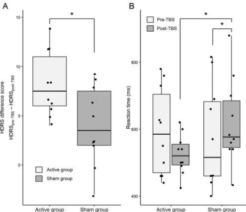
A significant effect of TBS was found in the difference scores of HDRS (HDRSpre-TBS – HDRSpost-TBS) between the active and sham group, t18=2.522, p= .021, Cohen’s d=1.128. In light of the collected data, Bayesian analysis indicated moderate evidence for a difference between the change of HDRS scores, BF10= 3.028. Based on our results, the data was 3 times more likely under H1 (i.e., TBS treatment results in affective changes in the active group) than H0 (i.e., TBS does not affect affective symptoms) (Supplementary Material S1). Fig. 1(A) shows that a higher reduction of HDRS scores was observed in participants receiving active TBS (mean ± SE scores:
active group 8.2 ± 3.360; sham group 4.2 ± 1.172).
ANCOVA controlling for baseline HDRS scores indicated that the effect of baseline HDRS was not significant, F1, 17= 1.118, p= .305, gp
2= 0.062, BFincl= 0.726, whereas a tendency towards the effect of stimulation type on HDRS scores persisted, F1,
17= 3.415, p= .082, gp2
= 0.167, BFincl= 2.372. The Bayesian model comparison yielded that the best model only included the type of stimulation, but not the covariate. Moderate evidence (BF10= 3.028) indicated that this model should be chosen over the null model (seeTable 2).
For the RTs of the 1-back task, significant TIMESTIMULATION interaction was found, F1, 18= 7.503, p= .013, gp2
= 0.294, BFincl= 4.501.
Pairwise comparisons revealed that the RTs of the active TBS group decreased significantly compared to the sham group, p= .031. There was a significant difference between the active and the sham group at the post-TBS time point, p= .046, while no difference was present at the pre-TBS time point, p> .05. The RTs of the active group dropped from (mean ± SE) 592.5 ± 45.3 to 524.5 ± 31.7, while the RTs of the sham group increased from 575.8
± 45.38 to 620.7 ± 31.7 (Fig. 1 (B)). The main effect of TIME, F1,
18= 0.318, p= .580, gp2
= 0.017, BFincl= 0.335, and STIMULATION, F1, 18= 0.597, p= .450, gp2
= 0.032, BFincl= 0.595, were not significant. The Bayesian analysis revealed that the null model slightly outpredicted the full
model (BF10= 0.908,
BF01= 1.101), indicating inconclusive evidence for the null model (Supplementary Material S2).
Regarding thed’scores of the 1- back task, the main effect of TIME, F1, 18= 0.051, p= .824, gp2
= 0.003, BFincl= 0.312, STIMULATION, F1, 18= 1.803,
p= .196, gp2
= 0.091, BFincl= 0.806, and the TIMESTIMULATION interaction, F1, 18= 0.006, p= .939, gp2< 0.001, BFincl= 0.381, was not statistically significant. The null model was the best-fitting model, i.e., it outperformed the full model of BF10= 0.095, BF01= 10.476.
The data were10 times less likely Fig. 1. Cognitive and affective changes in the active and sham group.(A)Box plot with individual
data points depicting the changes of HDRS difference scores (HDRSpre-TBS– HDRSpost-TBS).(B) Box plot with individual data points depicting the reaction time changes on the 1-back task.
Table 2.Model comparison results of Bayesian mixed-model ANOVA
Models P(M) P(M|data) BFM BF10 error %
Null model 0.250 0.144 0.504 1.000
Type of stimulation 0.250 0.436 2.315 3.028 4.367e -4
Type of stimulation + baseline HDRS 0.250 0.268 1.098 1.862 1.182
Baseline HDRS 0.250 0.153 0.541 1.061 0.001
P(M): prior model probabilities, P(M|data): updated probabilities, BFM: the degree change of the prior model odds after having observed the data, BF10: Bayes Factor in favor of H1
under H1than under H0which is considered a substantial evidence supporting the preference of the null model (Supplementary Material S3).
The average of the average RTs of the 2-back and 3- back tasks were entered into a mixed ANOVA which yielded a non-significant main effect of TIME, F1,
18= 0.520, p= .480, gp2
= 0.028, BFincl= 0.396, and STIMULATION, F1, 18= 1.798, p= .197, gp2= 0.091, BFincl= 0.710. The TIMESTIMULATION interaction, F1, 18= 1.422, p= .249, gp2= 0.073, BFincl= 0.630, was not significant either. The null model was the best model outperforming the full model of BF10= 0.180, BF01= 5.556. The data were5 times less likely to be observed under H1 than under H0. This evidence substantially supports that the null model should be preferred (Supplementary Material S4).
Considering thed’scores of the averaged 2-back and 3-back tasks, the main effect of TIME, F1, 18= 2.078, p= .167, gp
2= 0.104, BFincl= 0.712, STIMULATION, F1, 18= 0.098, p= .758, gp2
= 0.005, BFincl= 0.447, and TIMESTIMULATION interaction, F1, 18= 0.321, p= .578, gp2
= 0.018, BFincl= 0.433, was not significant. Bayesian analysis indicated that the best- fitting model was the null model. The results were 7 times less likely to be observed under H1 compared to H0 which is considered as a substantial weight of evidence supporting that the null model should be preferred over the full model, BF10= 0.146, BF01= 6.828 (Supplementary Material S5).
Attention network task
The mixed ANOVA of the overall RTs yielded that the main effect of TIME, F1, 18= 3.071, p= .097, gp
2= 0.146, BFincl= 0.908, the main effect of STIMULATION, F1, 18= 0.584, p= .455, gp2
= 0.031, BFincl= 0.551, and the TIMESTIMULATION interaction, F1, 18= 2.138, p= .161, gp2
= 0.106, BFincl= 1.164, were non-significant. The null model outpredicted the full model (BF10= 0.501, BF01= 1.995); however, the data were2 times less likely to be observed under H1 compared to H0 which only indicates anecdotal evidence in support of the null model (Supplementary Material S6).
Results on the alerting attention ratio indicated a non- significant main effect of TIME,F1, 18= 0.001,p= .973, gp2
< 0.001, BFincl= 0.306, STIMULATION, F1,
18= 0.233, p= .635,gp2
= 0.013, BFincl= 0.463, and a non-significant interaction of TIMESTIMULATION,F1,
18= 0.767, p= .393, gp2
= 0.041, BFincl= 0.500. The full model (BF10= 0.073, BF01= 13.718) was outpredicted by the null model. Strong evidence supported the preference of the null model as the data were 14 times less likely to be observed under H1
than under H0(Supplementary Material S7).
Regarding the orientating attention ratio, we found that the main effect of TIME, F1, 18= 0.961, p= .340, gp2= 0.051, BFincl= 0.495, the main effect of STIMULATION, F1, 18= 0.576, p= .458, gp2
= 0.031, BFincl= 0.450, and the TIMESTIMULATION interaction, F1, 18= 0.173, p= .682, gp2
= 0.010, BFincl= 0.430, were not significant. The full model,
BF10= 0.095, BF01= 10.545, was outperformed by the null model. The likelihood of the data being observed under H1 was 10 times less likely than under H0 indicating a strong evidence for the null model (Supplementary Material S8).
The mixed ANOVA of the executive attention ratio revealed a non-significant main effect of TIME, F1, 18= 0.336, p= .570, gp2= 0.018, BFincl= 0.378, STIMULATION, F1, 18= 3.320, p= .085, gp2
= 0.156, BFincl= 0.581, and a non-significant interaction of TIMESTIMULATION, F1, 18= 0.017, p= .897, gp
2< 0.001, BFincl= 0.373. The full model (BF10= 0.083, BF01= 12.042) was outperformed by the null model i.e. its interpretation is limited. Compared to H0, the likelihood of the data being observed under H1 was 12 times lower indicating strong evidence favoring null model (Supplementary Material S9).
DISCUSSION
Therapeutic effects of rTMS over the DLPFC on depressive symptoms are steadily gaining recognition.
Our results of improved affective symptoms in this randomized, sham-controlled study after ten sessions of bilateral TBS (cTBS over the right DLPFC + iTBS over the left DLPFC) support this notion. Bayesian analysis further corroborated the presence of substantial evidence in support of the antidepressive effects of TBS. However, targeting DLPFC – which is a widely preferred region for non-invasive brain stimulation (Holczer et al., 2020) and a strongly implicated area in MDD (Fitzgerald et al., 2008; Grimm et al., 2008) – might not only affect the affective symptoms but also the cogni- tive functioning (Diener et al., 2012). Strikingly, the cogni- tive effects of NIBS in MDD are rarely investigated with inconclusive preliminary results ranging from no effect (Wajdik et al., 2014) to limited efficacy in some subdo- mains (Iimori et al., 2019; Martin et al., 2017; Scho et al., 2019). Our results indicate that TBS has no or lim- ited effects on the working memory and attentional domains.
The only cognitive measurement on which we found a potential effect of TBS was the overall RT of the 1-back tasks. After active TBS, the frequentist analysis suggested an RT decrease similar to the practice effects experienced in healthy participants (Soveri et al., 2018).
On the contrary, in the sham group, pre-TBS and post- TBS RTs were comparable. The perceived shortening of RTs independently of the cognitive load may occur due to improved psychomotor processing speed. Psychomo- tor speed is often slower in MDD compared to healthy individuals (Liu et al., 2019; Semkovska et al., 2019;
Tian et al., 2016) and is associated with reduced cerebral blood flow in the motor cortex in MDD (Yin et al., 2018).
However, the Bayesian analysis indicated inconclusive results regarding the reaction time measures of the ANT and the 1-back tasks. Thus, more investigations are required to further verify this finding.
The improvement of psychomotor speed, if replicable, might stem from the fact that TBS effects are propagated to remote brain areas (Singh et al., 2020; Tang et al.,
2015). Furthermore, TBS may modulate motor cortex excitability (Cao et al., 2018) and cerebral blood flow (Cho et al., 2012). Another possible explanation can be that TBS might reduce frontal alpha asymmetry (Pellicciari et al., 2017), which is linked to psychomotor retardation (Cantisani et al., 2015).
More pronounced cognitive changes after TBS were hypothesized as single-session stimulation with identical protocols to ours resulted in TBS-induced theta power modulation (Chung et al., 2017). Although theta power increase is associated with improved working memory performance (Jensen and Tesche, 2002; Lisman, 2010) and cognitive control (Cavanagh and Frank, 2014), in our study, TBS did not lead to such cognitive enhance- ment. This result is in contrast with previous promising results (Cheng et al., 2016; Scho et al., 2019). However, in the study of Cheng et al. (2016), patients with treatment-resistant depression were recruited, and a higher dose of stimulation with 1800 pulses/session were delivered.Scho et al. (2019) who have found improved working memory performance, administered unilateral TBS to the left DLPFC. Higher doses of TBS have been proposed to exert more pronounced effects (Nettekoven et al., 2014); however, other results have not fully sup- ported this notion (Volz et al., 2013; Williams et al., 2018). Therefore, it is not clear whether the differences across results can be attributed to the difference in dosing TBS or other factors such as sample characteristics. It is also possible that the antidepressive and cognition- enhancing effects of TBS might be independent.
In the present study, several methodological decisions were based on reports of enhanced antidepressant effects (in the lack of similar methodological recommendations on enhancing cognition). For example, TBS was administered as add-on therapy, since concomitant pharmacotherapy might enhance the development of more stable TBS effects on depressive symptoms (Kedzior et al., 2012). However, cognition and affective symptoms might benefit from different stim- ulation parameters. Distinct patterns of metabolic changes may follow iTBS, cTBS and bilateral TBS (Li et al., 2018). Some TBS effects affecting regions outside the DLPFC relevant to the implementation of executive function (e.g., the medial prefrontal cortex and ACC for cognitive control (Alexander and Brown, 2011)) may be canceled out after bilateral TBS (Li et al., 2018). Thus, it is possible that iTBS, but not the combination of iTBS and cTBS might improve executive functions (Cheng et al., 2016).
One limitation of the present study includes the sham method chosen. While elevating the coil from the scalp hinders significant cortical stimulation (Siebner et al., 2009), other characteristic experiences such as scalp sensations and peripheral nerve stimulation are mostly abolished as well. Although the clicking sounds of the machine and some mechanical vibration can be experi- enced, the use of a more sophisticated sham method (e.g., a sham coil that produces shallow magnetic fields or weak electrical currents) would further improve the blinding of the participants.
Of note, our results may be slightly underpowered in some cognitive domains, as indicated by the BFincl
values. However, BFincl values should be interpreted as a continuous measure (Wagenmakers et al., 2018), and for the ANT indexes and the d’ scores of the n-back task, BFincl values of the interactions approached the cut-off score. This indicates that the conclusions drawn are less likely to be misleading regarding executive functions.
Importantly, we did not find evidence for any immediate cognitive adverse effects of TBS. In comparison, electroconvulsive therapy is associated with impaired executive functioning, episodic memory deficit, and deterioration of global cognition (Andrade et al., 2016; Ren et al., 2014) that reverse in a few months (Bodnar et al., 2016), we show that TBS has the advan- tage of not causing similar temporary impairments while exerting antidepressive effects in patients with MDD.
Taken together, the present study suggests that 10 sessions of bilateral TBS have evident antidepressive effects but have limited cognition-enhancing efficacy.
We found that executive functions were not affected by TBS. Hence, TBS might be a good alternative to electroconvulsive therapy as it does not cause transitory cognitive impairment. However, a systematic comparison of the antidepressant and pro-cognitive features (including the magnitude and the duration of the effects) of different brain stimulation paradigms is necessary. Further research is encouraged on the effects of TBS regarding psychomotor speed, as our results suggested a potential effect of TBS on RTs for visual stimuli. Several questions are yet to be answered regarding the optimal parameters of TBS and whether antidepressant and cognitive-enhancing effects require different parameters; thus, comparative studies of bilateral and unilateral stimulation are warranted.
Nevertheless, bilateral TBS seems to be an acceptable add-on therapy with promising antidepressant effects, a possible effect on psychomotor speed, and no adverse effects impacting attention or working memory.
FUNDING
The preparation of this manuscript was supported by the University of Szeged Open Access Fund (5101). AH was supported by the New National Excellence Programme.
LV received support from the grants GINOP-2.3.2-15- 2016-00034 and GINOP 2.3.2-15-2016-00048. PK was supported by the Hungarian Brain Research program:
2017-1.2.1-NKP-2017-00002. AM received support from the Bolyai Scholarship Programme of the Hungarian Academy of Sciences and from the EU-funded Hungarian grant EFOP-3.6.1-16-2016-00008.
DECLARATION OF INTEREST
The authors declare that the research was conducted in the absence of any commercial or financial relationships that could be construed as a potential conflict of interest.
AUTHORS’ CONTRIBUTIONS
AH: Formal analysis, Writing - original draft, review &
editing, Visualization, Investigation; VLN:
Conceptualization, Methodology, Investigation; TV:
Investigation, Writing - Review & Editing; KK:
Investigation, Resources; AK: Resources; ZsTK:
Conceptualization; Resources; LV: Conceptualization;
PK: Conceptualization; AM: Conceptualization, Methodology, Supervision.
REFERENCES
Alexander WH, Brown JW (2011) Medial prefrontal cortex as an action-outcome predictor. Nat Neurosci 14(10):1338–1344.
https://doi.org/10.1038/nn.2921.
Andrade C, Arumugham SS, Thirthalli J (2016) Adverse effects of electroconvulsive therapy. Psychiatric Clinics 39(3):513–530.
https://doi.org/10.1016/j.psc.2016.04.004.
Behera P, Gupta SK, Nongkynrih B, Kant S, Mishra AK, Sharan P (2017) Screening instruments for assessment of depression.
Indian J Med Specialities 8(1):31–37. https://doi.org/10.1016/j.
injms.2016.11.003.
Berlim MT, Van den Eynde F, Daskalakis ZJ (2013a) A systematic review and meta-analysis on the efficacy and acceptability of bilateral repetitive transcranial magnetic stimulation (rTMS) for treating major depression. Psychol Med 43(11):2245–2254.
https://doi.org/10.1017/S0033291712002802.
Berlim MT, Van FdenE, Daskalakis ZJ (2013b) High-frequency repetitive transcranial magnetic stimulation accelerates and enhances the clinical response to antidepressants in major depression: a meta-analysis of randomized, double-blind, and sham-controlled trials. J Clin Psychiatry 74(2):e122–e129.https://
doi.org/10.4088/JCP.12r07996.
Blumberger DM, Mulsant BH, Fitzgerald PB, Rajji TK, Ravindran AV, Young LT, Levinson AJ, Daskalakis ZJ (2012) A randomized double-blind sham-controlled comparison of unilateral and bilateral repetitive transcranial magnetic stimulation for treatment-resistant major depression. World J Biol Psychiatry 13 (6):423–435.https://doi.org/10.3109/15622975.2011.579163.
Bodnar A, Krzywotulski M, Lewandowska A, Chlopocka-Wozniak M, Bartkowska-Sniatkowska A, Michalak M, Rybakowski JK (2016) Electroconvulsive therapy and cognitive functions in treatment- resistant depression. World J Biol Psychiatry 17(2):159–164.
https://doi.org/10.3109/15622975.2015.1091501.
Boroojerdi B, Meister IG, Foltys H, Sparing R, Cohen LG, To¨pper R (2002) Visual and motor cortex excitability: a transcranial magnetic stimulation study. Clin Neurophysiol 113 (9):1501–1504.https://doi.org/10.1016/s1388-2457(02)00198-0.
Cantisani A, Koenig T, Horn H, Mu¨ller T, Strik W, Walther S (2015) Psychomotor retardation is linked to frontal alpha asymmetry in major depression. J Affect Disord 188:167–172.https://doi.org/
10.1016/j.jad.2015.08.018.
Cao N, Pi Y, Liu K, Meng H, Wang Y, Zhang J, Wu Y, Tan X (2018) Inhibitory and facilitatory connections from dorsolateral prefrontal to primary motor cortex in healthy humans at rest—An rTMS study. Neurosci Lett 687:82–87. https://doi.org/10.1016/j.
neulet.2018.09.032.
Cavanagh JF, Frank MJ (2014) Frontal theta as a mechanism for cognitive control. Trends Cognitive Sci 18(8):414–421.https://doi.
org/10.1016/j.tics.2014.04.012.
Chen J, Liu Z, Zhu D, Li Q, Zhang H, Huang H, Wei Y, Mu J, Yang D, Xie P (2014) Bilateral vs. unilateral repetitive transcranial magnetic stimulation in treating major depression: a meta- analysis of randomized controlled trials. Psychiatry Res 219 (1):51–57.https://doi.org/10.1016/j.psychres.2014.05.010.
Cheng C-M, Juan C-H, Chen M-H, Chang C-F, Lu HJ, Su T-P, Lee Y- C, Li C-T (2016) Different forms of prefrontal theta burst stimulation for executive function of medication- resistant
depression: evidence from a randomized sham-controlled study.
Prog Neuro-Psychopharmacol Biol Psychiatry 66:35–40.https://
doi.org/10.1016/j.pnpbp.2015.11.009.
Chistyakov AV, Kreinin B, Marmor S, Kaplan B, Khatib A, Darawsheh N, Koren D, Zaaroor M, Klein E (2015) Preliminary assessment of the therapeutic efficacy of continuous theta-burst magnetic stimulation (cTBS) in major depression: a double-blind sham- controlled study. J Affect Disord 170:225–229. https://doi.org/
10.1016/j.jad.2014.08.035.
Cho SS, Pellecchia G, Ko JH, Ray N, Obeso I, Houle S, Strafella AP (2012) Effect of continuous theta burst stimulation of the right dorsolateral prefrontal cortex on cerebral blood flow changes during decision making. Brain Stimulation 5(2):116–123.https://
doi.org/10.1016/j.brs.2012.03.007.
Chung SW, Lewis BP, Rogasch NC, Saeki T, Thomson RH, Hoy KE, Bailey NW, Fitzgerald PB (2017) Demonstration of short-term plasticity in the dorsolateral prefrontal cortex with theta burst stimulation: a TMS-EEG study. Clin Neurophysiol 128 (7):1117–1126.https://doi.org/10.1016/j.clinph.2017.04.005.
Demirtas-Tatlidede A, Vahabzadeh-Hagh AM, Pascual-Leone A (2013) Can noninvasive brain stimulation enhance cognition in neuropsychiatric disorders? Neuropharmacology 64:566–578.
https://doi.org/10.1016/j.neuropharm.2012.06.020.
Diener C, Kuehner C, Brusniak W, Ubl B, Wessa M, Flor H (2012) A meta-analysis of neurofunctional imaging studies of emotion and cognition in major depression. NeuroImage 61(3):677–685.
https://doi.org/10.1016/j.neuroimage.2012.04.005.
Drevets WC, Savitz J, Trimble M (2008) The Subgenual anterior cingulate cortex in mood disorders. CNS Spectr 13(8):663–681.
Fan J, McCandliss BD, Sommer T, Raz A, Posner MI (2002) Testing the efficiency and independence of attentional networks. J Cognit Neurosci 14(3):340–347. https://doi.org/10.1162/
089892902317361886.
Fitzgerald PB, Brown TL, Marston NAU, Daskalakis ZJ, de Castella A, Kulkarni J (2003) Transcranial magnetic stimulation in the treatment of depression: a double-blind, placebo-controlled trial.
Arch Gen Psychiatry 60(10):1002–1008.https://doi.org/10.1001/
archpsyc.60.9.1002.
Fitzgerald PB, Hoy K, Daskalakis ZJ, Kulkarni J (2009) A randomized trial of the anti-depressant effects of low- and high-frequency transcranial magnetic stimulation in treatment-resistant depression. Depression Anxiety 26(3):229–234. https://doi.org/
10.1002/da.v26:310.1002/da.20454.
Fitzgerald PB, Laird AR, Maller J, Daskalakis ZJ (2008) A meta- analytic study of changes in brain activation in depression. Hum Brain Mapp 29(6):683–695.https://doi.org/10.1002/hbm.20426.
Fox MD, Buckner RL, White MP, Greicius MD, Pascual-Leone A (2012) Efficacy of transcranial magnetic stimulation targets for depression is related to intrinsic functional connectivity with the subgenual cingulate. Biol Psychiatry 72(7):595–603.https://doi.
org/10.1016/j.biopsych.2012.04.028.
Ga¨rtner M, Ghisu ME, Scheidegger M, Bo¨nke L, Fan Y, Stippl A, Herrera-Melendez A-L, Metz S, Winnebeck E, Fissler M, Henning A, Bajbouj M, Borgwardt K, Barnhofer T, Grimm S (2018) Aberrant working memory processing in major depression:
Evidence from multivoxel pattern classification.
Neuropsychopharmacology 43(9):1972–1979. https://doi.org/
10.1038/s41386-018-0081-1.
Gorwood P, Richard-Devantoy S, Bayle´ F, Cle´ry-Melun ML (2014) Psychomotor retardation is a scar of past depressive episodes, revealed by simple cognitive tests. Eur Neuropsychopharmacol 24(10):1630–1640. https://doi.org/10.1016/j.
euroneuro.2014.07.013.
Grimm S, Beck J, Schuepbach D, Hell D, Boesiger P, Bermpohl F, Niehaus L, Boeker H, Northoff G (2008) Imbalance between left and right dorsolateral prefrontal cortex in major depression is linked to negative emotional judgment: an fMRI study in severe major depressive disorder. Biol Psychiatry 63(4):369–376.https://
doi.org/10.1016/j.biopsych.2007.05.033.
Haatveit BC, Sundet K, Hugdahl K, Ueland T, Melle I, Andreassen OA (2010) The validity of d prime as a working memory index:
Results from the ‘‘Bergen n-back task”. J Clin Exp Neuropsychol 32(8):871–880.https://doi.org/10.1080/13803391003596421.
Halper J, Manevitz A, Nishimoto K, Yagi C, Onishi A, Ahmed S (2019) Is there role for TMS delivered at intensities at or below 100%
resting motor threshold (RMT)? Brain Stimulation 12(4). https://
doi.org/10.1016/j.brs.2019.03.043e135.
Hamilton M (1960) A rating scale for depression. J Neurol Neurosurg Psychiatry 23(1):56–62.https://doi.org/10.1136/jnnp.23.1.56.
He X, Lan Y, Xu G, Mao Y, Chen Z, Huang D, Pei Z (2013) Frontoparietal regions may become hypoactive after intermittent theta burst stimulation over the contralateral homologous cortex in humans. J Neurophysiol 110(12):2849–2856. https://doi.org/
10.1152/jn.00369.2013.
Hecht D (2010) Depression and the hyperactive right-hemisphere.
Neurosci Res 68(2):77–87. https://doi.org/10.1016/j.
neures.2010.06.013.
Holczer A, Ne´meth VL, Ve´kony T, Ve´csei L, Klive´nyi P, Must A (2020) Non-invasive brain stimulation in Alzheimer’s disease and mild cognitive impairment—a state-of-the-art review on methodological characteristics and stimulation parameters.
Front Hum Neurosci 14. https://doi.org/10.3389/
fnhum.2020.00179.
Huang Y-Z, Edwards MJ, Rounis E, Bhatia KP, Rothwell JC (2005) Theta burst stimulation of the human motor cortex. Neuron 45 (2):201–206.https://doi.org/10.1016/j.neuron.2004.12.033.
Iimori T, Nakajima S, Miyazaki T, Tarumi R, Ogyu K, Wada M, Tsugawa S, Masuda F, Daskalakis ZJ, Blumberger DM, Mimura M, Noda Y (2019) Effectiveness of the prefrontal repetitive transcranial magnetic stimulation on cognitive profiles in depression, schizophrenia, and Alzheimer’s disease: a systematic review. Prog Neuro-Psychopharmacol Biol Psychiatry 88:31–40. https://doi.org/10.1016/j.
pnpbp.2018.06.014.
Isenberg K, Downs D, Pierce K, Svarakic D, Garcia K, Jarvis M, North C, Kormos TC (2005) Low frequency rTMS stimulation of the right frontal cortex is as effective as high frequency rTMS stimulation of the left frontal cortex for antidepressant-free, treatment-resistant depressed patients. Ann Clin Psychiatry 17(3):153–159. https://
doi.org/10.3109/10401230591002110.
Jensen O, Tesche CD (2002) Frontal theta activity in humans increases with memory load in a working memory task. Eur J Neurosci 15(8):1395–1399. https://doi.org/10.1046/j.1460- 9568.2002.01975.x.
Kaiser RH, Andrews-Hanna JR, Wager TD, Pizzagalli DA (2015) Large-scale network dysfunction in major depressive disorder: a meta-analysis of resting-state functional connectivity. JAMA Psychiatry 72(6):603–611. https://doi.org/
10.1001/jamapsychiatry.2015.0071.
Kaminski JA, Korb FM, Villringer A, Ott DVM (2011) Transcranial magnetic stimulation intensities in cognitive paradigms. PLoS ONE 6(9).https://doi.org/10.1371/journal.pone.0024836.
Kedzior KK, Rajput V, Price G, Lee J, Martin-Iverson M (2012) Cognitive correlates of repetitive transcranial magnetic stimulation (rTMS) in treatment-resistant depression—A pilot study. BMC Psychiatry 12:163.https://doi.org/10.1186/1471-244X-12-163.
Lefaucheur J-P, Aleman A, Baeken C, Benninger DH, Brunelin J, Di Lazzaro V, Filipovic´ SR, Grefkes C, Hasan A, Hummel FC, Ja¨a¨skela¨inen SK, Langguth B, Leocani L, Londero A, Nardone R, Nguyen J-P, Nyffeler T, Oliveira-Maia AJ, Oliviero A, Ziemann U (2020) Evidence-based guidelines on the therapeutic use of repetitive transcranial magnetic stimulation (rTMS): an update (2014–2018). Clin Neurophysiol 131(2):474–528.https://doi.org/
10.1016/j.clinph.2019.11.002.
Li C-T, Chen M-H, Juan C-H, Huang H-H, Chen L-F, Hsieh J-C, Tu P- C, Bai Y-M, Tsai S-J, Lee Y-C, Su T-P (2014) Efficacy of prefrontal theta-burst stimulation in refractory depression: a randomized sham-controlled study. Brain 137(Pt 7):2088–2098.
https://doi.org/10.1093/brain/awu109.
Li C-T, Chen M-H, Juan C-H, Liu R-S, Lin W-C, Bai Y-M, Su T-P (2018) Effects of prefrontal theta-burst stimulation on brain function in treatment-resistant depression: a randomized sham-
controlled neuroimaging study. Brain Stimulation 11 (5):1054–1062.https://doi.org/10.1016/j.brs.2018.04.014.
Lisman J (2010) Working memory: the importance of theta and gamma oscillations. Curr Biol: CB 20(11):R490–492.https://doi.
org/10.1016/j.cub.2010.04.011.
Liu T, Zhong S, Wang B, Liao X, Lai S, Jia Y (2019) Similar profiles of cognitive domain deficits between medication-naı¨ve patients with bipolar II depression and those with major depressive disorder. J Affect Disord 243:55–61. https://doi.org/10.1016/
j.jad.2018.05.040.
Lowe CJ, Manocchio F, Safati AB, Hall PA (2018) The effects of theta burst stimulation (TBS) targeting the prefrontal cortex on executive functioning: a systematic review and meta-analysis.
Neuropsychologia 111:344–359. https://doi.org/10.1016/j.
neuropsychologia.2018.02.004.
Martin DM, McClintock SM, Forster JJ, Lo TY, Loo CK (2017) Cognitive enhancing effects of rTMS administered to the prefrontal cortex in patients with depression: a systematic review and meta-analysis of individual task effects. Depression Anxiety 34(11):1029–1039. https://doi.org/10.1002/da.2017.34.
issue-1110.1002/da.22658.
Martin DM, McClintock SM, Forster J, Loo CK (2016) Does therapeutic repetitive transcranial magnetic stimulation cause cognitive enhancing effects in patients with neuropsychiatric conditions? A systematic review and meta-analysis of randomised controlled trials. Neuropsychol Rev 26(3):295–309.
https://doi.org/10.1007/s11065-016-9325-1.
Mendlowitz AB, Shanbour A, Downar J, Vila-Rodriguez F, Daskalakis ZJ, Isaranuwatchai W, Blumberger DM (2019) Implementation of intermittent theta burst stimulation compared to conventional repetitive transcranial magnetic stimulation in patients with treatment resistant depression: a cost analysis. PLoS ONE 14 (9).https://doi.org/10.1371/journal.pone.0222546e0222546.
Nebes RD, Pollock BG, Houck PR, Butters MA, Mulsant BH, Zmuda MD, Reynolds CF (2003) Persistence of cognitive impairment in geriatric patients following antidepressant treatment: a randomized, double-blind clinical trial with nortriptyline and paroxetine. J Psychiatr Res 37(2):99–108. https://doi.org/
10.1016/S0022-3956(02)00085-7.
Nettekoven C, Volz LJ, Kutscha M, Pool E-M, Rehme AK, Eickhoff SB, Fink GR, Grefkes C (2014) Dose-dependent effects of theta burst rTMS on cortical excitability and resting-state connectivity of the human motor system. J Neurosci 34(20):6849–6859.https://
doi.org/10.1523/JNEUROSCI.4993-13.2014.
Nyffeler T, Mu¨ri RM, Bucher-Ottiger Y, Pierrot-Deseilligny C, Gaymard B, Rivaud-Pechoux S (2007) Inhibitory control of the human dorsolateral prefrontal cortex during the anti-saccade paradigm a transcranial magnetic stimulation study. Eur J Neurosci 26(5):1381–1385. https://doi.org/10.1111/j.1460- 9568.2007.05758.x.
O’Reardon JP, Solvason HB, Janicak PG, Sampson S, Isenberg KE, Nahas Z, McDonald WM, Avery D, Fitzgerald PB, Loo C, Demitrack MA, George MS, Sackeim HA (2007) Efficacy and safety of transcranial magnetic stimulation in the acute treatment of major depression: a multisite randomized controlled trial. Biol Psychiatry 62(11):1208–1216. https://doi.org/10.1016/j.
biopsych.2007.01.018.
Pan Z, Grovu RC, Cha DS, Carmona NE, Subramaniapillai M, Shekotikhina M, Rong C, Lee Y, McIntyre RS (2017) Pharma- cological treatment of cognitive symptoms in major depressive disorder. CNS Neurol Disord: Drug Targets 16(8):891–899.https://
doi.org/10.2174/1871527316666170919115100.
Pellicciari MC, Ponzo V, Caltagirone C, Koch G (2017) Restored asymmetry of prefrontal cortical oscillatory activity after bilateral theta burst stimulation treatment in a patient with major depressive disorder: a TMS-EEG study. Brain Stimulation 10 (1):147–149.https://doi.org/10.1016/j.brs.2016.09.006.
Plewnia C, Pasqualetti P, Große S, Schlipf S, Wasserka B, Zwissler B, Fallgatter A (2014) Treatment of major depression with bilateral theta burst stimulation: a randomized controlled pilot trial. J Affect Disord 156:219–223.https://doi.org/10.1016/j.jad.2013.12.025.