The novel indications of cardiac CT Ph.D. Thesis
Andrea Bartykowszki M.D.
Doctoral School of
Basic and Translational Medicine Semmelweis University
Supervisor:
Pál Maurovich-Horvat M.D., Ph.D., Official reviewers:
Zsuzsanna Földes-Lénárd M.D., Ph.D.;
Attila Pálinkás M.D., Ph.D.,
Head of the Final Examination Committee Viktor Bérczi M.D., Ph.D.,
Members of the Final Examination Committee:
Attila Doros M.D., Ph.D., László Sallai M.D., Ph.D.;
Budapest
2018
1 1. Introduction
In recent years, modern therapeutic innovations have prolonged the life of patients living with different cardiac diseases leading to an increasing prevalence of heart failure. However, despite improvements in therapy, the mortality rate in patients with HF has remained high. Heart transplantation (HTx) remains the gold standard definitive surgical approach in the treatment of refractory heart failure.
According to the registry of the International Society of Heart and Lung Transplantation the 1 year survival after HTx is 85%, the 5-year survival is 73%. During the first year after heart transplantation cardiac allograft vasculopathy (CAV) is one of the leading causes of death. The overall prevalence of CAV at 1, 5, and 10 years after transplantation is 8%, 30%, and 50%, respectively. The mortality of patients with 3-vessel CAV can be up to 90% in the first year from diagnosis. In addition, more than 0.5 mm change in the maximal intimal thickness within the first post-transplantation year is associated with increased mortality.
CAV is characterized by diffuse concentric intimal hyperplasia which leads to ischemic consequences, including graft failure, arrhythmias, and sudden death. Because of the denervated nature of transplanted hearts, patients do not experience symptoms related to ischemia, therefore, early diagnosis of CAV is challenging. International guidelines recommend annual or biannual invasive coronary angiography (ICA) for the assessment of coronary status. However, ICA has limited diagnostic accuracy to detect CAV because of the diffuse and concentric manifestation of the disease. Furthermore, ICA does not provide information regarding the coronary wall; therefore, intravascular ultrasound or optical coherence tomography is suggested as a complementary imaging test.
2
The combination of ICA with intravascular imaging techniques increases sensitivity, but their routine use increases costs and rates of procedural complications; therefore, it is considered optional for CAV assessment. In addition, the International Society for Heart and Lung Transplantation consensus statement does not recommend the routine use of intravascular ultrasound for CAV assessment.
Coronary CT angiography (CTA) allows noninvasive visualization of the coronary artery wall and lumen with a high diagnostic accuracy, and it can detect 1.5–2 times more coronary segments with coronary atherosclerotic plaques than invasive coronary angiography does.
Contemporary CT scanners have a very good temporal resolution of 0.5-0.625 mm.
Notably, the absence of parasympathetic and sympathetic innervation of the transplanted hearts results in higher resting heart rates (HRs), which may compromise the diagnostic performance of coronary CTA in this patient population. Moreover, because of their higher HRs, retrospective ECG-gating has been used for HTx recipients, which results in higher radiation dose. These concerns precluded the widespread use of coronary CTA in HTx recipients. Prospectively ECG-triggered coronary CTA would be desirable because of its low radiation dose, but it requires a low HR (generally < 65 beats/min).
The HTx recipients have higher but steady HR with minimal HR variability because of the lack of autonomic innervation. The steady HR of HTx recipients might provide a unique opportunity to scan these patients with low radiation dose and achieve good image quality.
Beyond ruling out significant coronary artery disease (CAD), coronary CT can be a useful tool to assess cardiovascular risk. Calcified plaque burden can be evaluated on the non-enhanced, low radiation dose, ECG-gated images; the resulting coronary calcium score is an important predictor of cardiovascular morbidity and mortality. Calcium score assessment is possible in asymptomatic and in symptomatic patient population as well. According to the guidelines, chest pain evaluation
3
should be based on the probability assessment of CAD. Risk assessment is traditionally performed using the Diamond and Forrester model or the Duke clinical score, however, these models may overestimate the probability of obstructive coronary lesions yielding to additional unnecessary testing.
2. Aims
Our primary aim was to assess the image quality and the radiation dose of prospectively ECG-triggered coronary CTA in HTx recipients.
The secondary aim of our study was to assess the feasibility of quantitative coronary wall volume change assessment in HTx patients using coronary CTA. Furthermore, we aimed to compare the performance of quantitative coronary CTA assessment versus conventional qualitative clinical reading to rule out progressive coronary vessel wall thickening, indicative for CAV.
We perceived a need for an updated and stepwise approach to estimate the probability of coronary artery disease in patients with new onset of chest pain in a low prevalence population as clinical information and test results become available, in particular because implementation of the guidelines needs calculation of the pretest probability. Therefore, we aimed to estimate the probability of obstructive coronary artery disease on the basis of clinical presentation and cardiovascular risk factors, and to determine the incremental diagnostic value of exercise electrocardiography and the coronary calcium score.
4 3. Methods
3.1. The image quality of coronary CTA in heart transplant recipients
In a retrospective matched case-control cohort study, we evaluated the image quality of coronary CTA performed of HTx recipients. During a 4-year period, 97 coronary CTAs were performed of 57 HTx recipients to rule out CAV. If a patient underwent more than one scan, the scan obtained with the highest HR was selected. In total, 50 HTx recipients (HTx group) were included in the study. The image quality of the scans of the HTx recipients was compared with that of scans of a control group of patients who did not undergo HTx. The control group was selected from our institutional cardiac CT registry. We selected the control group according to matching criteria that may influence image quality: age, sex, body mass index (weight in kilograms divided by the square of height in meters), HR, data acquisition phase (systole or diastole), and coronary dominance.
All patients underwent imaging with a 256-MDCT scanner (Brilliance iCT 256, Philips Healthcare). Tube voltage was 100–120 kV, and the tube current was set to 100–300 mA depending on the body mass index of the patients. Collimation was 2 × 128 × 0.625 mm, with a gantry rotation time of 270 ms. Both the HTx recipients and the control group were scanned with a prospectively ECG-triggered acquisition mode. When the HR was over 80 beats/min, systolic triggering was used at 40% of the cardiac cycle with 3% padding (37–
43% of the R-R interval); in all other cases, diastolic triggering was used at 78% of the cardiac cycle with 3% padding (75–81% of the R- R interval). The same coronary CTA scan protocol and settings were used for each patients’ baseline and follow-up scans.
Reconstructed images were evaluated by two readers (with 5 and 3 years of experience in coronary CTA) using the 18-segment model of the Society of Cardiovascular CT. Coronary segments with a diameter
5
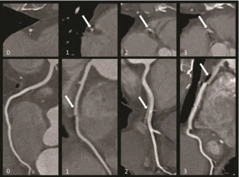
greater than 1.5 mm were assessed. Motion artifacts were described in every coronary segment using a 4-point Likert scale (Fig. 1).
To quantify the total amount of motion artifacts on a per-patient level, we defined the segment motion score, which describes how many segments had motion artifact, and the segment Likert score, which is the sum of the motion severity Likert score of the patient. Because the number of coronary segments affects the total obtainable score, we normalized the scores by dividing them by the number of segments present, which resulted in the segment motion score index and segment Likert score index. To describe how many non-diagnostic segments were present, we defined the segment non-diagnostic score and also divided it by the number of the evaluated segments, which yielded the segment non-diagnostic score index.
Figure 1. Examples of 4-point Likert scale of motion artifacts in heart transplant recipients: 0, excellent image quality with no artifacts; 1, good image quality with
6
minor artifacts; 2, moderate image quality, acceptable for routine clinical diagnosis; 3, not evaluable, with severe artifacts impairing accurate evaluation
3.2. Qualitative and quantitative image analysis
We included 35 consecutive HTx patients who underwent coronary CTA examination at one year and at two years after HTx as part of the routine clinical workflow at our institution. The first-year and second- year follow-up CTA scans were analyzed retrospectively and no data acquisition was performed in addition to routine examinations.
The Society of Cardiovascular CT guidelines were used for conventional data analysis. Coronary CTA reading was performed by a cardiologist with 6 years of experience in coronary CTA imaging.
Luminal stenosis was categorized into the following classes: (0) normal – absence of any plaque, no luminal stenosis; (1) minimal – plaque causing <25% stenosis; (2) mild – 25% to 49% stenosis; (3) moderate – 50% to 69% stenosis; (4) severe − 70% to 99% stenosis;
(5) occluded. Calcified, non-calcified and partially calcified lesions were distinguished. Baseline and follow-up coronary CTA datasets were loaded side by side for comparison. Progression was defined as the appearance of any novel coronary lesion and/or the classification of a previously described coronary lesion into a higher stenosis category on the follow-up scan.
A dedicated offline workstation (QAngioCT, version 2.1; Medis Medical Imaging Systems, Leiden, The Netherlands) was used for semi-automated lesion quantification. Two experienced observers evaluated the images in a random order, blinded to acquisition date and the results of the clinical read. Fix window settings of 1400/500 Hounsfield Units (HU) were used for segmentations. We used fixed threshold parameters to distinguish lesion components on different HU strata: calcified lesion volumes (>350 HU), non-calcified lesion volumes with high attenuation (131–350 HU), non-calcified lesion
7
volumes with intermediate attenuation (75–130 HU) and non-calcified lesion volumes with low attenuation (<75 HU). Lumen volume, overall lesion volume and overall lesion burden (total vessel volume minus the lumen volume, divided by the total vessel volume) were assessed on a per vessel basis. Progression was defined as more than 10% increase in overall lesion volume on the follow-up CTA, as compared to the baseline scan.
Statistical analysis
The Shapiro-Wilk test was used to assess normality. Because all continuous variables showed non-normal distribution, continuous variables are expressed as median and interquartile range (IQR).
Categoric variables are expressed as numbers and percentages. The Mann-Whitney U test was used to compare continuous data of the HTx and non-HTx groups. Categoric data were compared using the chi-square test. Intrareader and interreader reproducibility was assessed on the basis of 20 randomly selected individuals’ images using Cohen kappa, interpreted as follows: 1.00–0.81, excellent; 0.80–
0.61, good; 0.60–0.41, moderate; 0.40–0.21, fair; and 0.20–0.00, poor.
Categorical variables are expressed as numbers and percentages.
Wilcoxon signed-rank test was used to compare the plaque volumes of the baseline and follow-up CTA. Categorical data was compared using the McNemar test. The inter-reader reproducibility between quantitative plaque measurements was calculated using the intra-class correlation coefficient (ICC). The following descriptive scale was used for values of the ICC: <0.40 poor, 0.40–0.59 fair, 0.60–0.74 good and 0.75–1.00 excellent. All statistical calculations were done using SPSS software (version 23, IBM). A p < 0.05 was considered significant.
8
3.3. Prediction model to estimate presence of coronary artery disease
Researchers from Europe and the US formed a consortium. An existing database of at least 80 eligible patients was required for participation. Patients were eligible for the analysis if they presented with stable chest pain and were referred for catheter based or CT based coronary angiography (≥64 slice).
We collected data for age, sex, symptoms, cardiovascular risk factors, test results, and presence of coronary artery disease. Chest pain symptoms were classified as typical, atypical, or non-specific. Typical chest pain was defined as all of the following criteria: (1) substernal chest pain or discomfort that is (2) provoked by exertion or emotional stress and (3) relieved by rest or nitroglycerine (or both). We defined atypical chest pain as two of these criteria. If one or none of the criteria was present, symptoms were classified as non-specific. Primary outcome was obstructive coronary artery disease, defined as at least one vessel with at least 50% diameter stenosis found on catheter based coronary angiography.
We defined three prediction models: a basic model including age, sex, symptoms; a clinical model including age, sex, symptoms, diabetes, hypertension, dyslipidaemia, smoking, and body mass index; and an extended model including all clinical variables and the coronary calcium score. Since all clinical variables are known to be associated with coronary artery disease, all predictors were entered simultaneously in a multivariable, random effects, logistic regression model.
We quantified diagnostic performance by calculating the area under the receiver operating characteristics curve (c statistic).
Reclassification was assessed by use of the continuous net reclassification improvement. We regarded p < 0.05 to be statistically significant. Analyses were performed using Stata/SE 11 (StataCorp).
9 4. Results
4.1. The image quality of coronary CTA in heart transplant recipients
In total, 50 HTx recipients were included in our study. Every HTx recipient had a matched control subject who did not undergo HTx;
therefore, 100 subjects in total were evaluated. In the HTx group (11 women [22%]; 4.3 years after transplantation), the median age was 57.9 years (IQR, 46.7–59.9 years) and the median HR was 74.0 beats/
min (IQR, 67.8–79.3 beats/min), compared with 73.0 beats/min (IQR, 68.5–80.0 beats/ min) in the matched control group (p = 0.58). We found no significant difference between the HTx and control groups regarding anthropometric data and scan characteristics. The effective radiation dose was relatively low in both groups (3.7 mSv [IQR, 2.4–
4.3 mSv] in the HTx group vs 4.3 mSv [IQR, 2.6–4.3 mSv] in the control group; p = 0.24).
In total, 1270 coronary segments were evaluated, 662 segments in the HTx group and 608 segments in the control group. The distribution of motion scores between the two groups is shown in Figure 2. In the HTx group, more segments (624; 94.3%) had diagnostic image quality compared with the control group (504; 82.9%) (p < 0.001) (Fig. 2).
10
Figure 2. Proportions of coronary segments with non-diagnostic, moderate, good and excellent image quality in heart transplantation (HTx) recipients and control subjects.
In the HTx group, more segments had excellent image quality than in the control group (442 [66.7%] vs 271 [4.5%]; p < 0.001).
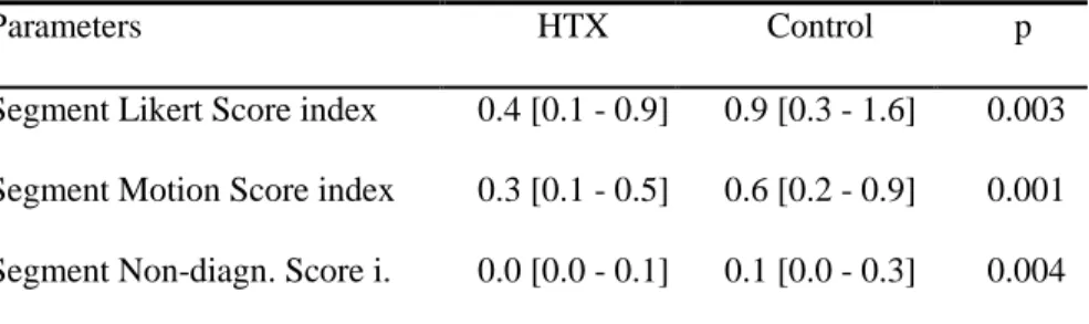
Furthermore, in the HTx group the number of non-diagnostic segments was approximately one-third of that of the control group (38 [5.8%] vs 104 [17.1%]; p < 0.001). We a found a significant difference between the two groups regarding the segment Likert score, the segment motion score, and the segment non-diagnostic score indices.
(Table 1.)
Table 1. Segment motion score indices in the heart transplantation group (HTx) and in the control group. Data are median and interquartile range [IQR]
Parameters HTX Control p
Segment Likert Score index 0.4 [0.1 - 0.9] 0.9 [0.3 - 1.6] 0.003 Segment Motion Score index 0.3 [0.1 - 0.5] 0.6 [0.2 - 0.9] 0.001 Segment Non-diagn. Score i. 0.0 [0.0 - 0.1] 0.1 [0.0 - 0.3] 0.004
11
Intrareader and interreader agreement for image quality scores was good (κ = 0.72 and κ = 0.62, respectively). Dichotomization of image quality scores to excellent and non-excellent image quality scores resulted in excellent intrareader (κ = 0.83) and good interreader (κ = 0.69) reproducibility. Dichotomization to diagnostic and non- diagnostic image quality scores also showed excellent intrareader (κ
= 0.82) and good interreader (κ = 0.73) reproducibility.
4.2. Qualitative image analysis
We included 35 consecutive HTx patients (age 58 [50–61] years, 66%
male). Coronary lesions were detectable in 74% (26/35) of the patients at baseline standard CT read, while on the follow-up CTs 80% (28/35) of the patients had at least one lesion present (p = 0.48). At baseline 19% (82/427), whereas at the follow-up 27% (116/427) of the coronary segments showed any lesion (p < 0.001). The distribution of the segments containing non-calcified, calcified and partially calcified lesions were 61% (50/82), 22% (18/82) and 17% (14/82) at the first CT, and 64% (74/116), 20% (23/116) and 16% (19/116) at the follow- up CT (p < 0.001, p = 1.00 and p = 0.26, respectively). At the baseline CT scan 55% (45/82) of the lesions caused minimal stenosis, 39%
(32/82) mild and 6% (5/82) moderate luminal narrowing. No severe coronary stenosis was revealed. At the follow-up CT scan, 50%
(58/116) of the coronary lesions caused minimal, 36% (42/116) mild, 10% (12/116) moderate luminal narrowing and in 4% (4/116) of the segments severe stenosis was detected (p < 0.001, p = 0.14, p = 0.08, p = 0.25, respectively). Patients with severe stenosis were referred for ICA, which confirmed the severe luminal stenosis in all cases.
4.3. Quantitative image analysis
The total length of analyzed coronary arteries did not differ between the baseline and follow-up CTAs, 248 [IQR: 213;295] mm versus 250
12
[IQR: 213;296] mm, (p = 0.18). Total lumen volume did not change between baseline and follow-up studies (2237 [IQR: 1610;2783] vs.
2197 [IQR: 1677;2527] mm3, p = 0.59). Total vessel wall volume showed significant increase during the follow-up period (464 [IQR:
338; 571] vs. 563 [IQR: 345; 718] mm3, p < 0.001). Accordingly, overall lesion burden increased from 17% [IQR: 14; 19] to 20% [IQR:
15; 24], p < 0.001.
The volume of high-, intermediate and low-attenuation non-calcified coronary vessel wall components showed significant increase (332 [IQR:217;425] vs. 385 [IQR: 238;489], 40 [IQR: 12;48] vs. 59 [IQR:
16;83] and 18 [IQR: 4;21] vs. 46 [IQR: 6;41] mm3, respectively, p < 0.05 all), while calcified volume did not change between baseline and follow-up CTAs (72 [IQR: 16;127] vs. 72 [IQR: 29;102] mm3, p = 0.73). Lesion volumes are summarized in Table 2.
4.4. Progressive vessel wall thickening
Based on conventional coronary CTA reading, progression was present in 11 of 35 (31%) patients, whereas quantitative analysis revealed progression in twice as many patients, 22 of 35 (63%), p = 0.01. Individual lesion volumes showed very good inter-observer agreement for calcified, high-, intermediate-, and low-attenuation non-calcified volumes (ICC: 0.93 [95%CI 0.65; 0.98], 0.92 [95%CI 0.66; 0.98], 0.92 [95%CI 0.66; 0.98] and 0.88 [95%CI 0.53; 0.97], respectively). These resulted in an excellent reproducibility for overall lesion volume and overall lesion burden (ICC: 0.87 [95%CI 0.48;
0.97] and 0.85 [95%CI 0.43; 0.96], respectively.)
13
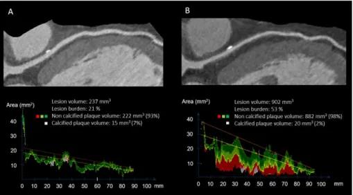
Figure 3. Coronary lesion tissue volumes at baseline (A) and at follow-up (B) of the same HTx patient quantified with dedicated software. The excessive lesion volume progression was mainly attributable to the increase of non-calcified lesions (from 222 mm3 to 882 mm3), while calcified components remained practically unchanged (15 mm3 and 20 mm3).
14
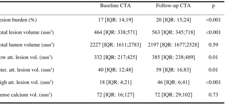
Table 2. Coronary wall thickness progression between baseline and follow-up coronary CTA as quantified with semi- automated software
Baseline CTA Follow-up CTA p
Lesion burden (%) 17 [IQR: 14;19] 20 [IQR: 15;24] <0.001
Total lesion volume (mm3) 464 [IQR: 338;571] 563 [IQR: 345;718] <0.001 Total lumen volume (mm3) 2227 [IQR: 1611;2783] 2197 [IQR: 1677;2528] 0.59 Low att. lesion vol. (mm3) 332 [IQR: 217;425] 385 [IQR: 238;489] 0.01 Inter. att. lesion vol. (mm3) 40 [IQR: 12;48] 59 [IQR: 16;83] 0.01 High att. lesion vol. (mm3) 18 [IQR: 4;21] 46 [IQR: 6;41] <0.001 Dense calcium vol. (mm3) 72 [IQR: 16;127] 72 [IQR: 29;102] 0.73
15
4.5. Prediction model to estimate presence of coronary artery disease
We retrieved databases from 18 hospitals. The study population included 5677 patients (3283 men, 2394 women; mean age 58 and 60 years, respectively). Nearly all patients (5190, 91%) underwent CT based coronary angiography, which revealed obstructive coronary artery disease in 1634 (31%). Of these 1634 patients, 1083 (66%) underwent catheter based coronary angiography, which showed positive results in 886 (82%). Of the 3556 patients without obstructive disease on CT based coronary angiography, 526 (15%) underwent catheter based coronary angiography, which showed negative results in 498 (95%). Overall, 2062 (36%) patients underwent catheter based coronary angiography, with 1176 (57%) diagnosed with obstructive coronary artery disease.
In the clinical model, all predictors except body mass index were significantly associated with obstructive coronary artery disease. The clinical model improved the prediction, compared with the basic model (cross validated c statistic improved from 0.77 to 0.79).
Whereas an abnormal exercise electrocardiography had limited predictive value in the multivariable prediction model the coronary calcium score was a major predictor, which increased the c statistic from 0.79 to 0.88. Most predictor effects decreased after addition of the coronary calcium score; dyslipidaemia and smoking were no longer significant.
5. Conclusions
In our retrospective matched case-control study, we found that scans of HTx recipients had better coronary CTA image quality than did scans of a matched control group with similar HRs. Despite the relatively high HR of HTx recipients, the number of non-diagnostic
16
segments was low (5.8%), suggesting that coronary CTA with prospective ECG triggering is a robust diagnostic tool with low radiation dose in this patient population. Based on our results the radiation dose does not exceed that what is associated with diagnostic invasive coronary angiography.
Our results demonstrate progression of coronary vessel wall volume already within the first two years after HTx. This progression was mainly attributable to non-calcified lesions causing only mild luminal narrowing. When evaluating lesion characteristics, the main components of non-calcified lesions showed high-attenuation (131–
350 HU) corresponding to fibrous tissue, while smaller percentage of the lesions showed intermediate attenuation (75–130 HU) consistent with fibro-fatty tissue, and low-attenuation (<75 HU) analogous with lipid-rich content. These findings are characteristic for CAV, in which coronary lesions are rather diffuse, and focal significant luminal narrowing develops only in a small number of patients.
Based on these results quantitative vessel wall assessment is feasible with coronary CTA in HTx patients. When using quantitative analysis with coronary CTA, CAV is detected in significantly more patients using ≥10% cut-off than detected with standard CT read. This finding suggests that invasive coronary angiography could be replaced by coronary CTA in experienced centers to diagnose CAV. With the use of coronary CTA in the clinical routine, the burden of invasive investigations could be reduced in this vulnerable patient population.
With recently collected data and modern statistical methods, we developed a prediction model that performed well in estimating the probability of coronary artery disease. The need for an updated model was evident by our results showing that the Duke clinical score significantly overestimated the probability of coronary artery disease.
Age, sex, symptoms, and coronary calcium score were strong predictors for disease.
17
6. Bibliography of the candidate's publications 6.1. Publications closely related to the presented
thesis
1. Bartykowszki A, Kolossvary M, Jermendy AL, Karady J, Szilveszter B, Karolyi M, Balogh O, Sax B, Merkely B, Maurovich- Horvat P. Image Quality of Prospectively ECG-Triggered Coronary CT Angiography in Heart Transplant Recipients AMERICAN JOURNAL OF ROENTGENOLOGY 210:(2) pp. 314-329. (2018) IF: 3.125
2. Karolyi M, Kolossváry M, Bartykowszki A, Kocsmár I, Szilveszter B, Karady J, Merkely B, Maurovich-Horvat P. Quantitative CT assessment identifies more heart transplanted patients with progressive coronary wall thickening than standard clinical read.
JOURNAL OF CARDIOVASCULAR COMPUTED
TOMOGRAPHY doi: 10.1016/j.jcct.2018.11.006. (2018) IF:3.095 3. Genders TSS, Steyerberg E, Hunink MGM, Nieman K, Galema TW, Mollet NR, de Feyter PJ, Krestin GP, Alkadhi H, Leschka S, Desbiolles L, Meijs MFL, Cramer MJ, Knuuti J, Kajander S, Bogaert J, Goetschalckx K, Cademartiri F, Maffei E, Martini C, Seitun S, Aldrovandi A, Wildermuth S, Stinn B, Fornaro J, Feuchtner G, de Zordo T, Auer T, Plank F, Friedrich G, Pugliese F, Petersen SE, Davies LC, Schoepf UJ, Rowe GW, van Mieghem CAG, van Driessche L, Sinitsyn V, Gopalan D, Nikolaou K, Bamberg F, Cury RC, Battle J, Maurovich-Horvat P, Bartykowszki A, Merkely B, Becker D, Hadamitzky M, Hausleiter J, Dewey M, Zimmermann E, Laule M. Prediction model to estimate presence of coronary artery disease: retrospective pooled analysis of existing cohorts BRITISH MEDICAL JOURNAL (BMJ) 344:(7862) Paper e3485. 13 p. (2012) IF: 17.215
18
6.2. Publications not related to the present thesis
1. Giannopoulos AA, Mitsouras D, Bartykowszki A, Merkely B, Chatzizisis YS, Buechel RR, Kaufmann PA, Gaemperli O, Maurovich-Horvat P. High-Risk Plaque Regression and Stabilization: Hybrid Noninvasive Morphological and
Hemodynamic Assessment CIRCULATION-
CARDIOVASCULAR IMAGING 11:(7) p. E007888. (2018) IF:6.221
2. Karady J, Panajotu A, Kolossvary M, Szilveszter B, Jermendy AL, Bartykowszki A, Karolyi M, Celeng C, Merkely B, Maurovich- Horvat P. The effect of four-phasic versus three-phasic contrast media injection protocols on extravasation rate in coronary CT angiography: a randomized controlled trial. EUROPEAN RADIOLOGY 27:(11) pp. 4538-4543. (2017) IF: 4.027
3. Karolyi M, Szilveszter B, Kolossvary M, Takx RA, Celeng C, Bartykowszki A, Jermendy AL, Panajotu A, Karady J, Raaijmakers R, Giepmans W, Merkely B, Maurovich-Horvat P. Iterative model reconstruction reduces calcified plaque volume in coronary CT angiography. EUROPEAN JOURNAL OF RADIOLOGY 87: pp.
83-89. (2017) IF: 2.843
4. Edes IF, Hajas A, Sax B, Bartykowszki A, Becker D, Merkely B.
Cardiac allograft vasculopathy: optical coherence guided innovative treatment options with the bioresorbable vascular scaffold - proof of concept. MINERVA CARDIOANGIOLOGICA 64:(4) pp. 487-493. (2016)
5. Szilveszter B, Elzomor H, Karolyi M, Kolossvary M, Raaijmakers R, Benke K, Celeng C, Bartykowszki A, Bagyura Z, Lux A, Merkely B, Maurovich-Horvat P. The effect of iterative model reconstruction on coronary artery calcium quantification.
19
INTERNATIONAL JOURNAL OF CARDIOVASCULAR
IMAGING 32:(1) pp. 153-160. (2016) IF: 1.896
6. Celeng C, Szekely L, Toth A, Denes M, Csobay-Novak C, Bartykowszki A, Karolyi M, Vago H, Szoke S, Coelho Filho OR, Andreka P, Merkely B, Maurovich-Horvat P. Multimodality Imaging of Giant Right Coronary Aneurysm and Postsurgical Coronary Artery Inflammation. CIRCULATION 132:(1) pp. E1-e5.
(2015) IF: 17.202
7. Maurovich-Horvat P, Karolyi M, Horvath T, Szilveszter B, Bartykowszki A, Jermendy AL, Panajotu A, Celeng C, Suhai FI, Major GP, Csobay-Novak C, Huttl K, Merkely B. Esmolol is noninferior to metoprolol in achieving a target heart rate of 65 beats/min in patients referred to coronary CT angiography: A randomized controlled clinical trial. JOURNAL OF CARDIOVASCULAR COMPUTED TOMOGRAPHY 9:(2) pp.
139-145. (2015) IF: 2.472
8. Bartykowszki A, Celeng C, Károlyi M, Maurovich-Horvat P. High Risk Plaque Features on Coronary CT Angiography CURRENT CARDIOVASCULAR IMAGING REPORTS 7:(8) p. Article Number: 9279. 12 p. (2014)
20 6.3. Hungarian publications
1. Bartykowszki A, Tóth L, Kerecsen G, Jermendy ÁL, Kolossváry M, Karády J, Szilveszter B, Károlyi M, Suhai FI, Panajotu A, Kolozsvári R, Balázs Gy, Hüttl K, Thury A, Batthyány I, Kiss RG, Merkely B, Maurovich-Horvat P. A koronária-CT-angiográfia értelmezése és leletezése. A Magyar Kardiológusok Társasága Kardiovaszkuláris Képalkotó Munkacsoportjának ajánlása.
CARDIOLOGIA HUNGARICA 47:(1) pp. 2-9. (2017)
2. Maurovich-Horvat Pál, Bartykowszki Andrea, Kerecsen Gábor, Thury Attila, Károlyi Mihály, Balázs György, Várady Edit, Tóth Levente, Pintér Nándor, Szukits Sándor, Kolozsvári Rudolf, Hoffer Krisztina, Király István, Nagy Lajos, Hüttl Kálmán, Préda István, Palkó András, Kiss Róbert Gábor, Battyány István, Merkely Béla.
A coronariák CT-angiográfiás vizsgálatának leletezése. MAGYAR RADIOLÓGIA ONLINE 4:(11) Paper a_coronariak.html. 11 p.
(2013)
3. Maurovich-Horvat Pál, Bartykowszki Andrea, Kerecsen Gábor, Thury Attila, Károlyi Mihály, Balázs György, Várady Edit, Tóth Levente, Pintér Nándor, Szukits Sándor, Kolozsvári Rudolf, Hoffer Krisztina, Király István, Nagy Lajos, Hüttl Kálmán, Préda István, Palkó András, Kiss Róbert Gábor, Battyány István, Merkely Béla.
A koronária-CT-angiográfia leletezése. A Magyar Kardiológusok Társasága Szív-CT Munkacsoportjának és a Magyar Radiológusok Társasága Szív Képalkotó Diagnosztikai Szekciójának közös ajánlása. CARDIOLOGIA HUNGARICA 43:(5) pp. 275-281.
(2013)
4. Bartykowszki A, Maurovich-Horvat P. A szív-CT vizsgálat és indikációja. MAGYAR CSALÁDORVOSOK LAPJA 2012:(1) pp.
47-51. (2012)