Scopolamine-based pharmacological MRI model for testing procognitive agents
Ph.D. thesis
dr. Nikolett Heged ű s
Semmelweis University
Doctoral School of Pharmaceutical Sciences
Supervisor: Dr. Károly Tihanyi, C.Sc
Official reviewers:
Dr. Tamás Tábi, Ph.D.
Dr. István Tarnawa, Ph.D.
Head of the Final Examination Committee:
Dr. György Bagdy, D.Sc
Members of the Final Examination Committee:
Dr. Ildikó Világi, Ph.D.
Dr. Viktor Gál, Ph.D.
Budapest
2016
1
TABLE OF CONTENTS
TABLE OF CONTENTS ... 1
LIST OF ABBREVIATIONS ... 5
I INTRODUCTION ... 8
I.1 Cognition and Alzheimer’s disease (AD) ... 9
I.1.1 Cognitive disorders ... 9
I.1.2 Alzheimer’s disease... 9
I.1.3 The pathophysiology and disease-modifying drugs of AD ... 11
I.1.3.1 Amyloid hypothesis... 11
I.1.3.1.1 Anti-amyloid β agents ... 11
I.1.3.1.2 Vaccination... 12
I.1.3.1.3 Selective Aβ42-lowering agent (SALA) ... 12
I.1.3.1.4 β- and γ-secretase inhibitors ... 12
I.1.3.1.5 α-secretase potentiator ... 13
I.1.3.2 Tau hypothesis... 13
I.1.3.3 Neurotransmitter-based model ... 13
I.1.3.4 Genetic-based model ... 14
I.1.3.5 Other environmental and human-related risks ... 15
I.2 A new approach with high translational power is required in drug development ... 15
I.3 The basis of suitable disease-modifying treatments in the future ... 17
I.4 Cognition and the cholinergic system ... 18
I.4.1 Acetylcholine ... 18
I.4.1.1 Nicotinic acetylcholine receptors (nAchRs) ... 19
I.4.1.2 Muscarinic acetylcholine receptors (mAchRs) ... 19
I.4.2 Scopolamine ... 20
I.4.3 Butylscopolamine ... 21
2
I.5 Cognitive enhancers in therapy and in active development phases ... 21
I.5.1 Donepezil ... 22
I.5.2 Piracetam ... 23
I.5.3 Vinpocetine ... 24
I.5.4 PNU-120596... 24
I.5.5 EVP-6124 ... 25
I.5.6 RG2260 ... 25
I.5.7 Neostigmine ... 25
I.6 Overview of Magnetic Resonance Imaging ... 26
I.6.1 What is Magnetic Resonance Imaging? ... 26
I.6.2 Basic physical background ... 27
I.6.2.1 The introduction of an external magnetic field (B0) ... 27
I.6.2.2 Excitation ... 28
I.6.2.3 Relaxation... 29
I.6.2.3.1 T1 relaxation ... 29
I.6.2.3.2 T2 relaxation ... 30
I.6.2.3.3 T2* relaxation ... 30
I.6.2.4 Image formation ... 31
I.6.2.5 MRI contrast mechanism ... 32
I.6.2.5.1 Proton-density contrast ... 33
I.6.2.5.2 T1 contrast ... 33
I.6.2.5.3 T2 contrast ... 34
I.6.3 Functional Magnetic Resonance Imaging ... 35
I.6.3.1 Physiological background of fMRI measurements ... 35
I.6.3.1.1 Neurovascular coupling... 35
I.6.3.1.2 The blood-oxygenation-level dependent (BOLD) contrast ... 37
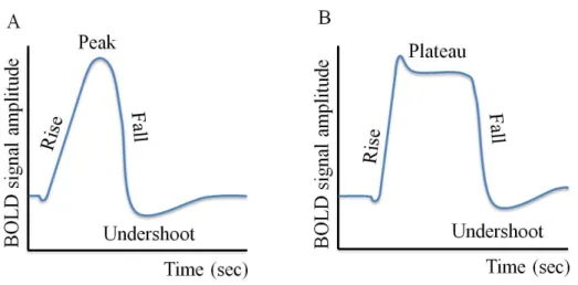
I.6.3.1.3 The shape of hemodynamic response (HDR) curve ... 37
3
II OBJECTIVES ... 39
III MATERIALS AND METHODS ... 41
III.1 fMRI measurement ... 41
III.1.1 Parameters of anatomical and functional images ... 42
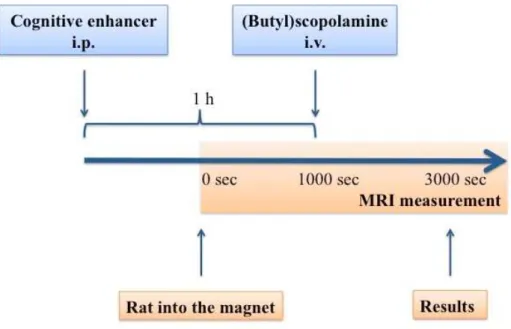
III.1.2 The experimental protocol ... 42
III.1.3 Data analysis ... 43
III.2 Water labyrinth test ... 45
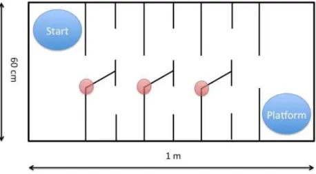
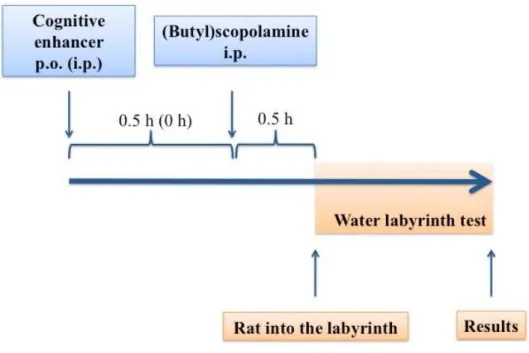
III.2.1 The experimental protocol ... 45
III.2.2 Data analysis ... 48
IV RESULTS ... 49
IV.1 fMRI measurement ... 49
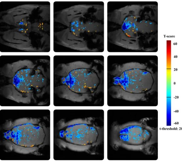
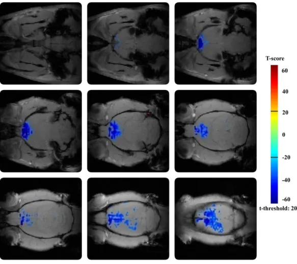
IV.1.1 The effect of amnestic agent scopolamine ... 49
IV.1.2 The effect of amnestic agent buscopan ... 50
IV.1.3 The effects of cognitive enhancers – direct administration ... 51
IV.1.4 The effects of cognitive enhancers in provocation model ... 52
IV.2 Water labyrinth test ... 65
V DISCUSSION ... 71
V.1 The neurovascular connection ... 71
V.2 The role of pericytes in microcirculation... 72
V.3 Why is BOLD fMRI a good choice to measure neuronal activities? ... 72
V.4 How do amnestic agents work? ... 73
V.5 The advantage of provocation model ... 74
V.6 How do cognitive enhancers work? ... 74
V.7 The water labyrinth tests confirm the result of fMRI studies ... 77
V.8 Looking to the future ... 78
VI CONCLUSIONS ... 79
VII SUMMARY ... 81
4
VIII ÖSSZEFOGLALÁS ... 82
IX BIBLIOGRAPHY ... 83
X PUBLICATIONS RELATED TO THE THESIS ... 103
XI OTHER PUBLICATION ... 104
XII ACKNOWLEDGEMENTS ... 105
5
LIST OF ABBREVIATIONS
Aβ amyloid beta
ABCA7 ATP-binding cassette transporter A Ach acetylcholine
AchEi acetylcholinesterase inhibitor AchR acetylcholine receptor
AD Alzheimer’s disease
ADRDA Alzheimer's Disease and Related Disorders Association ANOVA Analysis of variance
APP amyloid precursor protein ATP adenosine triphosphate
BACE beta-side amyloid precursor protein cleaving enzyme BBB blood-brain barrier
BIN1 bridging integrator 1
BOLD blood-oxygenation-level dependent CAA cerebral amyloid angiopathy cAMP cyclic adenosine monophosphate cAVA cis-apovincaminic acid
CBF cerebral blood flow CBV cerebral blood volume CD2AP CD2-associated protein CD33 CD33 molecule
ChAT choline acetyltransferase
CMRO cerebral metabolic rate of oxygen CNS central nervous system
CLU clusterin
CR1 complement component (3b/4b) receptor 1 CSF cerebrospinal fluid
CT computedtomography
CTF C-terminal fragment DMN default mode network
6 DTPA diethylene triamine pentaacetic acid EPHA1 EPH receptor A1
EPI echo planar imaging
fMRI functional magnetic resonance imaging FOV field of view
GABA gamma-aminobutyric acid GAG glycosaminoglycan
GEMS gradient echo multi slice sequence GM gray matter
GSK3β glycogen synthase kinase 3β GWAS genom-wide association study HDR hemodynamic response i.v. intravenous
i.p. intraperitoneal
LRP1 lipoprotein receptor related protein 1
M net magnetization
mAchR muscarinic acetylcholine receptor MCI mild cognitive impairment MED minimal effective dose
mGluR2 metabotropic glutamate receptor 2 mPFC medial prefrontal cortex
MRI magnetic resonance imaging MRS magnetic resonance spectroscopy
MS4A6A membrane-spanning 4-domains, subfamily A MTC methylthioninium chloride
nAchR nicotinic acetylcholine receptor nbM nucleus basalis of Meynert
NIFTI neuroimaging informatics technology initiative
NINCDS National Institute of Neurological and Communicative Disorders and Stroke
NMDA N-methyl-D-aspartate NFT neurofibrillary tangle
7 NSAID nonsteroidal anti-inflammatory drug PAM positive allosteric modulator
PCP phencyclidine PD Parkinson’s disease PEG polyethylene glycol
PET positron emitting tomography PFC prefrontal cortex
phMRI pharmacological magnetic resonance imaging
PICALM phosphatidylinositol-binding clathrin assembly protein p.o. per os
PSEN1 presenilin-1 PSEN2 presenilin-2
RF radiofrequency pulse ROI region-of-interest
SALA selective Aβ42 lowering agent sAPP soluble amyloid precursor protein
SIPP superparamagnetic iron-platinum particle SORL1 sortilin-related receptor-1
SP senile plaque
SPIO superparamagnetic iron oxide
TE echo time
TR repetition time
USPIO ultrasmall superparamagnetic iron oxide VSMC vascular smooth muscle cell
WM white matter
8
I INTRODUCTION
The brain is the most energy-demanding organ in the body. The high energy requirement is met exclusively by the oxidative breakdown of glucose. The biological oxidation of carbon and hydrogen in glucose demands a great amount of molecular oxygen. Some 20-25 % of the body-wide consumed oxygen is used by the brain.
Glucose is not stored in the brain tissue (apart from approximately 10 min supply in astrocytes) and no storage capacity exists for oxygen at all. The function of the brain depends on the availability of glucose and oxygen and therefore, it is greatly susceptible to minor changes in blood supply, which is the carrier of nutrients. An intact cerebral circulation is a precondition for satisfactory glucose and oxygen supply (Wang et al., 2013).
Whenever the presence of these resources is below optimal, the most delicate and most complex functions will be harmed first. Therefore, the deficiency of energy supply is inevitably followed by the deficiency of cognition. Nowadays, it is commonly recognized that ischemic deprivation (oxygen and glucose restriction) exerts detrimental effects on the cognitive function. A small fluctuation in cerebral oxygen delivery also has an impact on cognitive performance. With age, the cortical blood supply is reduced by up to 30 %. Further reduction of regional blood flow was demonstrated in patients with memory impairment. Wang et al. (2013) concluded from a multicenter study that decreases in cerebral blood flow is the first step towards Alzheimer’s disease.
As a consequence of insufficient ATP production, neurons depolarize which render membrane ion-transports inefficient. The increased calcium influx leads to the release of various neurotransmitters among others glutamate. The high calcium concentration activates destructive enzymes causing cellular degradation. In this way the cognitive deficit is not the only sign of inefficient energy production: destructive pathways will be soon activated as a consequence of energy restriction.
This vicious circle may be prevented by the maintenance of good cerebral blood circulation. Most importantly, improving blood circulation, especially in regions associated with cognitive processes, may reverse the cognitive impairment caused by insufficient cellular energy production. This may happen with or without the direct modulation of neurotransmitters or their receptors. The restoration of blood supply will
9
restore cognitive functions and also prevents secondary damage (Palmer and Love, 2011).
I.1 Cognition and Alzheimer’s disease (AD)
Cognition is a complex mental process, which requires attention, problem-solving, decision-making, and working memory. Several brain areas are involved in cognitive processes; among others prefrontal cortex (PFC), hippocampus, temporal and parietal cortex have prominent roles. Neurological (AD, Parkinson’s disease – PD) and psychiatric disorders (schizophrenia, depression) are frequently accompanied by cognitive deficits (Heidbreder and Groenewegen, 2003).
I.1.1 Cognitive disorders
Cognitive disorders are classified by DSM-V as delirium, dementia, amnestic disorder, and other cognitive disorders. Delirium is a disturbance of consciousness and a change in cognitive function that develops over a short period of time. Dementia is characterized by long lasting multiple cognitive deficits with memory impairment. The amnestic disorder is characterized by memory impairment in the absence of other significant accompanying cognitive impairments (DSM-V, 2013). While all of the above-mentioned disorders could be due to various etiologies, it is generally assumed that more or less common reasons, namely degenerative processes and neuronal dysfunctions are in the background of these diseases. Despite the intense research and development of memory enhancer drugs, no robustly effective agents have reached the market. Unfortunately, the prevalence of dementia is continuously rising in parallel with the increasing life span.
I.1.2 Alzheimer’s disease
The most common cause of dementia is Alzheimer’s disease or AD. AD is an irreversible, progressive neurodegenerative disease that adversely affects cortical functions: memory, thinking, and orientation. An estimated 44 million people worldwide have Alzheimer’s disease and other dementias. So economically, AD is a major public health problem (Prince et al., 2014). Postmortem studies reported that
10
60-90 % of patients with Alzheimer’s disease show varying degrees of cerebrovascular pathology at autopsy. Furthermore, it is speculated that vascular lesions contribute to the development of clinical symptoms in patients with AD and may lead to more rapid clinical decline (Kalaria, 2000).
AD includes the interaction of amyloid β (Aβ) aggregation with plaque development and hyperphosphorylation and aggregation of tau protein with tangles. The production of amyloid β is a critical step in AD pathogenesis. Aβ is a ~ 4kDa peptide, which is built up of 36 to 43 amino acids (Armstrong, 2009). It is the final product of cleavage of APP (amyloid precursor protein). The most common variety of Aβ (Aβ1-42) is predominantly found in plaques, whereas a more soluble form (Aβ1-40) is found in association with blood vessels (Armstrong, 1998).
Amyloid β forms insoluble and protease resistant fibrils, called senile plaques (SP). These plaques are lesions in the cerebral cortex. SPs have various types: diffuse (pre-amyloid), neuritic (primitive), classic (dense-cored), and compact-type (burned-out) plaques (Armstrong, 2009). These amyloid plaques differ from each other in the level of amyloid aggregation but there is no convincing evidence to support their transformation procedure to each other (Armstrong, 1998).
Neurofibrillary tangles (NFTs) consist of tau protein. In normal healthy subjects, tau is an essential component of the microtubule system, which plays a crucial role in intracellular transport. In patients with AD, tau protein is abnormally hyperphosphorylated and forms insoluble fibrils within the cell. These changes with additional inflammation and oxidative stress, such as a pathological cascade initiate the loss of synaptic integrity and progressive neurodegeneration (Galimberti and Scarpini, 2011).
Until now, the only way to make a precise diagnosis of AD was an autopsy or brain biopsy. In clinical practice, the diagnosis was usually based on the family history and results of on Mental Status Examination (e.g. NINCDS-ADRDA). The time from diagnosis to death varies from as little as 3 years to as long as 10 or more years. Patients with early-onset AD tend to have a more aggressive, rapid course than those with late-onset AD. Nowadays, there are other non-invasive, modern imaging techniques to detect AD. It is possible to diagnose cerebrovascular diseases with computed
11
tomography (CT), positron emitting tomography (PET), or magnetic resonance imaging (MRI) (DSM-V, 2013).
I.1.3 The pathophysiology and disease-modifying drugs of AD
The exact pathomechanism of Alzheimer’s disease is still not understood. Several theories have been proposed about the triggers or origin of the disease.
I.1.3.1 Amyloid hypothesis
In the last 20-25 years, „amyloid hypothesis“ was the leading scientific explanation of Alzheimer’s disease. This hypothesis proposed that the abnormally high level of accumulated amyloid β was the main cause of AD. The APP, as a precursor molecule of Aβ, plays a crucial role in AD pathogenesis, which was the heart of the amyloid hypothesis. Amyloid precursor protein is a type I membrane protein. Two enzymatic cleavages (β-secretase cleavage in the extracellular domain; γ-secretase cleavage in the transmembrane region) are needed to release the neurotoxic Aβ peptide.
Indeed, the transformation of APP to Aβ is a sequential proteolytic process: at first, the α-secretase (non-amyloidogenic) or β-secretase (amyloidogenic) cleaves the APP and generates the α- or β- C-terminal fragments (CTFs) of Aβ. The β-secretase, named BACE1 (β-site APP cleaving enzyme) cleaves the APP within the ectodomain and generates the N-terminus of Aβ. After that, the γ-secretase cleaves the α- or β-CTFs and releases the neurotoxic Aβ peptide into the extracellular space (Vassar, 2004).
Lots of studies and clinical trials with newly developed anti-AD agents tried to influence the progression of the disease assuming the above-mentioned pathomechanism of AD.
I.1.3.1.1 Anti-amyloid β agents
The glycosaminoglycan (GAG) mimetic agent, tramiprosate (AlzhemedTM, Neurochem Inc.) was able to decrease the Aβ level in human CSF by up to 70 % but did not show any significant effect on cognitive functions (Gervais et al., 2001;
Aisen et al., 2006). The other tested agent, colostrinin (O-CLN; ReGen Therapeutics) could prevent amyloid β aggregation and neurotoxicity in animal models but these
12
positive effects were only temporary during human trials (Leszek et al., 1999;
Bilikiewicz and Gaus, 2004).
I.1.3.1.2 Vaccination
Active immunization with Aβ showed some protective effects against AD in young transgenic mice, e.g. reduced Aβ aggregation and plaque formation, neuritic dystrophy and astrogliosis. In older animals, the antigen therapy was able to reduce the progression of the disease. Unfortunately, this active immunization therapy was not successful in human clinical trials. Some of the treated patients (6 %) had developed meningoencephalitis. In addition, the cognitive functions of the placebo group and antibody responders did not differ significantly (Gilman et al., 2005). To avoid complications due to the excess T-cell activation, passive immunization seemed to be a better therapy. However, the humanized monoclonal anti-Aβ antibodies – bapineuzumab, solanezumab – have not demonstrated the expected results (Grundman and Black, 2008).
I.1.3.1.3 Selective Aβ42-lowering agent (SALA)
Tarenflurbil (MPC-7869; Myriad Pharmaceuticals; FlurizanTM) is a pure R-enantiomer of the NSAID flurbiprofen. It can change the conformation of γ-secretase and make the final cleaved Aβ peptide shorter, more water soluble, and less toxic (Beher et al., 2004). Despite the promising results of preclinical studies (Kukar et al., 2007), tarenflurbil had failed in human phase III trial, so its development was stopped (Green et al., 2009).
I.1.3.1.4 β- and γ-secretase inhibitors
β- and γ-secretase play important roles in the formation of toxic Aβ plaques, thus their inhibition seemed to be an ideal therapeutic target in AD. These enzymes are responsible for the sequential cleavages of APP. Sadly, in human clinical trials, neither the BACE inhibitor nor the γ-secretase inhibitor was able to show any statistically significant difference in Aβ level or in cognitive functions (active treatment group vs.
placebo group) (Siemers et al., 2005; Siemers et al., 2006; Fleisher et al., 2008).
13 I.1.3.1.5 α-secretase potentiator
The α-secretase enzyme is the most important proteinase of the nonamyloidogenic pathway, which transforms the amyloid precursor protein to its soluble form (sAPPα).
Etazolate (EHT 0202; ExonHit Therapeutics) can stimulate α-secretase, protect cells from toxic Aβ plaques in the CNS, and prevent the neuronal death in rats (Marcade et al., 2008).
Altogether, the amyloid hypothesis has failed in several unsuccessful clinical trials. Nowadays, more and more authors have questioned this causality and suppose that Aβ accumulation in the brain is not the cause of AD but a consequence of it (Drachman, 2014).
I.1.3.2 Tau hypothesis
In regard to the above-mentioned pathophysiological changes in AD, the inhibition of tau hyperphosphorylation or oxidative processes seemed another good therapeutic choice.
Methylthioninium chloride (MTC; TauRx Therapeutics; Rember TM) or methylene blue, is a well-known indicator molecule in analytical chemistry. In human phase II trials, MTC was able to block the phosphorylation process of tau protein but further studies are required to gain convincing results (Wischik et al., 1996). Lithium, sodium valproate, pyrazolopyrazines, and pyrazolopyridines can inhibit some protein kinases (presumably glycogen synthase kinase 3 or GSK3β) which are responsible for tau hyperphosphorylation. These agents are still in development (Balaraman et al., 2006;
Martinez and Perez, 2008; Schneider and Mandelkow, 2008).
I.1.3.3 Neurotransmitter-based model
This model presumes some kind of injury in the neurotransmitter system (mostly in cholinergic and glutamatergic system). Based on this model, currently, the only available standard medical treatment in AD is the symptomatic therapy. It includes cholinesterase inhibitors (AchEi) – e.g. donepezil, galantamine, rivastigmine, tacrine –, a partial N-methyl-D-aspartate (NMDA) antagonist – memantine – or the combination
14
of these drugs. Acetylcholinesterase inhibitors were the first anti-Alzheimer drugs, which could increase the acetylcholine levels. From these, tacrine – the first drug approved for AD – is rarely used nowadays due to its hepatotoxicity; donepezil, galantamine, and rivastigmine are approved for mild-to-moderate AD. Donepezil is the most often prescribed anti-AD agent by patients with severe AD. In severe cases (for moderate-to-severe AD), memantine seems to be a good additional therapeutic option.
This is believed to protect neurons from the excitotoxic activity. But unfortunately, all of these therapies are only marginally effective and preserve cognitive function temporarily. Psychotropic medications are often used as adjuvants to treat secondary symptoms of AD, such as depression, agitation, and sleep disorders (Seltzer, 2005).
I.1.3.4 Genetic-based model
The genetic-based model assumes the presence of some alleles or genes in the human genome that can predict the onset of AD. Most people suffering from AD start showing symptoms after age 65 or later. But in rare cases, the disease develops much earlier, between ages of 30 and 60. This early-onset form of AD is caused by one of three genes mutations inherited from parents which cause aberrant protein formation.
Mutation of chromosome 21 causes abnormal APP formation, alteration of chromosome 14 induces abnormal PSEN1 (presenilin 1), while changes in chomosome 1 leads to abnormal PSEN2 (presenilin 2) formation (Van Cauwenberghe et al., 2015).
The more common late-onset form of AD usually develops after age 65. Its hereditary component is mounted because no single gene mutation is known to cause this late-onset form of the disease. The expression of the APOEε4 allele in an individual’s genome unambiguously classifies the risk of AD development into three groups. Individuals who are APOEε4 homozygotes have a very high risk to develop the disease while APOEε4 heterozygotes have a moderate chance to it. People who do not carry this gene have very low risk to suffer from this neurodegenerative disease (Hampel et al., 2010).
Researchers are hardly working to determine further genes that may influence the risk of late-onset AD. Until this time, numerous genes (PICALM, CLU, CR1, BIN1, MS4A6A locus, CD2AP, SORL1, CD33, EPHA1 and ABC7) have been identified by
15
GWAS (genom-wide association study) that may increase the people’s risks for AD.
These genes are clustered within three main groups depending on their roles in cholesterol and lipid metabolism, immune system and inflammatory response, and endosomal vesicle cycling (Van Cauwenberghe et al., 2015).
I.1.3.5 Other environmental and human-related risks
Environmental and other lifestyle factors may also contribute to the developing late-onset AD. Several researches promote that physical activity can slow down or prevent functional decline associated with aging, improves health in older individuals and reduces the risk for developing AD (Hakim et al., 1998; Middleton et al., 2008;
Scarmeas et al., 2009). Animal studies have been shown that even a small amount of physical activity decreases cortical amyloid burden in mice, possibly mediated by changes in the processing of amyloid precursor protein (Adlard et al., 2005).
People who regularly do some physical activities are often pay attention to their eating habits. Following mediterranean diet can further decrease the risk for developing AD or mild cognitive impairment (MCI) (Scarmeas et al., 2009).
I.2 A new approach with high translational power is required in drug development
In spite of the aforementioned scientific exploration, the successful treatment of Alzheimer’s disease is not yet developed, so there is a huge unmet medical need for an efficient disease-modifying treatment. The drug research has not been able to produce proven disease-modifying drugs since the understanding of AD pathomechanism and suitable disease models are still lacking. Therefore, the research and development of procognitive compounds are one of the most challenging areas in drug research. Until today, several disappointing clinical trials have disqualified targets that were believed to be in the main pathway of the disease, such as amyloid β (Aβ), tau, β-side amyloid precursor protein cleaving enzyme (BACE) etc. As a result, researchers have to face a relative shortage of relevant targets (Armstrong, 2011; Braak and Braak, 1991;
Drachman, 2006; Fjell and Walhovd, 2012; Herrup, 2010; Pimplikar, 2009;
Pimplikar et al., 2010; Reitz, 2012). These negative clinical trials draw attention to the
16
inefficient translational power of current animal models and call for the improvement of predictive value (Sarter et al., 2009).
Fortunately, the highly expanding field of small animal magnetic resonance imaging (MRI) seems to be a robust method, which may provide a new approach with higher translational power (Schwarz et al., 2006). The functional MRI (fMRI) blood-oxygenation-level dependent (BOLD) technique is suitable for the complex testing of brain functions and also the circulation (Marota et al., 2000). It is an important non-invasive tool for preclinical drug discovery with excellent temporal and spatial resolution (Leslie and James, 2000).
Rat BOLD pharmacological MRI (phMRI) is gaining popularity in studies aiming at the central effect of various pharmacological agents (Canese et al., 2009;
Chen et al., 1997; Sekar et al., 2011; Stark et al., 2006). There are several human phMRI studies as well that verify its translational role in neuroscience (Anderson et al., 2002; Deakin et al., 2008; Stein et al., 1998). The key advantage of MRI over other imaging modalities in neuroscience is its ability to follow changes in brain neurobiology after pharmacological, electrical, or environmental insult (Chang and Shyu, 2001; Reese et al., 2000; Stark et al., 2008). The reconstructed
‘fingerprint’ of the brain activity can characterize the activity and function of new psychotherapeutics in preclinical development and study the neurobiology of cognition (Ferris et al., 2011).
Unfortunately, the direct examination of drug effects is not always possible by fMRI. Frequently occurring problems are the weak or slowly evolving BOLD effects and the BOLD effect of pharmaceutical formulations that do not contain assumed active ingredients. Some of the studied procognitive agents intended for intravenous (i.v.) application have poor aqueous solubility; they are not soluble in an indifferent vehiculum. Since the BOLD readings coincide with the i.v. treatment of animals, the experimenter has to make sure that changes due to osmotic pressure, high dose volume, differing pH, or auxiliary pharmaceutical materials do not interfere with the BOLD effect of the test compound. As it is not always possible to formulate test materials in an indifferent formulation, pretreatment of animals with the test compound and testing its effect by a neutrally formulated challenging drug one hour later can solve the problem of unwanted formulation effect. This type of experimental approach was used in the
17
paper of Chin et al. (2011), where the effect of metabotropic glutamate 2/3 receptor agonist LY379268 was tested against ketamine. In another study, the effect of an allosteric potentiator or agonist of mGluR2 against phencyclidine (PCP) was measured (Gozzi et al., 2008; Hackler et al., 2010) and this sort of approach was used in our studies as well.
I.3 The basis of suitable disease-modifying treatments in the future
Nowadays, more and more publications emphasize the role of intact brain circulation and microcirculation in cognitive functions (Drachman, 2014;
Dotti and De Strooper, 2009; Hall et al., 2014; Hunter et al., 2012). Even a small fluctuation in cerebral oxygen or glucose delivery may have an impact on cognitive performance. Animal studies also prove that cerebral hypoperfusion by itself can cause increased oxidative stress, damage to cholinergic neurotransmitter system and cognitive impairment (Bink et al., 2013; Iadecola, 2013; Xi et al., 2014; Zlokovic, 2011).
Regional cerebral hypoperfusion is a pronounced sign of the cognitive failure and the clinical manifestation of AD (de la Torre, 2000; de la Torre and Stefano, 2000).
Furthermore, in advanced age, the cortical blood supply may be reduced by up to 30 %.
Altogether, it can cause critically low cerebral perfusion, when the energy demand exceeds the capacity of metabolic processes (de la Torre, 2000; Hunter et al., 2012;
Lu et al., 2011). The insufficient energy supply (e.g. due to a circulatory problem) may lead to amyloid β-peptide (Aβ) deposition in cerebral vessel walls and in brain parenchyma as well, termed cerebral amyloid angiopathy (CAA). Under intact energy supply, the low-density lipoprotein receptor-related protein 1 (LRP1) eliminates the soluble form of amyloid protein from the brain parenchyma to the blood. LRP1 is a potent amyloid β efflux transporter across the blood-brain barrier (BBB). It is easy to see that some kind of defects in vascular smooth muscle cells (VSMCs) lead to disrupted Aβ clearance. Hypoperfusion downregulates the clearance transporter which leads to disrupted Aβ clearance and amyloid deposition. The excess amyloid protein inhibits the synaptic transmission and basic neural functions; it worsens further the circulation and aggravates the energy deficit (Bell et al., 2009; Dotti and De Strooper,
18
2009). The Aβ deposition impairs the cholinergic system directly, which initiates a vicious circle (Olivero et al., 2014).
Improving blood circulation, especially in regions associated with cognitive processes, may reverse the cognitive impairment caused by insufficient cellular energy production. This may happen without direct modulation of the level of neurotransmitters or their receptors (Palmer and Love, 2011).
Following neuronal stimuli, increased blood flow and prompt relaxation of capillaries can be seen (Hall et al., 2014) due to the bidirectional neurovascular coupling (Peppiatt et al., 2006). The cerebral capillary flow is regulated mostly by pericytes. This type of cells covers mainly the brain microvessels; some 80 % of microvessels are enveloped in the brain while other organs contain only a few percent (Armulik et al., 2011; ElAli et al., 2014; Winkler et al., 2012). The abundance of pericytes seems to be proportional to the barrier function of the organ (Winkler et al., 2010; Winkler et al., 2012). Pericytes have lots of neurochemical receptors (Dore-Duffy, 2008; Hamilton et al., 2010) and receive paracrine stimuli either from other neurons through astrocytes and from endothelial cells that convey luminal signals (Peppiatt et al., 2006). Long-term hypoxia causes pericyte migration or its death around the capillaries (de la Torre and Stefano, 2000; Gonul et al., 2002). Therefore, the capillary system becomes unresponsive to physiological, neural, and luminal stimuli, which impairs irreversibly the neuronal system (Cleary et al., 2005).
These changes can also be observed in AD patients who did not suffer a pericyte degeneration (Baloyannis and Baloyannis, 2012; Farkas and Luiten, 2001; Sengillo et al., 2013). Targeting the pericytes – which could provide selective brain-specific action – in the prevention and therapy of dementias, such as AD is suggested in our and other studies as well (ElAli et al., 2014; Hamilton et al., 2010).
I.4 Cognition and the cholinergic system
I.4.1 Acetylcholine
Acetylcholine (Ach) is an important neurotransmitter, which undoubtedly plays a role in cognitive processes. It is located in synapses of peripheral nervous system, in vegetative ganglions, in parasympathetic postganglionic fibers, and in neuromuscular
19
junctions as well. Acetylcholine is considered to be a physiological agonist of nicotinic and muscarinic acetylcholine receptors (Brunton et al., 2005).
I.4.1.1 Nicotinic acetylcholine receptors (nAchRs)
Nicotinic acetylcholine receptors (nAchR) are ionotropic receptors with high sodium, potassium, and calcium permeability. nAchR has lots of subunits; the most important one related to cognitive functions is the α7 subunit (Brunton et al., 2005).
The α7 subunit containing nAchRs are highly expressed in brain regions implicated in cognitive processes, among others in the hippocampus, cortex, and several subcortical limbic regions (Gotti et al., 2006; Marks and Collins, 1982;
Rubboli et al., 1994). Low expression level can be seen in thalamic regions and in basal ganglia (Clarke et al., 1985; Poisik et al., 2008; Tribollet et al., 2004). Therefore, targeting α7 nAchRs with pharmacological agents seems to be an effective treatment in cognitive decline. Unfortunately, the uncommon properties of α7 nAchRs (low probability of channel opening and rapid desensitization) make the conventional agonist useless (Williams et al., 2011a,b). Positive allosteric modulators (PAMs) can solve these problems; they do not possess any direct agonist effects on the target but they increase the effect of classical orthosteric agonists (Gill et al., 2011). Type I PAMs increase the peak amplitude of Ach current while Type II PAMs increase the current response due to the inhibition of desensitization (Hu et al., 2009;
Schrattenholz et al., 1996; Timmermann et al., 2007).
I.4.1.2 Muscarinic acetylcholine receptors (mAchRs)
Muscarinic acetylcholine receptors (mAchR) are metabotropic G-protein coupled receptors, which have five subtypes (M1-M5). The neocortex, hippocampal, and striatal neurons are rich in M1 and M4 AchRs; M3 subtypes are located in thalamus and brainstem nuclei; M5 mAchRs are expressed in low levels in CNS, where they mediate the dilatation of arteries and arterioles; M2 receptor subtype can be found presynaptically, where it controls the transmitter release (Brunton et al., 2005).
20
I.4.2 Scopolamine
Pharmacological manipulation is a valuable technique to demonstrate the importance of the cholinergic system to memory functions in intact and diseased conditions. Scopolamine as a non-selective muscarinic receptor antagonist is a widely used memory disturbing amnestic agent in models of cognition in human and preclinical studies as well (Beatty et al., 1986; Caine et al., 1981; Drachman and Leavitt, 1974;
Flicker et al., 1990; Ghoneim and Mewaldt, 1975; Klinkenberg and Blokland, 2010;
Lenz et al., 2012). The effect of scopolamine might be mediated by the M1 and M5 receptor subtypes based on their special distribution within the brain (predominantly located in cortex, hippocampus and striatum) (Caulfield, 1993). First, scopolamine was used clinically as an adjunct to surgical or obstetric procedures to induce sedation and anterograde amnesia (Drachman and Leavitt, 1974). Nowadays, its application is less frequent due to the cholinergic cognitive deficit it evokes and which cholinergic deficit has a high resemblance to those associated with Alzheimer’s disease. This cholinergic blockade causes impairment in tasks of working memory, declarative memory, visual sustained attention, and psychomotor function (Wesnes and Revell, 1984;
Wesnes and Warburton, 1983, 1984). Older people are more sensitive to scopolamine treatment than younger people, which is in accordance with animal studies (Molchan et al., 1992). The reversible memory impairment property of scopolamine makes it a widely used model for the characterization of cognitive enhancers (Ebert and Kirch, 1998). Besides its negative cognitive effect, it also has a strong cerebrovascular action. Based on the fact that the cerebral vasculature is rich in cholinergic fibers and muscarinic receptors, scopolamine as an antimuscarinerg agent could act as a vasoconstrictor (Honer et al., 1988). Its peripherial administration mostly affects muscarinic receptors in the septo-hippocampal area, in the amygdala and in the parietal cortex (Klinkenberg et al., 2011). Scopolamine decreases the neuronal activity and metabolism in the indicated brain regions and consequently reduces the CBF (Honer et al., 1988). These statements are in accordance with the results of lesion studies related to the nucleus basalis of Meynert (nbM), which is the origin of cholinergic projections to the cerebral cortex. Lesioning of nbM leads to decreased
21
cerebral metabolism via the changed expression level of presynaptic choline acetyltransferase (ChAT) (Kiyosawa et al., 1987; Orzi et al., 1987).
All of the above-mentioned cerebrovascular effects are partially responsible for the negative cognitive effect of scopolamine as were proved by the non-brain penetrating analog butylscopolamine in our previous study.
I.4.3 Butylscopolamine
Butylscopolamine (buscopan) is a quaternary nitrogen-containing muscarinergic antagonist. Its use as premedication for gastrointestinal endoscopy reportedly causes anterograde amnesia in patients (Lee et al., 2007). This temporary memory impairment shows similarities to human dementias where vascular problems often precede cognitive deficit (Wang et al., 2013) or have prominent importance (Ortner et al., 2014). Recently, several studies have pointed out the importance of intact brain circulation in humans (Dotti and De Strooper, 2009) or even suggest its improvement as the only way to prevent Alzheimer’s disease (Drachman, 2014).
I.5 Cognitive enhancers in therapy and in active development phases
Unfortunately, there are no proven medication treatments to inhibit the developing of AD until this time. Various cholinergic and non-cholinergic agents have been tested clinically until today but the existing medication protocols have only modest potential.
The clinical relevance of the first-line medications AchE inhibitors are not clear. They positively influence the cognitive functions, behaviour and activities of daily living.
AchE inhibitors are often prescribed with additional cognitive enhancers, e.g. gingko biloba extractum, memantine, piracetam and vinpocetine to enhance their therapeutic effects (Atri et al., 2008; Stein et al., 2015)
There are further procognitive agents which are still in development phase.
PNU-120596 and EVP-6124, the two alpha 7 nicotinerg agents, can positively influence the cholinergic neurotransmission system through nicotinergic AchRs (McLean et al., 2012; Prickaerts et al., 2012; Wallace and Bertrand, 2013). The newly discovered
22
Richter compound, RG2260 can enhance the brain perfusion presumably via pericytes (results not published).
In the last years, our research group started to investigate the central effects of neostigmine. As a quaternary AchE inhibitor, its peripheral actions have been identified but its role in CNS-related processes was totally unknown (Brunton et al., 2005).
Based on the progressive nature of dementias and neurodegenerative disorders, there are no available pharmacological agents or complex medications today those can reverse the progession of AD. The applied medications are able to delay the progression of diseases but these positive effects are only temporary (Winslow et al., 2011).
I.5.1 Donepezil
Donepezil, a reversible, selective acetylcholinesterase inhibitor (AchEi) is the most prescribed medicine in Alzheimer’s disease (Burns et al., 1999;
Rogers and Friedhoff, 1996; Rogers and Friedhoff, 1998; Seltzer et al., 2004); often applied in vascular dementia (Black et al., 2003; Wilkinson et al., 2003) and in dementia associated with Parkinson’s disease (Aarsland et al., 2002; Leroi et al., 2004).
Its brain specificity was proved by preclinical studies (Seltzer, 2005).
Generally, it is administered orally, once a day at 5-10 mg dose in human therapy.
Donepezil has long absorption time; it reaches its plasma peak concentration within 3-5 hours (Rogers and Friedhoff, 1998). Its bioavailability is 100 %; no food effect or circadian effect are of known status (Tiseo et al., 1998a, b). It is a well-tolerated drug with few side effects: nausea, vomiting, diarrhea, increased hydrochloric acid secretion, insomnia, and vivid dreams (Burns et al., 1999; Galligan and Burks, 1986; Lewin, 1999;
Pratt et al., 2002; Rogers and Friedhoff, 1996). Its elimination is slow (half-life time
~70 hours, 96 % plasma albumin binding) via urine and feces (Tiseo et al., 1998a,b).
There are three other commercially available cholinesterase inhibitors, namely tacrine, rivastigmine, and galantamine. Unfortunately, none of them are able to stop or reverse the progression of AD (Galimberti and Scarpini, 2011).
23
I.5.2 Piracetam
Piracetam is a cyclic product of the neurotransmitter γ-aminobutyric acid (GABA) but its effect is totally independent of GABA neurotransmitter system. The exact mechanism of action has not been fully understood yet; presumably, piracetam influences positively the membrane fluidity. It has neural and vascular effects as well.
At neural level, it restores the normal neurotransmission – influences the cholinergic (Pilch and Müller, 1988; Stoll et al., 1992; Wurtman et al., 1981), serotonergic (Valzelli et al., 1980), noradrenergic (Olpe and Steinmann, 1982), and glutamatergic systems (Cohen and Müller, 1993) –, enhances the neuroplasticity (Brandão et al., 1995;
Brandão et al., 1996), has neuroprotective (Mingeot-Leclercq et al., 2003) and anticonvulsant effects (Hawkins and Mellanby, 1986; Kulkarni and Jog, 1983;
Mondadori and Schmutz, 1986; Mondadori et al., 1984). At vascular level, it decreases the erythrocyte adhesion to the endothelium (Nalbandian et al., 1983), stimulates prostacyclin synthesis, so in this way it causes vasodilatation (Moncada et al., 1976;
Moncada et al., 1977a,b; Moriau et al., 1993; Schrör et al., 1980), reduces the plasma fibrinogen and von Willebrand factor level in dose-dependent manner (Moriau et al., 1993), increases the cerebral blood flow (CBF) and the peripheral microcirculation (Herrschaft, 1978; Sato and Heiss, 1985). Piracetam was the first nootropic on the market without any sedative or stimulating side effect. Nowadays, its indication area involves cognitive disorders, dementia, vertigo, cortical myoclonus, dyslexia, and sickle cell anemia (Giurgea, 1972).
Generally, piracetam is administered orally; it achieves the peak plasma concentration within 30 min due to the rapid and full (its bioavailability is close to 100 %) absorption (Gobert and Baltès, 1977). Elimination of piracetam from the circulation is rapid (no metabolites and no bounding to plasma albumin) via urine (Gobert, 1972). Altogether, piracetam is a well-tolerated nootropic, which has no irreversible toxicity after single oral dose up to 10 g/kg in small animals. In human studies, the applied dose depends on the indication area and varies between 2.5-24 g (Winblad, 2005).
24
I.5.3 Vinpocetine
Vinpocetine (Cavinton®) is a synthetic derivative of Vinca Minor alkaloid, vincamine. This nootropic agent has been used for the treatment of various cerebrovascular diseases, such as cognitive decline and dementias for over 30 years (Szatmári and Whitehouse, 2003). Vinpocetine possesses a rather complex mechanism of action which has not been fully elucidated yet. Several studies emphasize that vinpocetine decreases the effect of hypoxia in the brain by enhancing the energy metabolism (Erdö et al., 1990). Furthermore, it improves the energy balance and the cerebral microcirculation due to the reduced cerebral vascular resistance (Kuzuya, 1985). Other scientists proved that vinpocetine points to molecular targets associated with voltage-dependent sodium and calcium channels, glutamate receptors, calcium- and calmodulin-dependent cGMP phosphodiesterases (Bönöczk et al., 2000;
Erdő et al., 1996; Kiss et al., 1991; Vas and Gulyás, 2005).
In human studies, vinpocetine is administered orally at 5-10 mg dose range; after rapid absorption process vinpocetine is hydrolyzed into its major metabolite, cis-apovincaminic acid (cAVA). This metabolite has been formed during the intense first pass metabolism (Chen et al., 2006; Miskolczi et al., 1987; Szakács et al., 2001).
Vinpocetine is undoubtedly responsible for several pharmacological effects, but its major metabolite, cAVA, also has pharmacological activity. Animal experiments proved that both compounds, parent and its metabolite as well, play important roles in neuroprotection (Nyakas et al., 2009).
I.5.4 PNU-120596
PNU-120596 (N-(5-chloro-2,4-dimethoxyphenyl)-N‘-(5-methyl-3-isoxazolyl)- urea) is an α7 selective nicotinic acetylcholine receptor (nAchR) positive allosteric modulator (Type II). It directly increases the cerebral blood flow (Si and Lee, 2002), the peak current, the opening time of the ion channels, and destabilizes the desensitized state of the receptors (Grønlien et al., 2007; Hurst et al., 2005). This agent is one of the most effective PAMs until this time but it is still in development phase (Williams et al., 2011a).
25
I.5.5 EVP-6124
EVP-6124 ((R)-7-chloro-N-quinuclidin-3-yl)benzo[b]thiophene-2-carboxamide) is a partial agonist of the neuronal α7 nicotinic acetylcholine receptor. After oral administration, it moderately binds to plasma proteins and easily passes through the blood-brain barrier (Bitner et al., 2007; Wallace et al., 2011; Wishka et al., 2006).
Several studies emphasize its memory improving property after short-term memory impairment in sub-to-low nanomolar dose range, which seems to be dose-dependent. It can directly enhance the cerebral blood flow as well (Si and Lee, 2002). EVP-6124 is still in development phase, but the interim results are encouraging. Therefore, EVP-6124 has a chance to become an effective cognitive enhancer in the future (Prickaerts et al., 2012).
I.5.6 RG2260
RG2260 is a newly invented, synthetic eburnane alkaloid by Gedeon Richter Plc.
It is still in preclinical phase. This cognitive enhancer candidate does not have any affinity to usual molecular targets that are associated with cognitive functions. RG2260 is not able to cross the blood-brain barrier, so its mechanism of action seems to be evoked only vascularly. It is presumed, that the endothelial pericytes are the targets of this drug, which are abundant in the proximity of cerebral capillaries. These findings explain its selectivity in the cerebral vascular system (not published data).
I.5.7 Neostigmine
Neostigmine is a reversible acetylcholinesterase inhibitor. The quaternary nitrogen-containing inhibitor exerts its effect only peripherally and it is unable to enter into the CNS. Thus, its therapeutic application is only peripheral; it improves the muscle tone in people with autoimmune myasthenia gravis (Brunton et al., 2005) and non-autoimmune congenital myasthenic syndromes. Neostigmine is used as a prophylactic agent for organophosphate poisoning (Yu et al., 2010) and it can reverse the effect of the non-depolarizing muscle relaxant agent at the end of anesthesia (Brunton et al., 2005).
26
Neostigmine has high aqueous solubility and usually administered orally. It has a moderate duration of action; its half-life time is approximately 50-90 min. After hepatic metabolism, it is excreted mostly via urine (Barber and Bourne 1974; Yu et al., 2010).
Since neostigmine does not cross the blood-brain barrier, its possible central effect has not been investigated before.
I.6 Overview of Magnetic Resonance Imaging
I.6.1 What is Magnetic Resonance Imaging?
Magnetic Resonance Imaging (MRI) is a widely used complex imaging modality in medicine. First of all, the most important features of MRI are its non-invasive and ionising radiation free character. Furthermore, most MRI measurements do not require any contrast agent or preparation.
There are only some unpleasant consequences of the measurements (claustrophobic feeling, dizziness, nausea, and headache) but altogether no harmful effects of MRI have been proved until now. (Huettel et al., 2008).
Modern human MRI scanners have a stable magnetic field in the range of 1.5 to 11 T while in animal studies up to 24 T scanners also appear. They have very good spatial (human scanner: 1-3 mm; small animal scanner: ~0.1 mm) and temporal (~1sec) resolution compared to the other imaging modalities.
MRI is a collective name of several main imaging modalities that use strong static magnetic field:
• Spectroscopy (MRS) measures the presence and concentration of several MR active metabolites or nuclei (1H, 13C, 19F, 23Na, 31P) in tissues.
• Structural MR imaging measures the static anatomical structure of the dedicated tissue.
• Functional MR imaging (fMRI) measures the activation or deactivation pattern in tissues (Jezzard et al., 2001).
27
I.6.2 Basic physical background
MRI records the absorption of radio frequency electromagnetic radiation by the atomic nuclei of the sample placed in a magnetic field. The behavior of a nucleus in magnetic field is determined by its spin. Spin is the intrinsic angular momentum of elementary particles. It is one of the basic physical parameters of the particle, among other parameters such as mass or charge. Charged particles with non zero spin have an intrinsic magnetic momentum. In living systems, the most abundant nucleus is that of the hydrogen atom: the proton. The magnetic momentum (MN) of the proton spin is:
) 1 ( +
⋅
= g S S
M
N Nµ
Nwhere gN is the nuclear factor, µN is the Bohr magneton, S=1/2 is the spin of the proton.
The value of the Bohr magneton can be calculated as:
p
N
m
h e
⋅
⋅
= ⋅ µ π
4
where e is the elementary charge, h is Planck’s constant, mP is the mass of the proton.
I.6.2.1 The introduction of an external magnetic field (B0)
After the introduction of an external magnetic field (B0), spins initiate a gyroscopic motion, known as precession (ν). They rotate along their axes with a well-determined frequency, called the Larmor frequency. This frequency depends on the type of the nucleus and the strength of the external magnetic field. The Larmor frequency can be calculated from the following equation for each type of nucleus:
28
ν = γ B
02 π
where γ is the gyromagnetic constant.
In the presence of an external magnetic field, the interaction between the magnetic moment of the particle and the external magnetic field causes the energy level of the proton to split into two levels, one of which will correspond to the ground state of the particle, and the other to the excited state. This splitting is called Zeeman effect. Some protons will be parallel, while others opposite (antiparallel) to the magnetic field. The energy difference between the two states depends linearly on the strength of the applied external magnetic field.
Under these conditions the net magnetization vector (M; it represents the sum of the magnetic momentums within the spin system) and the main magnetic field are in the same direction, so the imaging process is impossible. The net magnetization vector (M) needs to be turned out from longitudinal (z-direction) to transversal direction (x-y plane) to be measurable (Huettel et al., 2008).
I.6.2.2 Excitation
Excitation in MRI refers to the process in which atomic nuclei absorb electromagnetic energy to jump from a lower- to a higher-energy state and the spin populations of the two energy levels redistribute. Electromagnetic excitation happens at the same frequency as the frequency of the Larmor precession of the spins.
Consequently, the net magnetization vector rotates away from the z-direction induced by B0 static magentic field. The longitudinal component of the net magnetization vector tends to be zero and the transversal component will be measurable.
In general, 90-degree excitation pulse means a pulse of electromagnetic radiation which transfers the net magnetization vector from the z-direction into the x-y plane.
A 180-degree excitation pulse means an electromagnetic pulse that exactly inverses the direction of the net magnetization vector (Huettel et al., 2008).
29 I.6.2.3 Relaxation
The MR signal is not stable over time. After excitation, the spin system relaxes back to thermal equilibrium. The transversal magnetization (the projection of the magnetization vector in the x-y plane) slowly tips back into the longitudinal direction (z-direction) due to the rapid loss of phase coherence of spins. These changes are called relaxation. There are two major mechanisms which contribute to the loss of MR signal:
the longitudinal relaxation (T1 relaxation) and the transversal relaxation (T2 relaxation) (Huettel et al., 2008).
I.6.2.3.1 T1 relaxation
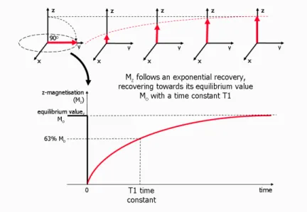
T1 relaxation time (tipically a few hundred milliseconds - a few seconds) is the time constant of the longitudinal relaxation (longitudinal recovery, or spin-lattice relaxation). It means that the net magnetization vector recovers to the longitudinal direction (Figure 1) (Huettel et al., 2008).
Figure 1 The overview of T1 relaxation process (T1 recovery). The net magnetization vector (M) has been tipped into the transversal plane with a 90-degree excitation pulse. The spins begin to relax after the excitation and release the excess energy into the surrounding environment. At the end, the net magnetization vector recovers to the longitudinal direction (adapted from Ridgway, 2010).
30 I.6.2.3.2 T2 relaxation
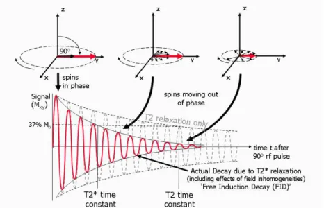
T2 relaxation time (a few hundred milliseconds) is the time constant of the transverse relaxation (transverse decay, or spin-spin relaxation). The precessing frequency of spins will be different from each other during T2 relaxation process, so they fall out of the same phase. This causes exponential reduction of the net magnetization in the transverse plane (Figure 2) (Huettel et al., 2008).
I.6.2.3.3 T2* relaxation
T2* relaxation is a type of T2 relaxation process which is sensitive to the inhomogeneity of the magnetic field as well. This additive inhomogeneity decreases the net magnetization vector faster than T2 relaxation. Thus, the T2* relaxation time is always shorter (a few tens of milliseconds) than the T2 relaxation time (Figure 2). This type of relaxation is the most important and usually used in functional measurements (Huettel et al., 2008).
Figure 2 The overview of T2 decay and T2* relaxation process. The net magnetization vector (M) has been tipped into the transversal plane with a 90-degree excitation pulse. After the excitation, this vector decays (T2 relaxation) or rapidly decays (T2* relaxation, including effects of field inhomogeneity) as a result of the loss of phase coherence (adapted from Ridgway, 2010).
31 I.6.2.4 Image formation
A basic one dimensional magnetic resonance spectroscopy (MRS) makes possible the chemical analysis of the sample without any spatial coding. The resulting spectrum carries qualitative and quantitative information. Increasing the number of dimensions with the application of gradient coils, the chemical analysis of every single unit (pixel, voxel) will be measureable.
The final goal of medical magnetic resonance imaging is image formation. Three dimesional MR images are the combinations of many 2D representations, which are basically spatial distribution maps of spins within planar cross-sections of the samples.
To allow 3D image formation, in addition to the main static magnetic field (B0), 3 gradient magnetic fields (x, y, z-gradients) are used. The strong static magnetic field (B0) arranges the atomic nuclei along the two determined directions while the gradient magnetic fields are used to gain spatially resolved information about the sample. The application of gradient coils ensures that atomic nuclei precess at different rates at various spatial location.
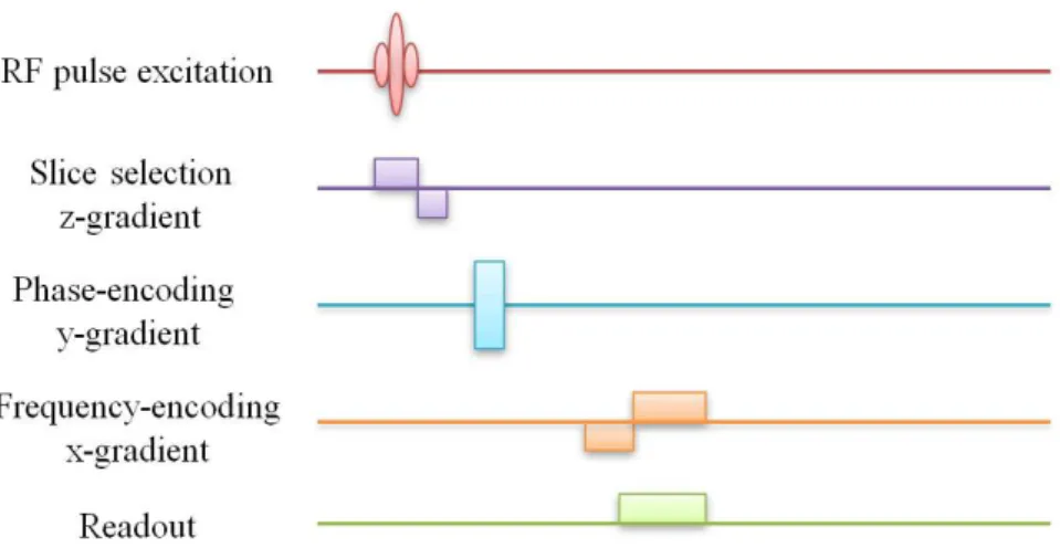
The slice selecting gradient (Gz - parallel with the scanner bore and the B0 magnetic field) is essential to index a slice within the 3D object in the z-direction. The frequency encoding gradient (Gx) changes the precession frequency of spins along the x-axis while the phase encoding gradient (Gy) has effect on spins along the y-axis. The acquired MR signal depends on the consecutive combination of the appropriate frequency and phase encoding gradients. The schematic overview of the applied radiofrequency (RF) pulse and magnetic gradients can be seen on the next diagram (Figure 3) (Huettel et al., 2008).
32
Figure 3 The illustration of a general pulse sequence which is necessary to obtain a two-dimensional image of a given slice. The application of slice selecting gradient determines the two-dimensional slices in the z-direction. An appropriate RF excitation pulse flips the net magnetization vector into the x-y plane within the determined slice. After the slice selection and the excitation procedure, the Gy and Gx
gradients change the precessing frequency of the spins. Altogether, these gradients ensure that the atomic nuclei precess at different rates at various spatial location (adapted from Huettel et al., 2008).
I.6.2.5 MRI contrast mechanism
MRI is a widespread and flexible neuroimaging technique, which uses various contrast mechanisms to create images. First of all, it is important to define two major factors: repetition time (TR) and echo time (TE). TR is the time interval between two consecutive excitations, expressed in seconds. TE is that time which goes by between the excitation and the data acquisition, usually expressed in milliseconds. These values depend on the strength and the homogeneity of the magnetic field (higher field strength causes slower longitudinal relaxation and longer T1 value, faster transverse relaxation and shorter T2 and T2* values), the type of atomic nuclei and several other factors.
There are four large contrast mechanism groups:
• Endogenous contrast represents the intrinsic properties of biological tissues.
The most frequently applied endogen contrast agent is the hemoglobin molecule.
During BOLD fMRI measurements the changes of oxy-deoxy hemoglobin ratio cause the determinable MR signal (Huettel et al., 2008).
• Exogenous contrast mechanism requires external contrast agent. These compounds increase the statical and motion contrast as well, so these are
33
frequently used in clinical MRI experiments. There are lots of agents applied in daily routine e.g. Gd-DTPA, superparamagnetic iron oxide (SPIO), ultrasmall superparamagnetic iron oxide (USPIO) (Nakamura et al., 2000), superparamagnetic iron-platinum particles (SIPPs) (Taylor et al., 2011), and manganese-based nanoparticles (Koretsky and Silva, 2004).
• Motion contrast depends on the diffusional (diffusion contrast) and perfusional motion (perfusion contrast) of the atomic nuclei within the sample (Huettel et al., 2008).
• Static contrast is a commonly used contrast mechanism. MRI measurements are based on this technique to determine the brain anatomy. It is sensitive to the type, number, relaxation time, and resonance properties of the spins (Huettel et al., 2008).
I.6.2.5.1 Proton-density contrast
It is the simplest static MR contrast based on the different numbers of spins within voxels. The suppression of T1 and T2 contrasts are required to maximize the proton-density contrast (very long TR and very short TE values). The most powerful signals come from the cerebrospinal fluid (CSF) and ventricles. Gray matter (GM) gives a less intensive signal while white matter is only barely distinguishable from the air (Huettel et al., 2008).
I.6.2.5.2 T1 contrast
This contrast mechanism is based on the different relaxation properties of atomic nuclei in tissues. Parallel with the above-mentioned proton-density contrast, varying the repetition time and echo time can create the ideal T1 contrast (intermediate TR and short TE). The most forceful signals come from the white matter (WM) and bone marrow, due to their short T1 values. The area of the gray matter (GM) seems to be less intense while the water content of the CSF provides no signal (dark area on the images) from the air (Huettel et al., 2008).
34 I.6.2.5.3 T2 contrast
The basis of this contrast type is similar to the T1 contrast. In this case, only the T2 or transverse relaxation is examined, so the T1 contrast must be suppressed (long TR and intermediate TE values). The maximal signals come from the water containing regions (CSF and ventricles), these are bright on the images. The gray matter (GM) seems to be moderately bright while the white matter (WM) is totally black.
T2* contrast is a special type of T2 contrast, that is susceptible to the magnetic field inhomogeneity. T2* contrast forms the basis of BOLD fMRI measurements because it is sensitive to the actual level of deoxyhemoglobin concentration in the blood (Huettel et al., 2008).
35
I.6.3 Functional Magnetic Resonance Imaging
In the 19th century, Angelo Mosso performed his famous 'human circulation balance' experiment. He measured the redistribution of blood in the brain noninvasively during emotional and intellectual activities (James, 1890; Sandrone et al., 2012).
In the further development of functional MR imaging, Charles Roy and Charles Sherrington played unquestionable roles (Roy and Sherrington, 1890). They were the first ones who could prove the connection between cerebral blood flow (CBF) and the brain functions (Raichle et al, 2001). In 1936, Linus Pauling and Charles Coryell realized that deoxygenated hemoglobin molecules can affect the homogeneity of the magnetic field.
After this revelation, Seiji Ogawa extended the usage of MRI from structural images to functional ones. He recognized the role of deoxyhemoglobin molecules as endogen contrast agent. Thereby, changes in neuronal activity in the brain could be seen as alteration of MR signal (Ogawa et al., 1990).
Presently, fMRI techniques rely on the fact that cerebral blood flow and neuronal activation are coupled (Huettel et al., 2008). Functional MRI is kept a modern, flexible, and indirect functional neuroimaging technique, which uses a high static magnetic field.
To create images about the physiological activity, the scanner uses continuously changing magnetic gradient fields and radiofrequency (RF) pulses, known as pulse sequence. The exact frequency of the RF excitation determines which MR active nucleus will be excited. Most scanners are tuned to the frequency of hydrogen nucleus (42.574 MHz/T) due to the high prevalence of water is biological system (Pykett et al., 1982).
I.6.3.1 Physiological background of fMRI measurements
I.6.3.1.1 Neurovascular coupling
The human brain consists of approximately 100 billion neurons, which have nearly 100 trillion synaptic connections with each other. Neurons are the basic information-processing units in the brain, which have complex roles in brain integration and signaling pathways. However, neurons and other brain structures do not have any
36
energy storing system. So, their energy demands (the maintenance and restoration of the ion concentration gradient consume lots of energy after neuronal activations) are directly covered by glucose and oxygen via the vascular system.
Currently, it is well known that the neural and the vascular system are coupled.
Neural activations generate strong determined alteration in the brain vasculature and increase the energy consumption in the given part of the brain tissue. The glucose and oxygenated hemoglobin concentration in the blood decreases (due to the increased cerebral metabolic rate of oxygen or CMRO) and the quantity of deoxygenated hemoglobin molecules increases. Fortunately, the brain is able to cover these temporary deficits via vasodilatation. The locally enhanced cerebral blood flow (CBF) and cerebral blood volume (CBV) are sufficient to increase the oxygenated-deoxygenated hemoglobin ratio. During this process, the blood volume remains unchanged within the brain, only the local blood distribution changes (Huettel et al., 2008). These relative oxygenated- and deoxygenated hemoglobin concentration changes are overviewed in Figure 4.
Figure 4 The changes of oxygenated- and deoxygenated hemoglobin concentration during neuronal activation. Neuronal activation causes a quick increase in the concentration of deoxygenated hemoglobin. 2 sec after the stimulus onset the time-curve achieves its maximum value and then rapidly declines (6 sec after the onset). This mechanism forms the basis of the BOLD hemodynamic response.
Meanwhile, the oxygenated hemoglobin concentration time-curve shows slight delay compared to the stimulus onset. It rises its peak much slower (5-6 sec) and recovers to the basic level in ~10 sec (adapted from Huettel et al., 2008).