INVESTIGATION INTO THE FACTORS INFLUENCING THE ONSET OF HEREDITARY ANGIOEDEMA ATTACKS
PhD thesis
Beáta Visy MD
Doctoral School of Basic and Translational Medicine Semmelweis University
Supervisor: Henriette Farkas, MD, D.Sc Official reviewers: Gabriella Gálffy, MD, Ph.D
Péter Holló, MD, D.Sc
Head of the Complex Examination Committee:
Péter Kempler, MD, D.Sc
Members of the Complex Examination Committee:
Gábor István, MD, Ph.D Adrienne Nagy, MD, Ph.D
Budapest
2019
1 1 Introduction
Hereditary angioedema due to the deficiency of the C1-inhibitor is an autosomal dominant disorder (C1-INH-HAE) characterized by recurrent angioedematous episodes involving the subcutis and/or the submucosa. HAE attacks affecting the mucosa of the upper airways may lead to suffocation, whereas oedema of the intestinal wall may raise the suspicion of a surgical emergency.
HAE can be diagnosed by complement testing. It is important to distinguish its clinical manifestations from those of other angioedema types with an allergic aetiology. In particular, the recurrent attacks of hereditary angioedema are unresponsive to the medications commonly used in the management of allergic disorders (such as epinephrine, antihistamines, and corticosteroids). In Hungary, human plasma-derived or recombinant human C1-inhibitor (C1- INH) and bradykinin B2-receptor antagonists are available as treatments – these agents prevent the release of bradykinin, a mediator involved in the pathomechanism of HAE attacks.
The onset of the manifestations of HAE may be induced by mechanical trauma, mental stress, surgical interventions, active infections, hormonal changes, and certain medicinal products.
As the acute oedematous episodes may progress into a potentially life- threatening condition, HAE has a substantial, negative impact on the patients’
quality of life. Notwithstanding the steady expansion of knowledge, the patients about to experience an attack cannot be identified in advance, as well as the time and the location of onset or the severity of the imminent HAE attack cannot be predicted.
2 Objective
Our objective was to explore the triggering factors of HAE attacks in as much detail as possible.
2.1
The relationship between sex hormones and the frequency of HAE attacks1 How do life situations associated with hormonal changes influence the frequency of the HAE attacks experienced by female patients?
2 What is the ratio between the levels of the individual sex hormones and their laboratory reference values?
3 Is there a difference between the sexes concerning the number of HAE attacks?
2
4 Is the frequency of oedematous HAE attacks correlated to the serum levels of sex hormones?
5 Do the various hormone levels have any predictive value?
2.2
The relationship between infection by Helicobacter pylori and the frequency of HAE attacksIn their study conducted in 2001 in Hungarian HAE patients, Farkas et al reported an increased frequency of oedematous abdominal attacks in patients infected with H. pylori. The number of HAE attacks decreased after the successful eradication of this pathogen.
1 In view of these findings, we initiated an international study to verify our hypothesis in a larger population of patients of different nationalities.
2 By following up on the subjects of the previous Hungarian study (by Farkas et al. 2001); we intended to ascertain whether the beneficial effect of H. pylori eradication lasts in the long term.
3 One of our patients who had undergone the eradication of H. pylori subsequently experienced an increase in the frequency of the occurrence of HAE manifestations. We investigated the possibility that this might have resulted from reinfection with H. pylori.
2.3
The relationship between vitamin D3 level and the frequency of HAE attacksWe wanted to ascertain whether there are differences between the vitamin D3 levels measured in, as well as the number of HAE attacks experienced and the quantity of pdC1-INH received by our patients during the summer-autumn and in the winter-spring periods. We also sought for possible correlations between vitamin D3 levels and other laboratory or clinical parameters.
3 Methods
3.1
Investigation of the possible relationship between sex hormone levels and the frequency of HAE attacksSeventy-eight patients registered at the Hungarian HAE Centre were enrolled (34 males and 44 females). The mean age of the study population was 30.3 years (range 4-70 years). At the time of the trial, 17 patients were of paediatric age (nine boys and eight girls, age range 4-15 years) and 25 were adult males (mean age 35.76±13.37 years). Among the female patients, 31 were of reproductive age (mean 32.29±11.07 years) and five were postmenopausal (59±7.84 years). Thirty-eight patients (4 children, 17 adult males, and 17 adult
3
females) underwent treatment with an attenuated anabolic steroid (danazol) during the trial.
A questionnaire was administered to explore the medical history of adult patients, in order to ascertain changes in the frequency of symptoms during 1) adolescence. 2) menstruation 3) pregnancy (as well as the consistency of changes during multiple pregnancies), and 4) the influence of oral contraceptive use on the clinical manifestations.
Serum levels of LH, FSH, progesterone, oestradiol, testosterone, PRL, and SHBG were measured in all patients. Serum levels of LH, FSH, progesterone, oestradiol, testosterone, and PRL were measured quarterly, on three occasions. Determinations were performed using 125I radioimmunoassay (RIA) (testosterone, progesterone, hFSH, hLH, and hPRL), and 125I immunoradiometric assay (IRMA) (SHBG) developed by the National Radioisotope Institute (Budapest. Hungary), and the ESTR-CTRIA kit (oestradiol) manufactured by CIS Bio International (Gif-Sur-Yvette Cedex, France). The free androgen index (FAI) is the quotient of the testosterone and SHBG concentration and it is expressed as percentage values.
C1-INH antigen concentration in serum was determined by radial immunodiffusion using antiserum to the C1-inhibitor, obtained from DiaSorin (Stillwater, MN, USA). The results were expressed as percentages of mean levels measured in normal specimens. Functionally active C1 inhibitor was determined using the Quidel C1-inhibitor enzyme immunoassay kit (Quidel.
San Diego, CA, USA).
Hormone levels were compared to the patients’ clinical symptoms – that is, the number of attacks experienced during the one-year period after blood sampling. Statistical analysis was undertaken with the SPSS 10.0 software (SPSS Inc., Chicago, IL, USA).
This study was supported by the following grants: ETT211/00, FKFP 0106/2000, OTKA T025449, OTKA T029044.
The study was performed in accordance with the Declaration of Helsinki.
3.2
Investigation of the possible correlation between infection by Helicobacter pylori and the frequency of HAE attacksBetween 2004 and 2005, 152 patients [64 males. 88 females; mean age: 34.41 years (1-77 years)] were enrolled from seven European countries collaborating in the study implemented within the framework of the PREHAEAT project (Novel Methods for Predicting Preventing and Treating Attacks in Patients with Hereditary Angioedema QLG1-CT-2002-01359) of the European Union. The inclusion criteria were as follows: HAE diagnosis, registered number of subcutaneous, abdominal, laryngeal attacks in last year,
4
and confirmed or excluded H. pylori infection. As exclusion criteria, we excluded from the international study those Hungarian patients, who participated in the previous Hungarian study in 2001. These patients were participants of the follow-up trial. No other patients were excluded from the study.
From Hungary, new cases identified after the conclusion of the previous study were entered into the international register; their results were analyzed together with those of the subjects of the international study.
Our report published in 2001 identified a subset of nine patients (out of 65 patients with HAE) who had had experienced frequent recurrences of abdominal oedematous attacks accompanied by dyspepsia, despite uninterrupted and previously effective long-term pharmacotherapy. All these nine patients were infected with H. pylori. Successful eradication of this pathogen reduced the cumulative number of abdominal attacks to one from 28 per year, whereas the total number of attacks decreased to 19 from 100 (Farkas et al, 2001). These patients were followed up for an additional 4 years. As one patient died in the meantime, only eight patients could be followed throughout the study.
The mean age (range) of the patients was 51 (41-69) years at the end of the 4- year follow-up; four of them were males and five were females. The deceased patient was the oldest women, who would have turned 69 years old at the end of follow-up.
This patient (H.J.) was 54 years old at the time of reinfection with H. pylori.
The principal investigators of the individual study centres obtained a detailed history from the patients managed at their institutions. Together with clinical and laboratory parameters, these historical data were uploaded to the international HAE Registry, using a password-protected webpage (www.haeregister.org). Epidemiological information on the patients enrolled into work package N° 4 (H. pylori susceptibility to abdominal attacks) was retrieved from the registry and subjected to statistical analysis along with the clinical data relevant to the study.
Active H. pylori infection was confirmed in the international population of HAE patients with the C14-urea breath test alone or in combination with testing for anti-H. pylori IgG antibodies.
Eradication was performed at each centre according to the local protocols and in conformity with the international guidelines.
A breath test was performed at the next follow-up visit – at least 6 weeks after the conclusion of eradication therapy. If eradication was successful (i.e. the positive breath test turned negative), we recorded the numbers of (subcutaneous. laryngeal, and abdominal) angioedematous episodes
5
experienced by the patient in the preceding year, along with the medication administered as prophylaxis. The same data were recorded again 12 months after the eradication.
The accumulated data were analyzed using the SPSS for Windows software package (version 13.0. SPSS Inc., Chicago, IL, USA).
This study was supported by the grants PREHAEAT QLG1-CT-2002-01359, ETT 194/2003, OTKA T046496, SBF 02.0347.
The study was performed in accordance with the Declaration of Helsinki.
3.3
Investigation of the possible relationship between vitamin D3 level and the frequency of HAE attacksIn 2013 and 2014, 118 patients (69 females 48 males, mean age 38.44 [2.1 to 83.25] years) out of the 175 C1-INH-HAE patients followed-up at the National Angioedema Reference Center were included in the study.
We determined the number of HAE episodes experienced and the type of the attacks, as well as the medication administered as a treatment in the given period using the entries of outpatient records and patient diaries.
The HAE attacks were categorized into three groups by their location. The first group was that of subcutaneous manifestations, whereas the second group comprised submucosal oedematous episodes involving the lips or the pharynx.
The third group consisted of the gastrointestinal events of submucosal oedema. In case angioedema manifested in multiple locations simultaneously, the clinically more severe occurrence was taken into account. (For example, the combination of subcutaneous and submucosal oedema was assigned to the group of submucosal events. as well as the joint occurrence of gastrointestinal and lip/laryngeal oedema was regarded as a lip/laryngeal episode.) Furthermore, the occurrence of erythema marginatum was also recorded.
Because the serum level of vitamin D3 is known to exhibit seasonal fluctuation, we determined vitamin D3 level on two occasions in two different seasons of the year, from freshly collected blood samples using a direct competitive chemiluminescent assay (LIAISON 25OH Vitamin D total assay;
DiaSorin, Stillwater MN, USA).
We divided the calendar year into two 6-month periods based on the physiological fluctuation of hormone level. The winter-spring period started on 1 December and ended on 31 May, whereas the summer-autumn period lasted from 1 June to 30 November. In all patients, one of the two blood samplings was performed at the annual follow-up visit, together with a complement screen, the checking of CBC along with the indices of hepatic and renal function, and inflammatory parameters (CRP, and occasionally ESR). The complement screen included the determination of C1q
6
(complement 1 protein q subunit), C3, C4, C1-INH concentration and functional activity, as well as anti-C1-INH antibodies.
Accordingly, vitamin D3 levels and clinical parameters were available from the winter-spring period for 111, and from the summer-autumn period for 105 patients. In the case of 98 patients, these values were available from both periods. Because complement testing was performed only at the annual follow-up visits, these were obtained in the winter-spring period in 57 and in the summer-autumn period in 61 patients.
The data were analyzed with version 7.00 of the GraphPad Prism statistical software. The Mann–Whitney test was applied to independent samples/groups, and the Wilcoxon’s was used to analyze related groups. Spearman’s test was performed to detect correlations.
The study was conducted with the approval of the Semmelweis University Regional and Institutional Committee of Science and Research Ethics (65/2008).
4 Results
4.1
The relationship between sex hormone levels and the frequency of HAE attacks4.1.1 Results of the survey conducted among female patients
Patients completed the questionnaire in the physician’s office. All 36 females after the menarche (age 14–69 years) submitted forms suitable for analysis, which yielded the following results. Twenty-one patients had been symptom free before menarche. Symptoms of 13/21 patients first occurred during adolescence, whereas the remaining eight patients had no attacks in the pubertal period. Oedema during the perimenstrual period was reported by 42.4%. According to data from 25 patients who had been pregnant, pregnancy was associated with a higher incidence of attacks in 36%. Oedema formation was less common in 56%, and 8% experienced no change in the frequency of symptoms. The pattern of attacks was consistent (i.e. characterized by increased or reduced frequency) in half of females, whereas it was variable in the remaining 50%. Genital oedema developed during labour in one patient, but resolved after the administration of C1-INH concentrate and did not interfere with delivery. Of the 11 patients using oral contraceptives, seven reported an increase in attack frequency.
4.1.2 Hormone levels
The results of these three measurements were strongly correlated, as demonstrated by the Spearman’s correlation test (mostly R>0.5 and
7
p<0.0001). Moreover, patients with low or high levels of a hormone at the initial measurement had almost identical concentrations at the second and third measurements as well. For example, PRL levels [median (interquartile range)] of the patients were 314 (194– 453) mU/L and 312 (187–472) mU/L, respectively, at the first and third determinations. Similarly, on the first occasion, we measured 112 (29.4–211.6) pmol/L oestradiol and 117.8 (27.6–
266.8) pmol/L on the third occasion. Therefore, the results of only the initial measurement were used in our subsequent statistical analysis. Any deviations of sex hormone levels from the normal ranges are summarized in Table 1.
Table 1 – Number of patients with low, normal, or high levels of sex hormones.
PRL T E2 FSH P LH
Low value 10 23 23 18 1 23
Normal value 52 54 46 46 36 51
Elevated value 15 1 9 14 41 4
All values were determined in consideration of the patient’s gender, age, and stage of the menstrual cycle. The serum level of sex hormones was normal in the majority of patients; however, more than half of the subjects had progesterone levels above the upper limit of the normal range.
4.1.3 Gender-related differences in the frequency of attacks in HANO patients
Significantly more attacks occurred in female than in male patients during the year that followed blood sampling for hormone measurements. The
differences were more pronounced with abdominal than with subcutaneous attacks.
4.1.4 Correlation between sex hormone levels and the frequency of HAE attacks with different location recorded during the 1-year period following the blood sampling for hormone measurements The potential relationship between sex hormone levels and the number of attacks recorded during the 1-year period after the trial were analysed by performing Spearman’s correlation. Considering the gender-related differences in the attack rates, the results were analysed separately for male and female patients (Table 2).
8
Table 2 – The number of patients with low, normal, or high levels of sex hormones.
Males Females
Subcutaneous Abdominal Total Subcutaneous Abdominal Total
PRL 0.25 (0.161) 0.071
(0.693)
0.208
(0.246) 0.078 (0.62) 0.443 (0.003)
0.204 (0.189) Oestradiol 0.119 (0.508) 0.003
(0.986)
0.109
(0.544) 0.308 (0.044) 0.105 (0.501)
0.332 (0.029) Progesterone 0.252 (0.158) 0.020
(0.911)
0.215
(0.229) 0.507 (0.001) -0.020 (0.9) 0.408 (0.007) SHBG -0.001 (0.996) -0.065
(0.726)
-0.076
(0.679) -0.509 (0.001) -0.29 (0.065)
-0.581 (<0.0001) Testosterone -0.089 (0.621) -0.221
(0.231)
0.010
(0.956) 0.147 (0.358) 0.059 (0.708)
0.214 (0.167)
FSH -0.076 (0.799) 0.030
(0.868)
0.091
(0.613) -0.015 (0.923) 0.180 (0.248)
0.077 (0.622)
LH -0.037 (0.837) 0.083
(0.647)
0.048
(0.790) 0.089 (0.572) 0.033 (0.836)
0.091 (0.560)
FAI -0.169 (0.381) 0.144
(0.457)
-0.194
(0.314) 0.373 (0.016) 0.192 (0.230)
0.446 (0.003)
A statistically significant correlation between hormone levels and the frequency of attacks was found only in females. A positive correlation was ascertained between the rate of subcutaneous attacks and oestradiol, as well as progesterone levels, whereas a significant, negative correlation was demonstrated for the SHBG level. By contrast, PRL levels correlated only with the frequency of abdominal attacks. When the frequency of all types of attacks (subcutaneous + abdominal + other) was considered, the attack rate correlated positively with oestradiol and progesterone levels, whereas a significant negative correlation was demonstrated for SHBG level. As FAI values were calculated from the SHBG levels, and in female patients a strong negative correlation (r = 0.822. p <0.001) was found between FAI values and SHBG concentrations, we found a positive correlation between FAI values and the number of oedematous attacks (Table 2). No correlation was found between progesterone and C1- INH concentration (OR 0.52; p=0.754) or C1- INH activity (OR 0.165; P = 0.317), and the same applied to oestradiol and PRL. During multiple linear regression, adjustment for age and danazol dose eliminated the statistical significance of the relationship between PRL level and abdominal attack rate in female patients (p=0.194), as well as the relationship between the oestradiol levels and the frequency of subcutaneous attacks (p=0.685). At the same time, the frequency of subcutaneous attacks correlated positively with progesterone levels (p=0.025), and negatively with SHBG levels (p=0.007) even after adjustment for age and danazol dose.
9
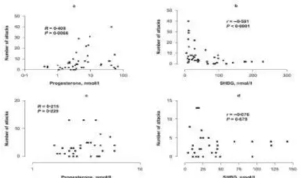
Figure 1. The correlation between serum progesterone (a, c) and SHBG (b, d) levels and the number of oedematous attacks occurring in the year after blood sampling in female (a, b) and in male (c, d) patients with HAE. Spearman’s correlation coefficients and their significance (p-values) are indicated.
Next, we plotted serum concentrations of the latter two variables against the frequencies of all types of attacks separately in female and male patients (Figure 1). Progesterone levels correlated with attack rate only in females (Figure 1 a,c). A significant, negative correlation between SHBG levels and attack rates was also found in females only (Figure 1b,d). However, when the plot of SHBG levels vs. attack rate in male patients (Figure 1,d) was analysed in detail, it turned out that high SHBG levels were associated with a low attack rate also in males.
4.1.5 Prediction by high progesterone and low SHBG levels of the high frequency of subcutaneous and all-type attacks recorded during the 1-year period following the blood sampling for hormone measurements
The study population was divided into two groups of patients with high or low levels of PRL, oestradiol, progesterone, and SHBG, using the following cut- off levels: PRL 450 mU/L (upper threshold of normal range); oestradiol 147 pmol/L (menopausal threshold); progesterone 4 nmol/L (menopausal threshold); SHBG 40 nmol/L (5% trimmed mean of the whole population).
Patients were further stratified into two subgroups according to attack rate below or equal to or above five per year. The data were analysed by multiple logistic regression, adjusted for the age of patients, the dosage of danazol and
10
(except SHBG) – in female patients – for the menstrual cycle at the time of blood sampling. A high progesterone level (≥4.0 nmol/L) was associated with a more than 13 times higher OR (p=0.0047) for a high subcutaneous attack rate, compared to patients with progesterone levels below 4.0 nmol/L. By contrast, neither high PRL levels, nor high oestradiol levels were significant predictors of a high rate of subcutaneous attacks. Similarly, the frequency of all types of attacks was predicted only with high progesterone levels, but both the odds ratio and the significance were found to be lower than in the case of subcutaneous attacks. High levels of SHBG seemed to be associated with low attack rates in both sexes (Figure 1). Therefore, we assessed the predictive value of the SHBG measurement in males, in females, and in all patients. A cut-off point – corresponding to the 5% trimmed mean (40 nmol/l) of the SHBG levels measured in the sera of all patients – was used for this calculation. High SHBG concentration predicted low attack rates in both sexes, although only with coefficients of marginal significance. When all patients were considered, a significant (p=0.034) and very low OR [0.25 (0.07–0.90)] was obtained. When we applied a different cut-off point (30 nmol/L) close to the median SHBG levels of the males (28.9 nmol/l) and of the females (30.2 nmol/l), a significant association was obtained only in females [OR 0.16 (0.03–0.82). p= 0.0281]. In male patients, high SHBG levels were not significantly associated with a low attack rate [OR 0.25 (0.02–2.58).
p=0.2469].
4.2
The relationship between H. pylori infection and the frequency of HAE attacks4.2.1 Screening for H. pylori infection in the international patient population
Oedematous abdominal attacks were identified in the history of 125 patients (82.2%); 39.5% (60/152) of these patients experienced five or more abdominal attacks per year.
Of the 152 patients, 139 were available for serological testing for H. pylori IgG, of which 61 (43.9%) yieldeld positive results. Thirty of the 61 H. pylori IgG-positive patients accepted eradication therapy after infection was confirmed by 14C-UBT. Eradication was successful in 28 patients, and the data recorded during their 12-month follow up were available. During the one-year follow up, as compared with the attack rate recorded during the 12 months before eradication, attack frequency decreased in 19 patients, did not change in seven patients, and increased in two patients. Wilcoxon’s nonparametric test revealed a statistically significant (p=0.0006) reduction in the number of
11
abdominal attacks (versus baseline) during the 1-year period following eradication, compared with data from the 12-month period preceding eradication. The total number of abdominal attacks decreased significantly (p=0.0006), whereas no significant change could be demonstrated in the occurrence of laryngeal (p=0.156) or subcutaneous (p=0.794) attacks.
4.2.2 Follow up of the patients who underwent H. pylori eradication during the previous Hungarian study
Neither the number, nor the location of the oedematous attacks changed in these patients during this period. No dosage adjustment of drug prophylaxis was necessary.
The numbers of the attacks sustained during the 12-month period before eradication was compared to the mean number of attacks occurring during the 4-year-long follow up. The statistical analysis performed using Friedman’s ANOVA test followed by Dunn’s post test revealed a significant reduction of total attack number (p<0.01), of the number of abdominal attacks (p<0.01), and of the dose requirements of prophylactic agents (p=0.05).
On the other hand, no differences were found when the period subsequent to eradication was compared with the follow-up period. In addition, C1-INH activity, which was found to be 25.8% (15.5–53.2) (median [interquartile range]) after eradication, increased significantly (p=0.039) to 75.6% (66.0–
85.0) during the follow-up period.
4.2.3 Case presentation
During follow up, a 54-year-old subject was re-infected with H. pylori, as demonstrated by the 14C-UBT. The incidence of oedematous manifestations increased during the period of active infection, but successful eradication reduced and stabilized the number of symptoms.
4.3
The correlation between vitamin D3 level and the frequency of HAE attacks4.3.1 Vitamin D3 levels in C1-INH-HAE patients
The subjects were assigned to the following three groups according to their vitamin D3 levels: definitely vitamin-deficient patients (<20 ng/ml);
presumably vitamin-deficient patients (20 to 30 ng/ml), and patients with normal vitamin D3 level (>30 ng/ml).
12
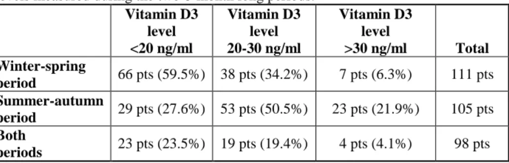
Table 3: The distribution of the 118 patients included in the study by vitamin D3 levels measured during the two 6-month long periods.
Vitamin D3 level
<20 ng/ml
Vitamin D3 level 20-30 ng/ml
Vitamin D3 level
>30 ng/ml Total Winter-spring
period 66 pts (59.5%) 38 pts (34.2%) 7 pts (6.3%) 111 pts Summer-autumn
period 29 pts (27.6%) 53 pts (50.5%) 23 pts (21.9%) 105 pts Both
periods 23 pts (23.5%) 19 pts (19.4%) 4 pts (4.1%) 98 pts During the winter-spring period, 59.5% of the subjects (n=66/111) was definitely vitamin D3-deficient, and only 6% (n=7/111) had serum levels corresponding to the recommended 30 ng/ml level. An additional 11.7%
(n=13/111) had severe vitamin D3 deficiency with a serum level lower than 10 ng/ml. During the summer-autumn period, the proportion of patients with vitamin D3 deficiency was still 27.6% (n=29/105), and only 21.9%
(n=23/105) had a normal serum level. Remarkably, 23.5% (n=23/98) of the study subjects had serum vitamin D3 levels below 20 ng/ml during both the winter-spring and the summer-autumn periods – that is, these patients may be considered vitamin D3-deficient throughout the year (Table 3).
4.3.2 The difference between vitamin D3 levels measured during winter-spring or summer-autumn periods
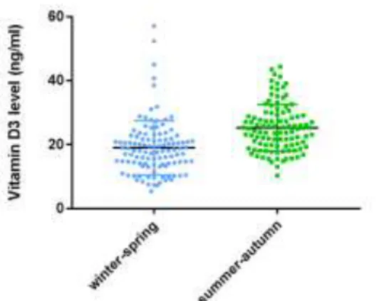
The comparison of data from the winter and from the summer periods reveals that in our study population, the serum levels of vitamin D3 were significantly (p<0.0001) higher during the summer than in the winter (Figure 2).
The number of vials containing pdC1-INH concentrate used by the patients during the summer-autumn period was significantly (p=0.1) smaller than during the winter-spring period. Comparing the levels of various complement parameters, there were no differences between the two periods with regard to the number of HAE episodes (neither by attack type nor altogether) experienced by the patients.
13
Figure 2. Vitamin D3 levels of patients with C1-inhibitor deficient hereditary angioedema during the winter-spring and the summer-autumn periods.
4.3.3 The relationship between vitamin D3 levels, as well as clinical and laboratory parameters
Our analyses did not detect any correlation between vitamin D3 level, and the cumulative dose of pdC1-INH required to relieve HAE episodes or the numbers of the oedematous attacks. Spearman’s correlation analysis was based on the vitamin D3 levels and clinical parameters shown in Table 4.
Subsequently, we analyzed the data from winter-spring and summer-autumn periods aggregately. This meant that the dataset included two data series for nearly every patient and hence the values measured in 216 samples in total were analysed. Clinical information could not be obtained for 9 samples and therefore, these were not included in the analysis. According to the analysis of the entire dataset, the median level of vitamin D3 was 20.8 ng/ml. The results were – in every respect – the same as those of the seasonal analyses. No correlation could be detected between the vitamin D3 level and the other parameters studied.
A significant correlation could not be confirmed between vitamin D3 level and the individual complement parameters – either during the winter-spring or during the summer-autumn period.
14
Table 4. The difference between the vitamin D3 levels and clinical indices measured in the same patients during the two seasonal periods (Wilcoxon’s test; N=98).
Winter median (25-75th pc)
Summer median (25-75th pc)
P-value (Wilcoxon’s teszt) Vitamin D3 level
(ng/ml)
18.1 (14-21.9)
24.9
(19.7-29.2) <0.0001 Erythema
marginatum
0 (0-0)
0
(0-0) NS
Subcutaneous angioedema
1 (0-4)
1
(0-3) NS
Laryngeal/lip angioedema
0 (0-0)
0
(0-0) NS
Gastrointestinal angioedema
0 (0-1)
0
(0-2) NS
All angioedema attacks
2 (0-6)
2
(0-5) NS
pdC1-INH treatment (vials)
0 (0-2)
0
(0-1) 0.01
Next, the samples from the summer-autumn period were assigned to two groups by vitamin D3 level lower than 20 ng/ml, or higher than 30 ng/ml. The samples corresponding to ‘borderline’ vitamin deficiency (i.e. with a vitamin D3 level between 20 and 30 ng/ml) were not taken into account in this analysis. This same comparison could be performed also during the winter- spring period between samples with a vitamin D3 level below 10 ng/ml or above 30 ng/ml. We sought for possible differences between the two groups with regard to the number of disease manifestations experienced (all, subcutaneous, submucosal, laryngeal/lip, and GI symptoms), the quantity of pdC1-INH supplementation administered, and the need for C1-INH supplementation during the week before blood sampling. We could not detect any differences between the study parameters of the two groups (Mann–
Whitney test).
4.3.4 Vitamin D3 supplementation habits
The analysis of the completed questionnaires showed that 31.17% (n=24/77) of the responders resorted to some form of vitamin D3 supplementation:
16.67% (n=4/24) used an oral solution, whereas 83.33% took tablets.
Concerning dosage frequency, vitamin D3 supplements were taken daily by 62.4% (n=15/24), every week by 16.67% (n=4/24), and 2 to 3 times a week by an additional four patients. As regards seasonal predilection 54.17% (n=13/24) takes vitamin D3 supplements all over the year, 37.5% (n=9/24) only during
15
the winter months, and 1-1 patients during the autumn, or the spring in addition to winter months. However, because the data gathered from anonymized questionnaires cannot be compared with clinical symptoms or laboratory findings, these results allow only limited interpretation.
5 Conclusions
1. In 42.4% of our patients, the frequency of HAE attacks increased during the perimenstrual period.
2. In patients taking oral (hormonal) contraceptive pills, the frequency and severity of the HAE attacks increased compared with the period before OC use.
3. The levels of the majority of sex hormones were within the physiologically normal range. However, progesterone levels exceeded the upper limit of the normal range in more than half of our C1-INH-HAE patients,
4. Compared with males, female patients experienced significantly more HAE attacks during the one-year period after blood sampling. This difference was more conspicuous in the case of abdominal oedematous episodes than with the instances of subcutaneous angioedema.
5. In female patients, high (4.0 nmol/l) progesterone levels were associated with a 13 times higher frequency of subcutaneous attacks than low progesterone levels. Accordingly, a progesterone serum concentration above the postmenopausal threshold was predictive of an increased attack frequency in female C1-INH-HAE patients.
6. Analysing male and female patients together, we found a significant correlation between the high SHBG level and the low number of HAE attacks.
7. The measurement of progesterone and SHBG levels in females, as well as of SHBG level in males might prove useful for the prediction of disease severity.
8. HAE attacks are more frequent among patients infected with H. pylori.
9. A significant reduction in the number of HAE attacks was observed following the successful eradication of H. pylori.
10. The beneficial effect of H. pylori eradication lasted in the long term and furthermore, this was reflected by an increase in C1-INH activity.
11. In 27.6% of C1-INH-HAE patients, vitamin D3 deficiency persists even in the summer-autumn period, and during the latter vitamin D3 levels reach the recommended value in only 21.9%. Considering the two seasonal periods together, the proportion of patients reaching the recommended 30 ng/ml vitamin D3 level was as low as 4.1% only.
16
12. In our study population, serum vitamin D3 levels were higher during the summer than during the winter.
13. The patients’ requirement for pdC1-INH concentrate was smaller during the summer-autumn than during the winter-spring period.
14. There was no correlation between vitamin D3 levels and the study parameters (complement screen and clinical manifestations).
17
6 Bibliography of the Candidate’s publications
6.1
Publications related to the PhD thesis1.
Visy B. Füst G. Varga L. Szendei G. Takács E. Karádi I. Fekete B. Harmat G.Farkas H. (2004) Sex hormones in hereditary angioneurotic oedema. Clin Endocrinol. 60: 508-515. IF: 3.023
2.
Visy B. Füst G. Bygum A. Bork K. Longhurst H. Bucher C. Bouillet L. Cicardi M. Farkas H. (2007) Helicobacter pylori infection as a triggering factor of attacks in patients with hereditary angioedema. Helicobacter. 12: 251-257. IF:2.423
3.
Visy B. Szilágyi T. Kőhalmi KV. Veszeli N. Varga L. Imreh É. Farkas H. (2019) D3-vitamin szint és a betegség súlyossága közötti kapcsolat vizsgálata herediter angioödémában. [Analysis of the relationship between vitamin D3 level and disease severity in hereditary angioedema]. Orv Hetil. 160: 987-993. (in Hungarian) IF: 0.564 in 20186.2
Other publications1. Farkas H. Harmat G. Füst G. Varga L. Visy B. (2000) Herediter angioneuroticus oedema gyermekkorban. [Hereditary angioneurotic edema in children] Orv Hetil.
141: 2541-2547. (in Hungarian)
2. Farkas H. Harmat G. Fáy A. Fekete B. Karádi I. Visy B. Varga L. (2001) Erythema marginatum preceding an acute oedematous attack of hereditary angioneurotic oedema. Acta Derm Venereol. 81: 376-377. IF: 1.477
3. Ozsváth L; Varga L. Nébenführer L. Visy B. Farkas H. (2002) Szerzett C1- esterase inhibitor hiány. Bőrgyógyászati és Venerológiai Szemle. 78: 203-206.
4. Farkas H. Harmat G. Füst G. Varga L. Visy B. (2002) Clinical management of hereditary angio-oedema in children. Pediatr Allergy Immunol. 13: 153-61.
*Review article. IF: 1.807 in the year of publication
5. Farkas H. Harmat G. Fekete B. Karádi I. Visy B. Varga L. (2002) Acute abdominal attack of hereditary angioneurotic oedema associated with ultrasound abnormalities suggestive of acute hepatitis. Acta Paediatr. 91: 971-974. IF: 1.260 6. Farkas H. Pintér J. Kalabay L. Jakab L. Visy B. Fekete B. (2002) Mometazon
furoat hatékonyságának vizsgálata. A gyógyszer hazai bevezetését követő első pollenszezon tapasztalatai. [Effectiveness of mometasone furoate nasal spray in seasonal allergic rhinitis. First experiences in practice after the introduction of the drug in Hungary]. Orv Hetil. 143: 1929-1933. (in Hungarian)
7. Farkas H. Visy B. Fekete B. Karádi I. Kovács JB. Kovács IB. Kalmár L. Tordai A. Varga L. (2002) Association of celiac disease and hereditary angioneurotic edema. Am J Gastroenterol. 97: 2682-2683. IF: 3.953
18
8. Visy B. Kocsis I. B. Kovács J. Kalmár L. Tordai A. Harmat Gy. Varga L. Fekete B. Karádi I. Farkas H (2003) Herediter angioneuroticus oedema és coeliakia együttes előfordulása. Gyermekgyógyászat. 54: 163-165.
9. Harmat Gy. Farkas H. Varga L. Visy B; Kincses L. (2003) Immunhiány – adathiány. Gyermekgyógyászat. 54: 201-204.
10. Visy B. Harmat Gy. Varga L. Farkas H. (2004) Herediter angioneuroticus oedema (HANO) gyermekkorban. Gyermekgyógyászat. 55: 193-198.
11. Agostoni A. Aygören-Pürsün E. Binkley KE. Blanch A. Bork K. Bouillet L.
Bucher C. Castaldo AJ. Cicardi M. Davis AE. De Carolis C. Drouet C.
Duponchel C. Farkas H. Fáy K. Fekete B. Fischer B. Fontana L. Füst G.
Giacomelli R. Gröner A. Hack CE. Harmat G. Jakenfelds J. Juers M. Kalmár L.
Kaposi PN. Karádi I. Kitzinger A. Kollár T. Kreuz W. Lakatos P. Longhurst HJ.
Lopez-Trascasa M. Martinez-Saguer I. Monnier N. Nagy I. Németh E. Nielsen EW. Nuijens JH. O'grady C. Pappalardo E. Penna V. Perricone C. Perricone R.
Rauch U. Roche O. Rusicke E. Späth PJ. Szendei G. Takács E. Tordai A.
Truedsson L. Varga L. Visy B. Williams K. Zanichelli A. Zingale L. (2004) Hereditary and acquired angioedema: problems and progress: proceedings of the third C1 esterase inhibitor deficiency workshop and beyond. J Allergy Clin Immunol. 114(3 Suppl): S51-131. IF: 7.205
S84: Visy B. Füst G. Varga L. Farkas H. Role of sexhormones in HAE part II
S104: Kollar T. Fekete B. Lakatos P. Visy B. Nemeth E. Farkas H. Investigation of bone turnover in patients with HAE
S108: Farkas H. Harmat Gy. Füst G. Varga L.Visy B. Clinical management of HAE in children
12. Felvinci R. Németh É. Visy B. Varga L. Jakab L. Farkas H. (2005) The effect of sexual hormon alterations on the frequency of oedematous attacks in patients with hereditary angioneurotic edema: A nemi hormonok változásának hatása a hereditaer angioneuroticus oedemában szenvedö betegek rohamainak gyakoriságára. Lege Artis Medicinae 15: 191-196.
13. Varga L. Széplaki G. Visy B. Füst G. Harmat G. Miklós K. Németh J. Cervenak L. Karádi I. Farkas H. (2007) C1-inhibitor (C1-INH) autoantibodies in hereditary angioedema. Strong correlation with the severity of disease in C1-INH concentrate naive patients. Mol Immunol. 44: 1454-1460. IF: 3.742
14. Blaskó B. Széplaki G. Varga L. Ronai Z. Prohászka Z. Sasvari-Szekely M. Visy B. Farkas H. Füst G. (2007) Relationship between copy number of genes (C4A.
C4B) encoding the fourth component of complement and the clinical course of hereditary angioedema (HAE). Mol Immunol. 44: 2667-2674. IF: 3.742
15. Farkas H. Varga L. Széplaki G. Visy B. Harmat G. Bowen T. (2007) Management of hereditary angioedema in pediatric patients. Pediatrics. 120:
e713-722. *Review article. IF: 4.473 in the year of publication
16. Farkas H. Jakab L. Temesszentandrási G. Visy B. Harmat G. Füst G. Széplaki G.
Fekete B. Karádi I. Varga L. (2007) Hereditary angioedema: a decade of human C1-inhibitor concentrate therapy. J Allergy Clin Immunol. 120: 941-947. IF:
19 8.115
17. Altorjai P. Visy B. Kormos Z. Harmat G. Fekete F. Farkas H. (2008) Pericardiac effusion complicating an acute abdominal attack of hereditary angioneurotic edema. American Journal of Case Reports. 9: 233-236.
18. Farkas H. Jakab L. Temesszentandrási G. Visy B. Harmat G. Füst G. Széplaki G.
Fekete B. Karádi I. Varga L. (2009) Hereditary Angioedema: A decade of human C1-inhibitor concentrate therapy 216-222. In: BL. Zuraw; MM. Frank (szerk.) A Selection of Important Papers in Hereditary Angioedema: A Compilation of Key Peer-Reviewed Papers From Leading Journals. Amsterdam. Hollandia: Excerpta Medica. (2009) Közlemény:1732575 (Passage in a book)
19. Kelemen Z. Moldovan D. Mihály E. Visy B. Széplaki G. Csuka D. Füst G.
Farkas H. Varga L. (2010) Baseline level of functional C1-inhibitor correlates with disease severity scores in hereditary angioedema. Clin Immunol. 134: 354- 358. IF: 3.932
20. Farkas H. Czaller I. Csuka D. Vas A. Valentin S. Varga L. Széplaki G. Jakab L.
Füst G. Prohászka Z. Harmat G. Visy B. Karádi I. (2010) The effect of long-term danazol prophylaxis on liver function in hereditary angioedema-a longitudinal study. Eur J Clin Pharmacol. 66: 419-426. IF: 3.032
21. Czaller I. Visy B. Csuka D. Füst G. Tóth F. Farkas H. (2010) The natural history of hereditary angioedema and the impact of treatment with human C1-inhibitor concentrate during pregnancy: a long-term survey. Eur J Obstet Gynecol Reprod Biol. 152: 44-49. IF: 1.764
22. Kelemen Z. Visy B. Csuka D. Czaller I. Füst G. Farkas H. (2010) Abdominal symptoms of hereditary angioedema and early weaning. Eur J Clin Nutr. 64:
1025-1027. IF: 2.563