The eukaryotic linear motif resource – 2018 update
Marc Gouw
1,*, Sushama Michael
1, Hugo S ´amano-S ´anchez
1, Manjeet Kumar
1, Andr ´as Zeke
2, Benjamin Lang
1, Benoit Bely
3, Luc´ıa B. Chemes
4,5,6, Norman E. Davey
7, Ziqi Deng
1,
Francesca Diella
1, Clara-Marie G ¨ urth
8, Ann-Kathrin Huber
8, Stefan Kleinsorg
8, Lara S. Schlegel
8, Nicol ´as Palopoli
9, Kim V. Roey
1, Brigitte Altenberg
1, Attila Rem ´enyi
2, Holger Dinkel
1,10and Toby J. Gibson
11Structural and Computational Biology Unit, European Molecular Biology Laboratory, Heidelberg 69117, Germany,
2Institute of Enzymology, Research Centre for Natural Sciences, Hungarian Academy of Sciences, Budapest H-1117, Hungary,3European Molecular Biology Laboratory, European Bioinformatics Institute (EMBL-EBI), Wellcome Trust Genome Campus, Hinxton, Cambridge CB10 1SD, UK,4Protein Structure-Function and Engineering Laboratory, Fundaci ´on Instituto Leloir and IIBBA-CONICET, Buenos Aires CP 1405, Argentina,5Departamento de Fisiolog´ıa y Biolog´ıa Molecular y Celular, Facultad de Ciencias Exactas y Naturales, Universidad de Buenos Aires, Buenos Aires CP 2160, Argentina,6Instituto de Investigaciones Biotecnolt ´ogicas, Universidad Nacional de General San Mart´ın, IIB-INTECH-CONICET, San Mart´ın, Buenos Aires CP 1650, Argentina,7UCD School of Medicine & Medical Science, University College Dublin, Belfield, Dublin 4, Ireland,8Ruprecht-Karls-Universit ¨at, Heidelberg 69117, Germany,
9Department of Science and Technology, Universidad Nacional de Quilmes, CONICET, Bernal B1876BXD, Buenos Aires, Argentina and10Leibniz-Institute on Aging, Fritz Lipmann Institute (FLI), Jena D-07745, Germany
Received October 01, 2017; Revised October 17, 2017; Editorial Decision October 17, 2017; Accepted October 23, 2017
ABSTRACT
Short linear motifs (SLiMs) are protein binding mod- ules that play major roles in almost all cellular pro- cesses. SLiMs are short, often highly degenerate, difficult to characterize and hard to detect. The eu- karyotic linear motif (ELM) resource (elm.eu.org) is dedicated to SLiMs, consisting of a manually curated database of over 275 motif classes and over 3000 motif instances, and a pipeline to discover candidate SLiMs in protein sequences. For 15 years, ELM has been one of the major resources for motif research.
In this database update, we present the latest addi- tions to the database including 32 new motif classes, and new features including Uniprot and Reactome in- tegration. Finally, to help provide cellular context, we present some biological insights about SLiMs in the cell cycle, as targets for bacterial pathogenicity and their functionality in the human kinome.
INTRODUCTION
Short linear motifs (SLiMs) are small functional protein modules that mediate protein–protein interactions and pro- tein sequence modifications (1,2). They play essential roles in almost all cellular processes, including cell signaling, traf- ficking, protein stability, cell-cycle progression and molec- ular switching mechanisms (2–5). SLiMs have also been
found to play an increasingly important role in human dis- ease, including viral pathogenicity (6) and are also emerg- ing as major players in cancer, especially the degron class of motifs (7,8).
SLiMs are short degenerate sequences, generally between 3 and 15 amino acids in length, and are typically formed by a few highly conserved residues located between more loosely conserved positions (1). As a result, an individual motif binds with relatively weak affinity, usually in the low micromolar range. However, multiple SLiMs often cooper- ate to create strong yet dynamic interfaces. They generally occur in intrinsically disordered regions, and (in the absence of a binding partner) have no stable three dimensional struc- ture. Although SLiMs are short and mostly participate in transient interactions, they are essential to a protein’s bind- ing specificity and proper functioning. Current estimates suggest there may be in the order of 1 000 000 different SLiMs in the human proteome (9). However, despite their abundance and importance, far fewer have been properly described. The eukaryotic linear motif (ELM) resource is a project dedicated to cataloging, characterizing and identi- fying these motifs.
THE ELM RESOURCE
The ELM resource is a database and web server focused on SLiMs (elm.eu.org). ELM was first released in 2003 (10), and has grown into one of the most widely used and reli- able resources for high quality SLiM annotations, mostly
*To whom correspondence should be addressed. Tel: +49 6221 387 8398; Fax: +49 6221 387 7517; Email: gibson@embl.de
C The Author(s) 2017. Published by Oxford University Press on behalf of Nucleic Acids Research.
This is an Open Access article distributed under the terms of the Creative Commons Attribution License (http://creativecommons.org/licenses/by/4.0/), which permits unrestricted reuse, distribution, and reproduction in any medium, provided the original work is properly cited.
focused on, but not limited to, eukaryotic proteins (11–13).
The resource consists of two main components: a manually curated database of SLiM definitions and an exploratory pipeline which uses these definitions to look for putative SLiMs in protein sequences.
The database component of ELM contains manually curated characterizations of over 275 SLiMs contributed by our community of biologists and biocurators. Each SLiM (named ‘motif class’ in ELM) is defined using a regular expression, a computational syntax that can ex- press complex patterns of letters (or single letter amino acid abbreviations). For example, the regular expression ...[ST]P[RK]is used to express an amino acid sequence that starts with any three amino acids...before either a serineSor threonineT, followed by a prolineP, and end- ing in an arginine R or lysineK. Curators annotate each motif class based on experimentally validated motif occur- rences (named ‘motif instances’ in ELM) from the scien- tific literature. Each motif class annotation is accompanied by a detailed description, links to the original studies and crosslinks to external databases and ontologies including the Gene Ontology (14), Proteomics standards initiative–
molecular interaction (PSI–MI; (15)), the NCBI taxonomy (16,17) and the Protein Data Bank (PDB; (18)).
The ELM exploration pipeline is used to detect matches to SLiMs in protein sequences. When a user submits a se- quence, it is matched against all regular expressions anno- tated in the ELM database. Since SLiM patterns are short and often highly degenerate, SLiM pattern matching alone is likely to generate many false positive predictions. Any mo- tifs likely to be non-functional are deprecated by applying structure and domain architecture filters based on protein disorder (from GlobPlot (19) and IUPred (20)), protein sec- ondary structure (21) and protein domains (from SMART (22) and Pfam (23)). The result contains putative SLiMs lo- cated in disordered regions that are accessible for making binding interactions. The motif occurrences are also given a conservation score to reflect how conserved this sequence is across aligned homologous proteins. The results of the ELM exploration pipeline are a useful starting point for in- ferring possible functions of a protein and selecting novel candidates for further examination with other bioinformat- ics resources. For example, the context of motifs in a se- quence alignment and other information such as intrinsic disorder prediction and disease mutations can be visualized with ProViz (24). To follow up interesting individual motifs a tool such as SLiMSearch (25) can query protein databases, providing a ranking for your protein of interest relative to other proteins containing the motif.
NEW CONTENT IN ELM New ELM classes
The main type of data curated in ELM are the motif classes.
Each motif class consists of the SLiM name and descrip- tion, its regular expression, and the complete set of motif instances and experimental data used to define the class.
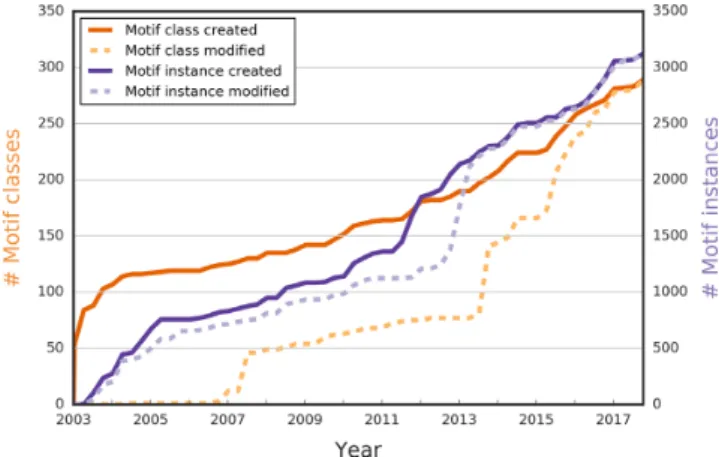
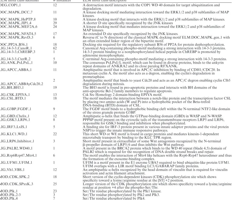
Currently there are over 275 motif classes, 32 more than in the lastNARpublication in 2016 (13) (Figure1and Ta- ble1). Most notably six variants of the mitogen-activated protein kinases (MAPK) docking D-motifs and additions
Figure 1. The number of SLiMs (motif classes and motif instances) cre- ated and modified in ELM. For the past 15 years, ELM has been steadily growing, and the total number of motif classes (dark orange) and motif in- stances (dark purple) continues to grow each year. As of September 2017, there are over 275 motif classes defined, and over 3000 motif instances annotated. Besides contributing new content, curators also updated exist- ing annotations to include new findings (last modification dates for motif classes in dashed light purple and for motif instances in dashed light or- ange).
and improvements to cell cycle regulatory motifs includ- ing relevant degrons and kinases such as the Polo-like ki- nases (Plks). We have tried to be comprehensive for de- gron motifs (recently reviewed in (8)) with the most re- cent addition being the pLxIS motif involved in immune re- sponse of interferon-regulatory factor IRF3 but which has degron-like properties for rotavirus hijacking (26). Another example of a hijacked motif is the tyrosine-kinase regulat- ing motif EPIYA, which is a common motif mimic used by pathogenic bacteria. Also, several existing motif entries have been redefined or expanded, including recent updates to the abundant and versatile class of 14-3-3-binding mo- tifs and to the cell cycle checkpoint retinoblastoma protein pRb-binding LxCxE motif.
New ELM instances
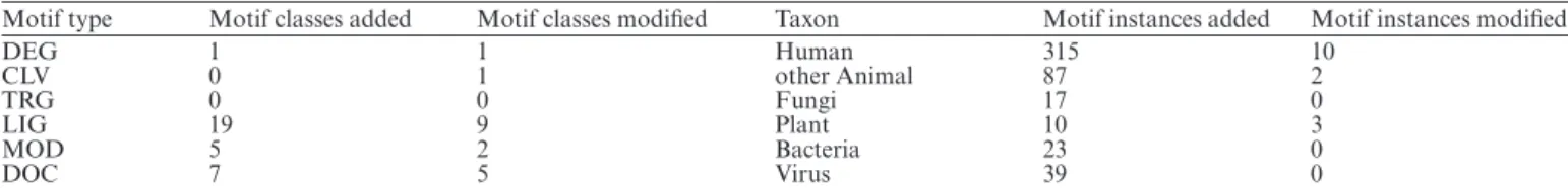
One of the principal types of data contained in ELM are the motif instances, i.e. experimentally validated occurrences of motif classes in proteins. As of September 2017, ELM has 3093 instances, having added 491 new motif instances since the lastNARdatabase publication (13) and also updated many existing entries (Figure1). Following previous years, the majority of new motif instances are for human proteins and other animals, although we have had a large increase in the number of viral motif mimics and we have begun the process of adding instances of bacterial motif mimicry from a systematic review of the literature (Table2).
NEW FEATURES IN ELM
In this release, we have further integrated ELM with other bioinformatics databases and resources. An important de- velopment is that UniProt (27) now includes ELM as a database cross-reference in the ‘protein–protein interaction databases’ section. We have also updated the experimental evidence codes used in ELM to the latest version of PSI–MI
Table 1. New ELM motif classes
ELM motif class identifier # Instances ELM motif class description
DEG COP1 1 12 A destruction motif interacts with the COP1 WD 40 domain for target ubiquitination and degradation.
DOC MAPK DCC 7 11 A kinase docking motif mediating interaction toward the ERK1/2 and p38 subfamilies of MAP kinases.
DOC MAPK HePTP 8 10 A kinase docking motif that interacts with the ERK1/2 and p38 subfamilies of MAP kinases.
DOC MAPK JIP1 4 29 A shorter D site specifically recognized by the JNK kinases
DOC MAPK MEF2A 6 24 A kinase docking motif that mediates interaction toward the ERK1/2 and p38 subfamilies of MAP kinases.
DOC MAPK NFAT4 5 17 An extended D site specifically recognized by the JNK kinases.
DOC MAPK RevD 3 6 Reverse (C to N direction) of the classical MAPK docking motif ELM:DOC MAPK gen 1 with an often extended linker region of the bipartite motif.
DOC PP2A B56 1 18 Docking site required for the regulatory subunit B56 of PP2A for protein dephosphorylation.
LIG 14-3-3 CanoR 1 62 Canonical Arg-containing phospho-motif mediating a strong interaction with 14-3-3 proteins.
LIG 14-3-3 ChREBP 3 1 14-3-3 protein binding to a nonphosphorylated helical peptide in ChREBP is promoted by adenosine monophosphate.
LIG 14-3-3 CterR 2 5 C-terminal Arg-containing phospho-motif mediating a strong interaction with 14-3-3 proteins.
LIG ANK PxLPxL 1 10 The consensus PxLPxI/L motif, which can be found in diverse proteins, binds to the ankyrin repeat domains of ANKRA2 and its close paralog RFXANK.
LIG APCC ABBA 1 11 Amphipathic motif that is involved in APC/C inhibition by binding of CDH1/CDC20. In metazoan cyclin A, the motif also acts as a degron, enabling the cyclin’s degradation in prometaphase.
LIG APCC ABBAyCdc20 2 2 Amphipathic motif that binds to yeast Cdc20 and acts as an APC/C degron enabling cyclin Clb5 degradation during mitosis.
LIG BH BH3 1 19 The BH3 motif is found in pro-apoptotic proteins and interacts with BH domains of the anti-apoptotic Bcl-2 family members to regulate apoptosis.
LIG CSK EPIYA 1 13 Csk Src Homology 2 domain-binding EPIYA motif.
LIG CSL BTD 1 18 The motif mediates the interaction between a notch-like protein and the transcription factor CSL by placing two amino acids (W and P) into a hydrophobic pocket of the Beta-trefoil
DNA-binding (BTD) domain of CSL.
LIG G3BP FGDF 1 9 The FGDF motif binds to a hydrophobic binding cleft within the N-terminal NTF2-like domain of the stress granule protein G3BP.
LIG GBD Chelix 1 12 Amphipatic␣-helix that binds the GTPase-binding domain (GBD) in WASP and N-WASP.
LIG GSK3 LRP6 1 8 PPPSP motif present on the cytosolic tails of the transmembrane receptors LRP5 and LRP6, responsible for GSK3 binding and inhibition when phosphorylated.
LIG IRF3 LxIS 1 5 A binding site for IRF-3 protein present in various innate adaptor proteins and the viral protein NSP1to trigger the innate immune responsive pathways.
LIG KLC1 WD 1 22 This short WD or WE motif is found in cargo proteins and mediates kinesin-1-dependent microtubule transport by binding to the KLC TPR region.
LIG LRP6 Inhibitor 1 3 Short motif present in extracellular of some Wnt antagonists recognized by the N-terminal
-propeller domain of LRP5/6 and thus inhibits the Wnt pathway.
LIG PALB2 WD40 1 1 A motif present in the BRCA2 protein which binds to the WD 40 repeat (blade 4,5) domain of PALB2 which is required for the recognition of DNA double strand breaks and repair.
LIG Rrp6Rrp47 Mtr4 1 6 The motif enables the interaction of Mtr4 like helicaes with the Rrp6-Rrp47 heterodimer and thus the formation of the exosome-binding complex.
LIG UFM1 UFIM 1 1 UFIM is a motif present in the E1 enzyme UBA5 required to bind ubiquitin-like protein UFM1.
UFIM overlaps with a LIR motif binding LC3/GABARAP family proteins.
LIG Vh1 VBS 1 12 An amphipathic␣-helix recognized by the head domain of vinculin that is required for vinculin activation and actin filament attachment.
MOD CDK SPK 2 18 Short version of the cyclin-dependent kinases (CDK) phosphorylation site which shows specificity toward a lysine/arginine residue at the [ST] +2 position.
MOD CDK SPxxK 3 25 Longer version of the CDK phosphorylation site which shows specificity toward a lysine/arginine residue at position +4 after the phospho-Ser/Thr.
MOD Plk 1 23 Ser/Thr residue phosphorylated by the Plk1 kinase.
MOD Plk 2-3 3 Ser/Thr residue phosphorylated by Plk2 and Plk3.
MOD Plk 4 7 Ser/Thr residue phosphorylated by Plk4.
Since the last NAR database issue publication 32 motif classes have been annotated to the database. (13)
version 2.5 (15). The most notable changes in PSI–MI are that terms ‘GST-pulldown’ and ‘HIS-pulldown’ have each been demerged into a combination of terms: ‘glutathione s transferase tag’ and ‘pull down’ and ‘his tag’ and ‘pull down’. We have also integrated ELM with the Reactome pathway database (28), and introduced programmatic ac- cess to the ELM exploration pipeline, both of which we de- scribe below in more detail.
Reactome
One way to gain additional insights into which biological processes a SLiM may be involved in, is to examine the cel- lular pathways that contain proteins with this motif. ELM already has links to pathways contained in the KEGG path- way database (12,29). In order to augment the cellular net-
work knowledge potential available in ELM, we have now integrated ELM with another pathway database: Reactome.
Reactome is a manually curated peer reviewed pathway database (28). Pathways are defined by reactions and the entities participating in them (nucleic acids, proteins, com- plexes and small molecules), and are supported by literature citations and expert curation. It is now possible to visualize and download all Reactome annotations for proteins avail- able in ELM. Every protein in ELM having a Reactome annotation now has a link to display a Reactome pathway diagram that highlights where this protein functions. The complete list of Reactome annotations can also be retrieved from the ELM downloads page. Later in this paper, we will illustrate how the ELM annotated Reactome data can be used to analyze the motifs involved in the cell cycle.
Table 2. New ELM motif classes and instances
Motif type Motif classes added Motif classes modified Taxon Motif instances added Motif instances modified
DEG 1 1 Human 315 10
CLV 0 1 other Animal 87 2
TRG 0 0 Fungi 17 0
LIG 19 9 Plant 10 3
MOD 5 2 Bacteria 23 0
DOC 7 5 Virus 39 0
Since the last NAR database issue publication in 2016 (13) a total of 32 motif classes and 491 motif instances have been added to the database. Most of the new motifs added are either Ligand (LIG) or Docking (DOC) motifs. Most of the new motif instances are Human, although motif instances for many other branches of life have also been added.
The ELM API
The ELM exploration pipeline is a useful tool to predict pu- tative SLiMs in protein sequences. Nevertheless, the graphi- cal user interface is not suitable for automated or large scale analyses. One of the latest updates to the ELM resource has been to include an application programmatic interface (API) to the ELM exploration pipeline (30). The ELM ex- ploration API allows users to submit either a protein se- quence or a UniProt ID to predict which SLiMs might exist in it. The protein sequence is matched against all of the reg- ular expressions annotated in ELM and each motif match is passed through a combination of structural context filters, which help to predict whether the motif is likely to be bi- ologically functional. Motif matches are filtered out of the predicted motifs if they occur in globular domains, trans- membrane regions or extracellular regions. The API also returns whether any of the motifs detected are already anno- tated in ELM, or whether the motif has been annotated in a homolog in ELM. The output is provided as atsv(tab sep- arated values) file, which is easy to read and analyze com- putationally. The API can be accessed using any program- ming framework that can process HTTP requests, for exam- plewget, curland the python ‘requests’ package. For more information on using the API, usage guidelines and how to interpret the results, see (30) and read the documentation onelm.eu.org/api/manual.html.
MOTIFS IN BACTERIAL PATHOGENS
Motifs are not unique to eukaryotes; they also exist in bac- teria and viruses. It has been known for some time that viruses use motif mimicry to interfere with biological pro- cesses of the host cell (6). This behavior is not limited to viruses, but the data for pathogenic bacteria are more lim- ited (31,32). In the latest version of ELM, we report in- stances from a handful of bacteria that are now known to use motif mimicry for pathogenicity.
Among the bacterial proteins with newly added motifs are OspF fromShigella flexneriand SpvC fromSalmonella Typhimurium, which use a D-motif to recognize MAPK proteins like ERK, JNK or p38 and irreversibly modify a phosphorylated residue to block downstream MAPK sig- naling, thus preventing the activation of the immune re- sponse (33,34). Enterohaemorrhagic Escherichia coli uses the multi-valency of a GBD domain-binding motif to ac- tivate up to seven WASP proteins with a single effector pro- tein, espFU (35,36). The same protein has five tandem PxxP motifs that bind to the SH3 domain of BAIAP2L1/IRTKS with the highest reported affinity for a motif-SH3 com- plex (500 nM) (35). Finally, the tyrosine-phosphorylated
EPIYA motif present in the cellular protein Pragmin is also used by CagA from Helicobacter pylori and LspA1 from Haemophilus ducreyi to recruit CSK and phospho- rylate Src-family kinases (37,38), interfering with cell fate and phagocytosis (39,40). Besides their role in pathogenic- ity, motif mimicry by bacteria also has implications for bac- terial oncogenicity, such as the oncogenic potential ofH.
pyloristrains (41).
MOTIFS IN THE CELL CYCLE
One of the new features included in ELM are the pro- tein’s pathway annotations from the Reactome database (28), which can be downloaded from the ELM ‘downloads’
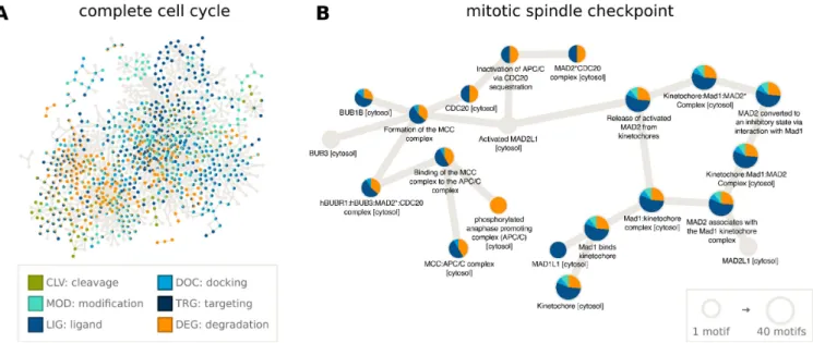
page. These annotations allow the construction of network diagrams to examine the roles of motifs within any signal- ing pathway in Reactome. As an example, we have anno- tated the motifs present in the cell cycle (R-HSA-1640170) (Figure2A, created with Cytoscape (42)), which consists of 610 proteins, 199 of which have motifs annotated in ELM.
In Figure2B, we highlight the 20 proteins involved in the mitotic cell cycle checkpoint (R-HSA-69618), almost all of which have multiple SLiMs (Figure2A). Degradation mo- tifs recognized by the APC/C complex, an E3 ubiquitin lig- ase, as well as LIG MAD2 motifs are prominent in these checkpoint proteins. Many proteins involved in the cell cy- cle contain one or more functionally important linear mo- tifs and combining SLiM annotation with pathway infor- mation will help unravel the roles SLiMs play in the cell.
THE KINOME IN ELM
In this release, we report an expansion of the portion of the human kinome annotated in ELM, including new motif en- tries for CDKs (not discussed in this article), MAPKs and Plks.
MAPKs form an important part of conserved signaling pathways involved in processes such as cell division, dif- ferentiation, growth and apoptosis (43–45). MAPKs are serine/threonine kinases that recognize substrates by the [ST]Pmotif, and for specificity rely on additional motifs (for example D-motifs) to bring the kinase and its substrate close together for phosphorylation. These motifs harbor one or two basic residues, a variable linker segment and usually three hydrophobic amino acids. Interestingly, the motif orientation can be from the N- to C-terminus where charged residues are followed by linker and hydrophobic residues (for example DOC MAPK NFAT4 5, Figure3A, produced using Chimera (46)) or C- to N-terminus, where hydrophobic residues precede the charged amino acids (e.g.
Figure3B).
Figure 2. SLiMs play major roles in many biological pathways, including those involved in the progression of the cell cycle. Using the Reactome pathway annotations downloaded from ELM and using Cytoscape (42) to visualize the data, we can see that many SLiMs (especially Ligand and Degradation motifs) are involved in the cell cycle (A) and specifically in the mitotic spindle checkpoint (B).
Figure 3. Surface representation showing two MAPK docking motifs bound to the MAPK docking groove. Negative charges and positive charges are shown in red and blue, respectively, on MAPK and the docking motif is rendered in yellow. (A) The N- to C-terminal orientation of the MAPK docking motif shown for the DOC MAPK NFAT4 5 motif (with regular expression:[RK][∧P][∧P][LIM].L.[LIVMF]). Here, charged amino acids[RK]
are followed by hydrophobic residues (PDB:2XS0; (43)). (B) The reverse MAPK docking motif shown for the DOC MAPK RevD 3 motif (with regular expression:[LIVMPFA].[LIV].1,2[LIVMP].4,6[LIV]..[RK][RK]), where the N-terminus has hydrophobic amino acids followed by charged residues (PDB:2Y9Q; (43)). Figures produced using Chimera (46).
Plks are central to the cell cycle and are often found re- stricted to cellular locations involved in mitosis (such as centrosomes, kinetochores and the spindle) (47). Humans have four functional Plks. The C-terminal parts of Plks 1–
3 have two polo-box domains that help target and recruit the kinase substrates by recognizing the short consensus se- quence (S[ST]) which, when phosphorylated on the sec- ond residue, acts as a docking/activation site (48,49). Speci- ficity is conferred by the Plk’s specific target motif: Plk1 re- quires an Asp or Glu two positions before the phosphosite, Plks 2 and 3 require an Asp or Glu either two positions be- fore or after the phosphosite and Plk4 has a varied motif re- quirement where hydrophobic residues are strongly favored after the phosphosite consensus sequence.
CONCLUSION AND FUTURE DIRECTIONS
Every year ELM continues to grow in terms of new con- tent and connectivity to other resources. As more content is added to ELM we also expect to characterize more and im- prove existing motif classes. Each addition to the database will allow researchers to uncover new biological insights involving motifs in protein–protein interactions, pathways and networks as well as better understanding the roles of SLiMs in disease and pathogenicity. One of the important aspects of this work will be not only to add new content to the database, but also to review and update the existing con- tent with new discoveries from the scientific literature. We will also continue integrating ELM with existing and emerg- ing bioinformatics resources for SLiM research and protein biology. In parallel we will further develop the ELM API to facilitate the integration of ELM with other bioinformatics tools and resources. We expect that ELM will continue to be a useful and unique resource for SLiM research and the life science community. Users are also encouraged to visit the ‘external links’ page (http://elm.eu.org/infos/links.html) which lists many other useful tools and databases for SLiM research such as QSLiMFinder (for motif discovery (50)) and ProViz (for motif exploration (24)). We also welcome any feedback you can give us that can help us improve ELM.
ACKNOWLEDGEMENTS
We would like to thank all of the users of ELM for their continued interest and support, as well as the community of annotators for their time and efforts in keeping the database up to date.
FUNDING
European Molecular Biology Laboratory (EMBL) Interna- tional PhD Program; Humboldt Foundation Postdoctoral Fellowship; National Research, Development and Innova- tion Office (NKFIH) OTKA Grants [NN114309, K108798, PD120973]; Argentinian National Science Ministry (AN- PCyT) [PICT 2013/1895 to L.B.C.]; National Science Re- search Council (CONICET, Argentina) (to L.B.C., N.P.);
Ministry of Science and Technology and German Academic Exchange Service (MinCyT-DAAD) [CyCmotif DA/16/05 to L.B.C., T.G.]; Erasmus Traineeships (A.A. 2016/2017) [Progetto 2016-1-IT02-KA103-023753]. Funding for open access charge: EMBL.
Conflict of interest statement.None declared.
REFERENCES
1. Davey,N.E., Van Roey,K., Weatheritt,R.J., Toedt,G., Uyar,B., Altenberg,B., Budd,A., Diella,F., Dinkel,H. and Gibson,T.J. (2012) Attributes of short linear motifs.Mol. Biosyst.,8, 268–281.
2. Van Roey,K., Uyar,B., Weatheritt,R.J., Dinkel,H., Seiler,M., Budd,A., Gibson,T.J. and Davey,N.E. (2014) Short linear motifs:
ubiquitous and functionally diverse protein interaction modules directing cell regulation.Chem. Rev.,114, 6733–6778.
3. Diella,F. (2008) Understanding eukaryotic linear motifs and their role in cell signaling and regulation.Front. Biosci.,13, 6580–6603.
4. Van Roey,K., Gibson,T.J. and Davey,N.E. (2012) Motif switches:
decision-making in cell regulation.Curr. Opin. Struct. Biol.,22, 378–385.
5. Van Roey,K., Dinkel,H., Weatheritt,R.J., Gibson,T.J. and Davey,N.E. (2013) The switches.ELM resource: a compendium of conditional regulatory interaction interfaces.Sci. Signal.,6, rs7.
6. Davey,N.E., Trav´e,G. and Gibson,T.J. (2011) How viruses hijack cell regulation.Trends Biochem. Sci.,36, 159–169.
7. Uyar,B., Weatheritt,R.J., Dinkel,H., Davey,N.E. and Gibson,T.J.
(2014) Proteome-wide analysis of human disease mutations in short linear motifs: neglected players in cancer?Mol. Biosyst.,10, 2626–2642.
8. M´esz´aros,B., Kumar,M., Gibson,T.J., Uyar,B. and Doszt´anyi,Z.
(2017) Degrons in cancer.Sci. Signal.,10, eaak9982.
9. Tompa,P., Davey,N.E., Gibson,T.J. and Babu,M.M. (2014) A million peptide motifs for the molecular biologist.Mol. Cell,55, 161–169.
10. Puntervoll,P. (2003) ELM server: a new resource for investigating short functional sites in modular eukaryotic proteins.Nucleic Acids Res.,31, 3625–3630.
11. Dinkel,H., Michael,S., Weatheritt,R.J., Davey,N.E., Van Roey,K., Altenberg,B., Toedt,G., Uyar,B., Seiler,M., Budd,A.et al.(2012) ELM–the database of eukaryotic linear motifs.Nucleic Acids Res., 40, D242–D251.
12. Dinkel,H., Van Roey,K., Michael,S., Davey,N.E., Weatheritt,R.J., Born,D., Speck,T., Kr ¨uger,D., Grebnev,G., Kuba ´n,M.et al.(2014) The eukaryotic linear motif resource ELM: 10 years and counting.
Nucleic Acids Res.,42, D259–D266.
13. Dinkel,H., Van Roey,K., Michael,S., Kumar,M., Uyar,B.,
Altenberg,B., Milchevskaya,V., Schneider,M., K ¨uhn,H., Behrendt,A.
et al.(2016) ELM 2016’data update and new functionality of the eukaryotic linear motif resource.Nucleic Acids Res.,44, D294–D300.
14. The Gene Ontology Consortium (2017) Expansion of the Gene Ontology knowledgebase and resources.Nucleic Acids Res.,45, D331–D338.
15. Kerrien,S., Orchard,S., Montecchi-Palazzi,L., Aranda,B., Quinn,A.F., Vinod,N., Bader,G.D., Xenarios,I., Wojcik,J., Sherman,D.et al.(2007) Broadening the horizon–level 2.5 of the HUPO-PSI format for molecular interactions.BMC Biol.,5, 44–55.
16. Sayers,E.W., Barrett,T., Benson,D.A., Bryant,S.H., Canese,K., Chetvernin,V., Church,D.M., Dicuccio,M., Edgar,R., Federhen,S.
et al.(2009) Database resources of the National Center for Biotechnology Information.Nucleic Acids Res.,37(Suppl. 1), 5–15.
17. Benson,D.A., Karsch-Mizrachi,I., Lipman,D.J., Ostell,J. and Sayers,E.W. (2009) GenBank.Nucleic Acids Res.,37, D26–D31.
18. Berman,H.M., Westbrook,J., Feng,Z., Gilliland,G., Bhat,T.N., Weissig,H., Shindyalov,I.N. and Bourne,P.E. (2000) The protein data bank.Nucleic Acids Res.,28, 235–242.
19. Linding,R., Russell,R.B., Neduva,V. and Gibson,T.J. (2003) GlobPlot: exploring protein sequences for globularity and disorder.
Nucleic Acids Res.,31, 3701–3708.
20. Doszt´anyi,Z., Csizmok,V., Tompa,P. and Simon,I. (2005) IUPred:
web server for the prediction of intrinsically unstructured regions of proteins based on estimated energy content.Bioinformatics,21, 3433–3434.
21. Via,A., Gould,C.M., Gem ¨und,C., Gibson,T.J. and
Helmer-Citterich,M. (2009) A structure filter for the eukaryotic linear motif resource.BMC Bioinformatics,10, 351–369.
22. Letunic,I., Doerks,T. and Bork,P. (2015) SMART: recent updates, new developments and status in 2015.Nucleic Acids Res.,43, D257–D260.
23. Finn,R.D., Coggill,P., Eberhardt,R.Y., Eddy,S.R., Mistry,J., Mitchell,A.L., Potter,S.C., Punta,M., Qureshi,M.,
Sangrador-Vegas,A.et al.(2016) The Pfam protein families database:
towards a more sustainable future.Nucleic Acids Res.,44, D279–D285.
24. Jehl,P., Manguy,J., Shields,D.C., Higgins,D.G. and Davey,N.E.
(2016) ProViz-a web-based visualization tool to investigate the functional and evolutionary features of protein sequences.Nucleic Acids Res.,44, W11–W15.
25. Krystkowiak,I. and Davey,N.E. (2017) SLiMSearch: a framework for proteome-wide discovery and annotation of functional modules in intrinsically disordered regions.Nucleic Acids Res.,45, W464–W469.
26. Zhao,B., Shu,C., Gao,X., Sankaran,B., Du,F., Shelton,C.L., Herr,A.B., Ji,J.-Y. and Li,P. (2016) Structural basis for concerted recruitment and activation of IRF-3 by innate immune adaptor proteins.Proc. Natl. Acad. Sci. U.S.A.,113, E3403–E3412.
27. Bateman,A., Martin,M.J., O’Donovan,C., Magrane,M., Alpi,E., Antunes,R., Bely,B., Bingley,M., Bonilla,C., Britto,R.et al.(2017) UniProt: the universal protein knowledgebase.Nucleic Acids Res.,45, D158–D169.
28. Fabregat,A., Sidiropoulos,K., Garapati,P., Gillespie,M.,
Hausmann,K., Haw,R., Jassal,B., Jupe,S., Korninger,F., McKay,S.
et al.(2016) The reactome pathway knowledgebase.Nucleic Acids Res.,44, D481–D487.
29. Kanehisa,M., Furumichi,M., Tanabe,M., Sato,Y. and Morishima,K.
(2017) KEGG: new perspectives on genomes, pathways, diseases and drugs.Nucleic Acids Res.,45, D353–D361.
30. Gouw,M., S´amano-S´anchez,H., Van Roey,K., Diella,F., Gibson,T.J.
and Dinkel,H. (2017) Exploring short linear motifs using the ELM database and tools. In: Bateman,A, Draghici,S, Khurana,E, Orchard,S and Pearson,WR (eds).Current Protocols in Bioinformatics. John Wiley & Sons, Inc., Hoboken, pp.
8.22.1–8.22.35.
31. Via,A., Uyar,B., Brun,C. and Zanzoni,A. (2015) How pathogens use linear motifs to perturb host cell networks.Trends Biochem. Sci.,40, 36–48.
32. Ruhanen,H., Hurley,D., Ghosh,A., O’Brien,K.T., Johnston,C.R. and Shields,D.C. (2014) Potential of known and short prokaryotic protein motifs as a basis for novel peptide-based antibacterial therapeutics: a computational survey.Front. Microbiol.,5, 1–18.
33. Zhu,Y., Li,H., Long,C., Hu,L., Xu,H., Liu,L., Chen,S., Wang,D.C.
and Shao,F. (2007) Structural insights into the enzymatic mechanism of the pathogenic MAPK phosphothreonine lyase.Mol. Cell,28, 899–913.
34. Li,H., Xu,H., Zhou,Y., Zhang,J., Long,C., Li,S., Chen,S., Zhou,J.-M.
and Shao,F. (2007) The phosphothreonine lyase activity of a bacterial type III effector family.Science,315, 1000–1003.
35. Aitio,O., Hellman,M., Kazlauskas,A., Vingadassalom,D.F., Leong,J.M., Saksela,K. and Permi,P. (2010) Recognition of tandem PxxP motifs as a unique Src homology 3-binding mode triggers pathogen-driven actin assembly.Proc. Natl. Acad. Sci. U.S.A.,107, 21743–21748.
36. Aitio,O., Hellman,M., Skehan,B., Kesti,T., Leong,J.M., Saksela,K.
and Permi,P. (2012) Enterohaemorrhagic escherichia coli exploits a
tryptophan switch to hijack host F-Actin assembly.Structure,20, 1692–1703.
37. Tsutsumi,R., Higashi,H., Higuchi,M., Okada,M. and Hatakeyama,M. (2003) Attenuation of Helicobacter pylori
CagA·SHP-2 Signaling by Interaction between CagA and C-terminal Src Kinase.J. Biol. Chem.,278, 3664–3670.
38. Dodd,D.A., Worth,R.G., Rosen,M.K., Grinstein,S., van Oers,N.S.C.
and Hansen,E.J. (2014) The Haemophilus ducreyi LspA1 protein inhibits phagocytosis by using a new mechanism involving activation of C-terminal Src kinase.Mbio,5, doi:10.1128/mBio.01178-14.
39. Tauzin,S., Starnes,T.W., Becker,F.B., ying Lam,P. and Huttenlocher,A. (2014) Redox and Src family kinase signaling control leukocyte wound attraction and neutrophil reverse migration.
J. Cell Biol.,207, 589–598.
40. Berton,G., M ´ocsai,A. and Lowell,C.A. (2005) Src and Syk kinases:
key regulators of phagocytic cell activation.Trends Immunol.,26, 208–214.
41. Jones,K.R., Joo,Y.M., Jang,S., Yoo,Y.J., Lee,H.S., Chung,I.S., Olsen,C.H., Whitmire,J.M., Merrell,D.S. and Cha,J.H. (2009) Polymorphism in the cagA EPIYA motif impacts development of gastric cancer.J. Clin. Microbiol.,47, 959–968.
42. Shannon,P. (2003) Cytoscape: a software environment for integrated models of biomolecular interaction networks.Genome Res.,13, 2498–2504.
43. Garai,A., Zeke,A., Gogl,G., Toro,I., Fordos,F., Blankenburg,H., Barkai,T., Varga,J., Alexa,A., Emig,D.et al.(2012) Specificity of linear motifs that bind to a common mitogen-activated protein kinase docking groove.Sci. Signal.,5, ra74.
44. Zeke,A., Bastys,T., Alexa,A., Garai, ´A., M´esz´aros,B., Kirsch,K., Doszt´anyi,Z., Kalinina,O.V. and Rem´enyi,A. (2015) Systematic discovery of linear binding motifs targeting an ancient protein interaction surface on MAP kinases.Mol. Syst. Biol.,11, 837–858.
45. Zeke,A., Misheva,M., Remenyi,A. and Bogoyevitch,M.A. (2016) JNK Signaling : regulation and functions based on complex protein- protein partnerships.Microbiol. Mol. Biol. Rev.,80, 793–835.
46. Pettersen,E.F., Goddard,T.D., Huang,C.C., Couch,G.S., Greenblatt,D.M., Meng,E.C. and Ferrin,T.E. (2004) UCSF Chimera––a visualization system for exploratory research and analysis.J. Comput. Chem.,25, 1605–1612.
47. Barr,F.A., Sillj´e,H.H.W. and Nigg,E.A. (2004) Polo-like kinases and the orchestration of cell division.Nat. Rev. Mol. Cell Biol.,5, 429–441.
48. Park,J.E., Soung,N.K., Johmura,Y., Kang,Y.H., Liao,C., Lee,K.H., Park,C.H., Nicklaus,M.C. and Lee,K.S. (2010) Polo-box domain: a versatile mediator of polo-like kinase function.Cell. Mol. Life Sci., 67, 1957–1970.
49. Lowery,D.M., Lim,D. and Yaffe,M.B. (2005) Structure and function of Polo-like kinases.Oncogene,24, 248–259.
50. Palopoli,N., Lythgow,K.T. and Edwards,R.J. (2015) QSLiMFinder:
improved short linear motif prediction using specific query protein data.Bioinformatics,31, 2284–2293.