C H A P T E R 8
The Chloroplast Inside and Outside the Cell
RACHEL M. LEECH
Department of Biology, University of York, Heslington, England
I. Introduction 137 II. The Structure of the Mature Chloroplast Inside the Cell . . . 1 3 9
A. The Chloroplast Envelope 139
B. The Stroma 139 C. The Plastoglobuli (Osmiophilic Globules) 140
D. The Lipoprotein Complex 141 III. The Chloroplast Outside the Cell 149 IV. The Chloroplast as an Autonomous Unit 155
A. Algae 156 B. Leaves of Higher Plants 157
Acknowledgements . 1 5 9
References . . . 159
I. INTRODUCTION
The chloroplast is unique among cell organelles in being restricted to green plant cells. Isolated chloroplasts have been shown to be "the complete photosynthetic unit" of the cell (Arnon et al, 1954; Jensen and Bassham, 1966). The ability of chloroplasts to conserve in photosynthesis the radiant energy of light in a biologically utilizable form and recent findings of D N A , R N A and ribosomal-mediated protein synthesis in chloroplasts (see Kirk and Tilney-Bassett, 1967 for a review) suggests that the mature chloroplast is, potentially at least, metabolically autonomous from the rest of the cell.
There are also convincing indications that the chloroplast is to some degree autonomous genetically. In lower plants mature chloroplasts are able to replicate and in lower plants and higher plants there are now several well- authenticated examples of determinants of plastid characteristics located within the plastid and apparently outside nuclear control. Considerable discussion and experimentation at the present time is concerned with investi
gating the actual degree of autonomy exhibited by chloroplasts in a wide variety of organisms.
The most characteristic feature of the chloroplast is its complex pigment- bearing lipoprotein membrane system. It is here that the light-dependent
Η 137
138 R. Μ. LEECH
FIG. 1 Unpublished electron micrograph by A. D. Greenwood of a chloroplast in a leaf cell of Vicia faba L. χ 29,000. Fixed with glutaraldehyde/osmium and stained with lead. Embedded in Epon. The chloroplast envelope (E), plastoglobuli (PG), grana (G)
and stroma (S) are clearly seen. A starch grain (SG) is also present.
8. THE CHLOROPLAST INSIDE AND OUTSIDE THE CELL 139 oxygen evolution and electron transport systems of photosynthesis are local
ized. The key to the understanding of the molecular basis of the unique properties of chloroplasts probably lies in a more detailed knowledge of its molecular structure, origin and development.
This review will be concerned with recent attempts to describe the three- dimensional molecular architecture of the chloroplast, particularly of the lipoprotein system, and to consider information about the preservation of chloroplast structure in isolation. Finally chloroplast autonomy will be discussed.
I I . THE STRUCTURE OF THE MATURE CHLOROPLAST INSIDE THE CELL
Electron microscope studies using sections of chloroplasts from a wide range of green plants have revealed that four distinct components are charac
teristic of all mature algal and phanerogamic chloroplasts. The mature chloro
plast consists of a lamellar (lipoprotein) system embedded in a granular stroma and surrounded by a double membrane. Within the stroma osmio- philic "plastoglobuli" are always present. These components are shown in Fig. 1.
A. THE CHLOROPLAST ENVELOPE
The chloroplast envelope is a continuous boundary of two osmiophilic unit membranes (in the sense of Robertson, 1964) and completely separates the contents of the chloroplast from the rest of the cell. Pores have never been demonstrated in this envelope. Recently Weier has suggested, from high resolution electron micrographs, that the envelope is made up of spherical subunits as are the internal chloroplast membranes (Weier et al, 1965a;
Weier et al, 1966).
B. THE STROMA
The stroma is a hydrophilic proteinaceous continuum within the chloroplast envelope and surrounding and containing the intra-chloroplast membrane system but not penetrating within it. Within the stroma are particles hetero
geneous in size and staining properties (Gunning, 1965; Gunning and Jagoe, 1967). One class of 175A particles which stain with uranyl acetate and are completely destroyed by ribonuclease are undoubtedly ribosomes (Jacobson et al, 1963; Kislev et al, 1965). These are frequently seen in groups resembling polysomes (Gunning, 1965). Using glutaraldehyde (or acrolein)/osmium fixations, Gunning (1965) has discovered in chloroplasts of Avena sativa a
"stromacentre" 1μ in diameter and consisting of aggregated fibrils 85A in diameter and of uncertain length. The stroma centre can be seen in the light microscope and from its staining properties would appear to be proteinaceous
140 R. Μ. LEECH
but has so far not been reported from other plants and its significance is unknown. It is possibly regularly aggregated stroma material which is more dispersed in other species and may indeed in part be Fraction I protein. It is not known how it is related to the strikingly similar pockets of fibrils induced in the stroma of chloroplasts by treatment with either ozone or peroxylacetyl nitrate (Thomson et al, 1965, 1966). Earlier, Perner (1962) described apparently crystalline structures in the stroma of isolated spinach (Spinacia oleraced) chloroplasts. The relationship of these structures to the stroma centre is not clear; it is possible they are different manifestations of aggrega
tions of stroma proteins. Starch grains, when present, are localized in the stroma between but not within the elements of the membrane system.
Pyrenoids are morphologically specialized parts of the stroma found in some algae and in the Anthocerotales (Menke, 1961; Gibbs, 1962a, 1962b;
Drawert and Mix, 1962; Kaja, 1966). They appear to be finely granular in the electron microscope but their composition and functions are unknown.
Manton (1966) has published a series of electron micrographs showing variations in pyrenoid structure.
C. THE PLASTOGLOBULI (OSMIOPHILIC GLOBULES)
The plastoglobuli are a constant morphological feature in sections of osmium-fixed chloroplasts from all types of plant. The circular profiles of plastoglobuli vary from 100-5000A in diameter and are embedded in the stroma between the lipoprotein membranes but never within a granum.
Plastoglobuli are labile in KMnC>4 fixation. In the literature they have been described by a variety of names: spherical granules (Steinmann and Sjostrand, 1955), lipid-like globules (Gibbs, 1962a, 1962c), osmiophilic granules (Murakami and Takamija, 1962), Elektronenstreuende Kugeln (Heitz, 1958), lipid droplets (Sager and Palade, 1957), magnoglobuli (Falk, 1960), osmiophilic globules (Greenwood et ah, 1963) and osmiophilous globules by Silaeva and Shiriaev (1966). However, the least ambiguous term would seem to be
"plastoglobuli" suggested by Lichtentahler and Sprey (1966) and it is hoped that this term will be used by all future workers.
In green actively photosynthesizing leaves, the plastoglobuli contain a variety of lipidic and lipophilic compounds particularly plastoquinone-45, α-tocopherol, α-tocopherolquinone and vitamin K i (Bailey and Whyborn, 1963; Lichtentahler, 1964; Lichtentahler and Sprey, 1966) and polyisoprenols (Wellburn and Hemming, 1966) but are completely devoid of chlorophyll and carotenoid pigments (Greenwood et ah, 1963; Lichtentahler and Sprey, 1966). The plastoglobuli vary in size and staining properties in chloroplasts of different ages (and therefore presumably in chemical composition) and would seem to be a store of surplus lipid in the chloroplast in the same way as starch is a store of surplus carbohydrate material. Sprey and Lichtentahler
8. THE CHLOROPLAST INSIDE AND OUTSIDE THE CELL 141 (1966) recently showed a decrease in plastoglobuli in greening etiolated leaves of barley seedlings during membrane formation and so suggest that the plastoglobuli, at this stage of development, are a pool from which lipids can be taken during membrane synthesis. Evidence that plastoglobuli are not artefacts of fixation is provided by their presence in freeze-etched chloroplasts (Miihletahler et al., 1965). There is no evidence that plastoglobuli have enzymic activity.
D. THE LIPOPROTEIN COMPLEX
The lipoprotein fretwork system of the chloroplast contains the chlorophyll and carotenoids and is suspended in but apparently not open to the stroma.
In recent years intensive studies of the three-dimensional architecture of this system have been undertaken in several laboratories using every preparative fixation and staining procedure available in the electron microscopist's armoury. The structure has been explored in a wide variety of algal and higher plant chloroplasts in sections of cells, and in whole mounts, of isolated chloroplasts and chloroplast fragments and also after freeze-etching.
It has been conclusively demonstrated by Trebst et al. (1958) that the light reactions and associated electron transport reactions of photosynthesis are localized in the internal chloroplast membrane system. It is now possible using modern electron microscopical techniques to preserve and resolve globular proteins of approximately 35A in diameter and fibrous proteins 5-10A in diameter and studies and ideas about the precise molecular arrangements responsible for the integrated metabolic activities have reached an exciting stage. However, terminology in the field of chloroplast structure is confused and confusing and makes communication difficult for in some cases apparently similar structure is described in several different ways. It would be very help- ful for those of us concerned with chloroplast function to have progress reports on current knowledge of chloroplast membrane structure but this is not easy to obtain for several apparently mutually exclusive models have been published.
These ideas and the investigations from which they were derived have recently been excellently reviewed by Kirk and Tilney-Bassett (1967). In our search for an underlying pattern in the structure it may be valuable to leave the models for a moment and to look again at the pictures and to see if it is possible to suggest, even naively, alternative possible explanations for the apparent contradictions in the models.
The term "disc" was originally introduced by Steinmann and Sjostrand in 1955 to describe the basic subunit of the chloroplast membrane system.
These discs were alternatively called "membrane-bound vesicles" (Sager and Palade, 1957; Sager, 1958; Miihletahler, 1960) or "thylakoids" (saclike) (Menke, 1960, 1962). Each disc consists of two parallel membranes joined at their margins. Each membrane is about 70A thick although dimensions
142 R. Μ. LEECH
vary greatly with different fixation techniques (Weier et al., 1965a). Each pair of membranes surrounds an intradisc space composed of a homogeneous material of low electron opacity after fixation. There is no chemical informa
tion about this component of low electron opacity. The arrangement of the discs is different in chloroplasts from different plants (see Menke, 1966 and Manton, 1966 for summaries). In red algae (Rhodophyceae) for example, the individual discs are characteristically scattered throughout the stroma of the chloroplast while in Euglena (Gibbs, 1960), 2-5 closely appressed discs form a band across the chloroplast. There are about 12 bands per chloroplast.
In the bands of Euglena the internal membranes are just less than twice as wide as the outside or terminal membranes.
The granum, a structure found in the chloroplast of higher plants and some algae, is a stack of regularly arranged discs. The width of the intradisc space in a granum is extremely variable both in vivo and in isolation varying from 40-700A but the electron dense region (2 adjacent disc membranes) between the intradisc spaces is remarkably constant being just less than twice as thick as the terminal membranes.
At the edge of grana, extensions of the flattened discs penetrate into the intergranal region. Menke used the terms "large" or "stroma" thylakoids to describe the larger discs which extend into the intergranal region and "small"
or "grana" thylakoids to describe the discs restricted to the grana. However, these are interpretations of three-dimensional structures derived from the appearance in a two-dimensional section. Weier and Thomson (1962) have now shown that in Nicotiana rustica and Phaseolus vulgaris, at least, the absence of projections from a granal disc in one section may be mislead
ing as from serial sections it can be calculated that each granal disc has, in fact, 4-8 projections from its margins. Wehrmeyer (1964) has come to similar conclusions. In view of these new findings the particular distinc
tion between large (stroma) and small (grana) thylakoids no longer seems necessary.
The pattern of the extensions into the intergranal regions is not well understood but certainly they often extend over at least two grana and possibly more (Wehrmeyer and Perner, 1962). In the intergranal regions some of the membranes may be perforated (Diers and Schotz, 1966).
Wehrmeyer (1964) has shown that at the edge of a granum a lamella may bifurcate or fold back on itself thus contributing two discs to the same granum. It is quite possible that the details of disc shape vary from one species to another and at different stages in development. Using serial sections cut at right angles, attempts have been made to see whether there are perfora
tions within the granal stack between one disc space and the next. Heslop- Harrison (1963) with Cannabis and Diers and Schotz (1966) with Oenothera have shown that such perforations do occur but are infrequent. Differences
8. T H E C H L O R O P L A S T I N S I D E A N D O U T S I D E T H E C E L L 143
in composition between grana and granal projections have not been reported but this is technically very difficult to investigate.
A picture emerges then of a highly complex interconnected membrane system closed off from the stroma which in particular does not penetrate into the intergranal spaces. However, Weier's group in Davis, California have recently emphasized that the concept of a membrane system made up of appressed discs does not adequately describe the structures seen in their very high resolution electron micrographs. They have particularly focused atten- tion on the electron opaque region between two intradisc spaces where, on the disc concept, two adjacent discs lie with membranes adjacent. As has already been mentioned, this region is consistently found to be less than twice as wide in section as the single outside membranes of the outer discs of the granum. Within the granum it is often difficult to resolve two distinct black lines in this region. This suggests that there is some degree of lateral fusion between adjacent intergranal membranes and the idea that the granum rather than the disc should be regarded as the unit of chloroplast structure has been evolved (Weier et al, 1963). In support of this concept Weier's group show pictures of isolated chloroplasts and chloroplasts within mineral- deficient leaves, i.e. partially damaged chloroplasts in both cases, but in which, despite considerable fracture of the intergranal connections, the grana persist as compartmented structures. It is certainly remarkable that although the intergranal spaces can swell to many times their normal width in isolation the granum remains as a unit. This was first shown by Jacobi and Perner (1961) and has since been verified in several laboratories, including our own (A. D . Greenwood and R. M. Leech, 1967; unpublished results; Leech, 1964; K a h n and von Wettstein, 1961; Wehrmeyer, 1961; Nobel etal, 1966a, 1966b).
Weier's group have introduced a new set of terms to describe more appositely the membrane structures revealed in their pictures of Aspidistra (a monocotyledon), Phaseolus vulgaris, Pisum sativum (dicotyledons) and the green alga Scenedesmus quadricauda (Weier et al, 1965a; Weier et al, 1966).
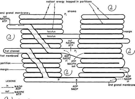
The thick dark membranes within the grana are termed partitions and are either connected at their margins or joined to other grana by frets (Fig. 2).
The light space enclosed by the partition and margin is termed the loculus and that enclosed by the fret membrane is the fret channel. The outside membranes are narrower (see below) than the internal partitions and are called the end granal membranes. On Weier's interpretation the "stack of coins" pictures, obtained by shadowing OsOi-fixed fragments of isolated, chloroplasts (Steinmann, 1952) and formerly interpreted as separated intact discs which have slid apart, would now appear to have arisen by breakage at the margin and across the loculus, leaving the partitions intact and exposing the intra-locular surfaces.
144 R. Μ . L E E C H
Perhaps the most exciting recent development has been the first evidence for the chloroplast membranes being composites of particulate subunits. A similar structure is currently under discussion for other biological membranes (Green and Perdue, 1966). Frey-Wyssling and Steinmann (1953) were the first to observe globular subunits in isolated and shadowed chloroplast membranes and Frey-Wyssling (1957) later pointed out how puzzling it was that they did not show up in sections of membranes. Park and Pon (1961)
radian t energ y trappe d in partitions ,
ou t t ˝˜—
carbo n cycl e
FIG. 2 Diagram showing the relationship between membranes and stroma within a higher plant chloroplast (reproduced with permission from Weier et al., 1967).
Possible relationships of components involved in photosynthetic reactions are also shown. The carbon cycle is located in the stroma and the light reactions in the membranes. Weier et al. (1967) postulate that energy transfer from the membranes occurs by movement of ATP, ADP, NADP and NADPH in the directions shown in
the diagram.
and Park and Biggins (1964) extended these studies and obtained pictures showing layers of particles 160-185A long, 155A wide and lOOA thick within the granal membranes. The name "quantasome" was coined for these sub- units ; this was perhaps a rather unfortunate term as the relationship of this
8. THE CHLOROPLAST INSIDE AND OUTSIDE THE CELL 145 structural unit to the photosynthetic unit or to the physiological quantum conversion unit is still obscure. The particles were originally (1961) conceived of as being partially embedded in and forming one component of the mem
brane but also filling the intradisc space. More recently, Bamberger and Park (1966) have suggested the particles are actually part of the membrane and do not occupy the intradisc space. The 175A particles are often in highly organized arrays. In addition, other granal surfaces have been shown to bear more scattered 9 0 A particles. The particles can also be visualized in negatively-stained preparations. Using the technique of freeze-etching intro
duced by M o o r et al. (1961), Miihletahler and his associates have again shown particles of two size categories but of smaller dimensions than those observed by Park and his colleagues. Miihletahler (1966) describes particles 120A wide, each probably of 4 subunits 6θΑ deep, and a second class of single 60A particles. Recently, Bamberger and Park (1966) have also shown their 9 0 A and 175A particles in freeze-etched preparations. This technique of freeze-etching involves "sectioning" frozen specimens followed by surface replication and would be expected to minimize fixation artefacts. However there is presently considerable disagreement between Miihletahler's group and Park's group concerning the location of the particles within the granal structure. The decision as to whether the particles lie outside but partially embedded in the membranes (Miihletahler) or actually constitute the mem
brane itself (Park) rests on knowledge of the plane of fracture along or within the membrane. This is extremely difficult to determine with certainty and neither group seems to have published convincing pictorial evidence that their interpretation of the plane of sectioning is the correct one. Branton (1966) has, however, published evidence supporting the contention that frac
ture is within the membrane and not along the surface. The pictures produced by the two groups by similar techniques are very similar (Miihletahler et al.,
1965; Bamberger and Park, 1966), both showing two categories of particles and differences in size which may reflect preparative rather than real differences in the particle sizes in vivo.
Whatever the location of these particular particles it seems possible that we may very well be observing several chemically different particles in chloro
plasts both within the membranes and also lying superficially on the mem
branes but originating in the stroma. Miihletahler (1966) himself suggests the possibility that one class of particles that he observes may be ribosomes.
Alternatively, some of the superficial 120A particles may be Fraction I protein which has been shown by Haselkorn et al. (1965) to measure 120A along its edge.
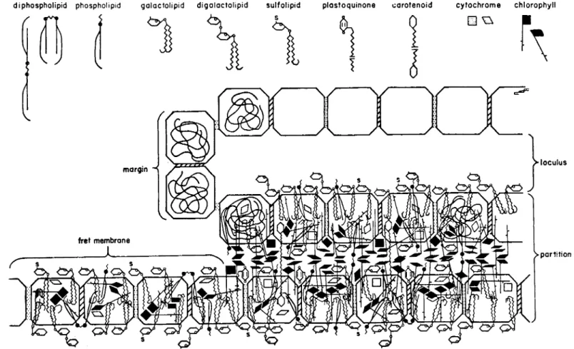
In sections of membranes, too, apparently spherical subunits have now been visualized, using sophisticated fixation procedures and high resolution electron microscopy (Weier et al., 1963,1965a, b, 1966). In micrographs, these
FIG. 3 Membrane structure as interpreted by Weier et al. (reproduced with permission from Weier et al. (1967).
di phospholipid phospholipid gaiactolipid d i g a l a c t o l i p i d sulfolipid plastoquinone carotenoid cytochrome chlorophyll
8. T H E C H L O R O P L A S T I N S I D E A N D O U T S I D E T H E C E L L 147
subunits in glutaraldehyde and permanganate fixations appear to consist of a light core about 37A in diameter with a dark rim about 28A wide. Partitions apparently consist of two rows of particles and fret membranes of a single row of subunits. However, again differences in interpretation exist; Bam
berger and Park (1966) would interpret their lipoprotein particles as being enclosed top and bottom by a continuous galactolipid layer but Weier (see Weier and Benson, 1966) would apparently conceive of the whole membrane consisting of mixed protein and lipid components. Again from X-ray scatter
ing studies Kreutz (1966) has suggested that the lipid and protein components are separate.
The membrane proteins presumably include the protein isolated by Criddle (1966), plastocyanin (a copper-containing protein) and the cytochromes / , be and 6559. The lipids, apart from chlorophylls a and b and the carotenoids, include the phytoquinones, plastoquinones A, B, C, D, plastochromanol, tocopherols and tocopherolquinones, vitamins Κ and phospholipids and glycolipids (see Kirk and Tilney-Bassett, 1967 for a summary).
It is of considerable interest to try fitting this variety of chemical molecules into positions within the three-dimensional membrane structure visualized by the electron microscope. Weier and Benson (1966) published the first detailed suggestions along these lines. They do not take into account the relative proportions of the different molecules but suggest that the molecules of protein are relatively hydrophobic in nature and that the hydrocarbon chains of chlorophyll, carotenoids and lipids are buried within the protein molecular band in hydrophilic association. This interesting formulation is shown in Fig. 3 and suggests further experimentation.
Another approach to structure/function relationships at the molecular level is to search for methods of altering specifically the morphological and biochemical properties of the membranes. Detergents have been used for this purpose. More recently solvents have also been used but microscopical resolution does not appear sufficient to identify specific changes in morpho
logical structures. Ninety-three per cent of plastoquinones A and C can be removed from isolated chloroplasts using heptane and petroleum ether and only 1 8 % chlorophyll but only minor modification in membrane structure (Magree et al., 1966) can be detected. Chloroplasts isolated from leaves prefixed in glutaraldehyde appear to be little changed morphologically but retain only 2 5 % of their Hill reaction activity. Acetone also destroys Hill reaction activity but no change in structure has yet been detected. By studying membrane structure after protease and lipase treatment, Bamberger and Park (1966) deduced that chlorophyll is located in the membrane particles.
There is no evidence that chemical differences exist between fret membranes and partitions and indeed this would be very difficult to explore.
148 R. Μ . L E E C H
8. T H E C H L O R O P L A S T I N S I D E A N D O U T S I D E T H E C E L L 149 I I I . THE CHLOROPLAST OUTSIDE THE CELL
To study quantitatively the chemical composition and biochemical capa
bilities of the parts of the chloroplast, isolation from the cell is necessary.
Electron microscope studies have shown that many isolated chloroplasts used in biochemical studies are considerably altered morphologically from their state in the cell. Two general types of isolation method are available, either (a), grinding the fresh material in an osmotically adjusted, buffered aqueous medium or (b), grinding lyophilized material in non-aqueous solvents, for example, petroleum ether/carbon tetrachloride mixtures (Thalacker and Behrens, 1959; Heber, 1957) or hexane/carbon tetrachloride mixtures (Stocking, 1959). By neither the aqueous nor the non-aqueous technique is it possible to separate the chloroplast cleanly from the other components of the cell using only differential centrifugation; density gradient centrifugation is always required (Leech et al., 1964). Retention of soluble proteins is good in non-aqueous preparations notably of the enzymes NADP-triose-phosphate de hydrogenase (1.2.1.13) and ribulose-l-5-diphosphate carboxylase (4.1.1.39.,1) essential in the first stages of photosynthetic carbon reduction (Smillie and Fuller, 1959; Heber et al., 1963) but Hill reaction activity is totally lost (Heber and Tyszkiewitz, 1962). In contrast, in chloroplasts isolated by aqueous techniques, the soluble enzyme systems are the most vulnerable.
Electron micrographs have shown that the loss is due to fracture of the envelope and physical washing out of the stroma. Perner (1965) has shown that free spinach chloroplast membranes devoid of stroma can only be maintained in isolation in a state equivalent to that in vivo in hypertonic sucrose with pyrophosphate and not in 0-2 % NaCl.
In Vicia faba loss of the stroma itself is considerably greater if the chloro
plasts are isolated in buffered 0*35M NaCl than if they are isolated in 0-3-0-8M buffered sucrose (compare Figs 4 and 6). Even if the envelope is still intact its properties may change in isolation. Isolated spinach (Spinacia oleracea) chloroplasts have been studied by light and electron microscopy by K a h n and von Wettstein (1961) who distinguished two types of chloroplast in their suspensions and suggested that the chloroplasts appearing opaque in the FIG. 4 Electron micrographs of chloroplasts isolated from Vicia fabia L. χ 8000.
Fixed in buffered (pH 7-0) 0s04: A and C embedded in Epon; Β embedded in araldite. (R. M. Leech and A. D. Greenwood, unpublished results).
A. A section of a 1000 χ g pellet showing intact membrane-bound chloroplasts (I) free lamella systems (L) mitochondria (M) and cell debris (D).
B. A section of a "chloroplast" pellet prepared according to James and Das (1957).
Only free lamella systems (L) are present.
C. A section of a chloroplast pellet prepared according to Leech (1964). Almost all the profiles are of intact membrane-bound chloroplasts.
FIG 5
150 R . Μ . L E E C H
8. THE CHLOROPLAST INSIDE AND OUTSIDE THE CELL 1 5 1 light microscope corresponded to those in the electron micrographs with intact envelopes and stroma. Chloroplasts, granular in appearance in the light microscope, would be those shown in the electron micrographs to be stripped of their bounding envelopes and a considerable portion of their stroma. However, the suggestion could not be vindicated until suspensions of chloroplasts consisting exclusively of one morphological category only could be prepared and examined. Using the method of James and Das, ( 1 9 5 7 ) , suspensions can be prepared containing only chloroplasts stripped of their outer membranes (James and Leech, 1964). By selecting a fraction from a sucrose density gradient, a suspension consisting virtually entirely of intact chloroplasts can be prepared (Leech, 1964). The latter method has recently been adopted for the Zonal centrifuge (C. A. Price, private communication). In Figs 4 and 5 are shown the appearances of the preparations containing only one type of chloroplast in both light and electron micrographs. A preparation containing both chloroplast types is also shown. It can be seen that at least for Vicia faba the suggestion of Kahn and von Wettstein is vindicated. A note of caution should be added here. A bio
chemist wishes to know the state of preservation of the chloroplast in the actual suspension he used for his biochemical assay. Any gross subsequent changes during fixation and embedding for electron microscopy will lead to a more pes
simistic view than is warranted. Isolated cell organelles appear to be very much less resilient to osmotic shock than are intact cells and osmotic adjustment of not only the fixative but also the dehydration stages is necessary (Leech, 1 9 6 4 ) . Despite their respective limitations, aqueous and non-aqueous techniques have been used with considerable success in the study of chloroplast enzymology. Hall and Whatley ( 1 9 6 7 ) have reviewed the information to date.
The state of integrity of the chloroplasts clearly affects their biochemical attributes in vitro. Walker ( 1 9 6 5 , 1 9 6 7 ) obtained higher C O 2 fixation rates and lower rates of A T P synthesis in suspensions containing a large proportion of intact chloroplasts than in suspensions of broken chloroplasts. Jeffrey et al. ( 1 9 6 6 ) using intact chloroplasts from the chrysomonad Hymenomonas obtained rates of C O 2 fixations equivalent to 2 5 % of the rate of fixation by the intact organism. This was the highest rate expressed as a percentage of whole plant fixation published at that time. The recent achievement of Jensen and Bassham ( 1 9 6 6 ) in obtaining in vitro rates of C O 2 fixation ( 2 4 8 μ η ι ο ^ CCVmg chlorophyll/hr) actually in excess of in vivo rates, can also FIG. 5 Light micrographs (χ 1750) of the same material as shown in Fig. 4 . A. A 1000 χ g "chloroplast" fraction. Both intact (opaque) (I) and envelope-free chloroplasts (L) (showing grana) are present.
B. A section of a "chloroplast" pellet prepared according to James and Das (1957).
Only free lamella systems (L) are present.
C. A section of a chloroplast pellet prepared according to Leech (1964). Almost all the profiles are of intact membrane-bound chloroplasts.
152 R . Μ . L E E C H
FI G 6
8. T H E C H L O R O P L A S T I N S I D E A N D O U T S I D E T H E C E L L 153 be attributed in part to their use of suspensions containing a high proportion of intact chloroplasts. The activity of multi-enzyme systems localized within the pigment-containing membrane system may be lower when assayed in intact chloroplasts than when using free membrane systems. This is presum
ably because of the impermeability of the chloroplast envelope and has been clearly shown for cyclic photophosphorylation in intact and broken pea chloroplasts by Walker (1967).
Much attention has been devoted to obtaining reliable quantitative values for the chemical composition of the chloroplast but this has proved very difficult. Clearly these values can only have significance if they are referred to chloroplasts in a particular morphological state isolated from one species of plant at one developmental stage (Smillie and Krotkov, 1961). Many of the earlier estimates, obtained before the considerable damage attendant on
TABLE I
Total protein and total lipid content of chloroplasts from Vicia faba leaves (3 weeks old).
jChloroplast
preparation •Total lipid
/*g//*g Chlorophyll
fTotal protein Lipid: Protein
tlntact 15-0Ί
15-9 V15-8 16-5 J
3-2Ί 3-7 ^3-4 3-2J
4-65
1000 x g fraction
3 - 8 /3'8 5 2-3^
2-6 ^2-3
2-0 J 1-67
* Lipid values Jarvis et al, 1967.
t Protein values and methods of chloroplast isolation from Leech, 1966.
Chlorophyll determined in 80% acetone (Arnon, 1949).
Protein determined by the method of Lowry et al. (1951).
Lipid determined gravimetrically after 3 extractions in 80% acetone.
% The bacterial count for this chloroplast preparation was 5 χ 103-103/ml.
isolation was discovered, are almost certainly too low. Protein loss from chloroplasts during aqueous isolation is more often allowed for (e.g. Kirk and Tilney-Bassett, 1967) and indeed can be correlated with the degree of integrity of the chloroplasts (Leech, 1966 and Table I.) For chloroplasts which are intact under the electron microscope from three-week-old Vicia faba plants the protein/chlorophyll value is 3 : 1 and about 35 % of the protein
FIG. 6 1000 χ g chloroplast fraction prepared in buffered 0-35 Μ NaCl (R. M.
Leech and A. D. Greenwood, unpublished results).
A. Electron micrograph of a section of the chloroplast pellet fixed in buffered Os04 and embedded in Araldite.
B. Light micrograph of the same suspension.
ι
154
k. Μ. it&mis in the membrane fraction. Loss of lipid during aqueous isolation had previously been suspected (Leech, 1966) but L. Jarvis, Η. M. Hallaway and R. M. Leech (1967, unpublished results) have shown this can be very consider
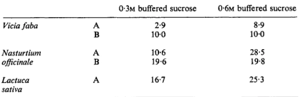
able and cannot be ignored. Since chlorophyll is restricted to the chloroplast lamellae, measurement of the lipid to chlorophyll ratio provides an index of the relative amount of lipid in the different types of preparation. Jarvis has clearly shown (Table II) that using a 1000 χ g fraction the ratio is affected considerably according to the species used, the isolation media and the type of homogenization. In the comparison of intact chloroplasts and a 1000 χ g preparation which contains a mixture of whole and broken chloroplasts, mitochondria etc., it can be seen that about 70 % of the chloroplast lipids have already been lost (Table I). Increasing the molarity of the sucrose in the
TABLE II
The lipid/chlorophyll 0*g//xg) ratios in 1000 x g fraction from leaves using different extraction techniques (data of Jarvis et al., 1967).
Lipid/chlorophyll (/Ag//*g)
0·3Μ buffered sucrose 0*6M buffered sucrose
Vicia faba A 2-9 8-9
Β 100 100
Nasturtium A 10-6 28-5
officinale Β 19-6 19*8
Lactuca A 16-7 25-3
sativa
A. Initial homogenization using a pestle and mortar.
B. Initial homogenization using a liquidizer.
isolation media increases the proportion of intact chloroplasts and, as would be expected, also the lipid/chlorophyll ratio. Intact chloroplasts have a ratio lipid/chlorophyll of 15-8 ± 0-7 compared with 3-4 ± 0-3 for protein/
chlorophyll and this would suggest a lipid/protein ratio of between 4 and 5 in the intact chloroplast. These chloroplasts contained only a very few small plastoglobuli and they could not have accounted for the high values of stroma lipid. To obtain quantitative values per chloroplast, an accurate method for determining the proportion of intact and envelope-free chloroplasts is necessary. Haemocytometer counting is tedious and not very precise but differences in resistance may be sufficient to enable the two classes of chloroplasts to be distinguished on the Coulter Counter (Ridley et al.,
1967).
8. THE CHLOROPLAST INSIDE AND OUTSIDE THE CELL 155 IV. THE CHLOROPLAST AS AN AUTONOMOUS UNIT
The mature chloroplast of the higher plant is the primary site of the whole complex of reactions leading to the capture of light and its eventual storage in chemical form in carbohydrates. Isolated chloroplasts from a variety of plants fix C O 2 and synthesize A T P and N A D P H 2 and recent work has demonstrated they are also able to synthesize sucrose (Bird et al., 1965), lipids (Stumpf and James, 1962; Stumpf, etal., 1963) and proteins (Spencer,
1965; Sissakian et al., 1965). They are also able to reduce nitrate and nitrite photochemically (Ramirez et al., 1964; Paneque et al., 1964; Losada and Paneque, 1966) and can probably carry out the complete biosynthesis of chlorophylls and carotenoids. Evidence for this wide range of synthetic reactions, taken together with very good evidence that higher plant chloroplasts contain D N A (see Gibor and Granick, 1964) and R N A (see Kirk and Tilney- Bassett, 1967 for a critical assessment of the evidence) and are also able to synthesize their own R N A (Kirk, 1964), demonstrates very clearly that chloro- plasts have a high degree of biochemical autonomy. It is pertinent to ask just how independent is the chloroplast of the rest of the cell not only bio- chemically but also genetically as a self-determining self-duplicating structure.
Gibor and Granick published a very comprehensive review of the subject in 1964 and Kirk and Tilney-Bassett (1967) have reviewed the additional information u p to 1966.
In studies of this kind the green algae Euglena and Acetabularia have been used with considerable success as experimental material. It is not difficult to see why the biological characteristics of these organisms made them such valuable experimental objects. I n Euglena gracilis var. bacillaris formation of the chloroplast is light-dependent and by keeping dark grown cells on "rest- ing" medium and exposing them to the light it is possible to study chloroplast development uncomplicated by cell division. Acetabularia is a giant cell with only a single nucleus and if the nucleus is removed the rest of the cell is able to survive for about three months in the light. Using this organism one has the unique possibility of studying the behaviour of chloroplasts in vivo in the presence and in the absence of the nucleus. When assessing the degree of autonomy of the chloroplast, observations on Euglena and Acetabularia are usually taken in conjunction with evidence derived from studies of higher plants. This could be misleading for a highly determinate heterogeneous organ such as a leaf is very different biologically from a haploid organism such as Euglena reproducing by binary fission and different again from the vegetative stage of a diploid giant unicell such as Acetabularia. In the leaf the immature chloroplasts can be regarded as the end-products of differentia- tion from proplastids of the meristem and there is no evidence that these chloroplasts are able to divide in the mature state. In many algae, on the
156 R. Μ. LEECH
other hand, chloroplasts have frequently been seen in division (e.g. the recent cine film of chloroplast division in Nitella by Green, 1964) and the products of the division are the first chloroplasts of the next cell generation. Recent evidence from algae and from higher plants will, therefore, be considered separately here.
Gibor (1967) has pointed out that the perpetuation of a genetic system requires the presence of D N A and R N A and of enzymes for D N A synthesis, for R N A synthesis and for protein synthesis. These attributes will be con
sidered in turn.
A. ALGAE 1. Acetabularia
Gibor and Izawa (1963) and Balthus and Brachet (1963) demonstrated that in bacteria-free enucleate Acetabularia extranuclear D N A is present to the extent of 1 χ 1 0 "16 g/chloroplast (Gibor and Granick, 1964). This D N A is localized in the chloroplast and has a buoyant density of 1-695 in a CsCl gradient (Gibor, 1965). Gibor (1967) has fed 1 4C 02 to enucleated Acetabu
laria in the light and has shown clearly that synthesis of deoxynucleosides, their phosphorylation and polymerization can proceed in the absence of the nucleus. The location of these synthetic reactions is not known but may be in the chloroplast. Actinomycin D-sensitive R N A synthesis has been demon
strated in isolated Acetabularia chloroplasts and Janowski (1965) has shown
3 2P incorporation into R N A of more than one kind in the chloroplast fraction isolated from enucleated Acetabularia. Shepherd (1965) has used autoradio
graphic techniques to show that chloroplast R N A and protein synthesis proceeds at the same rate and D N A synthesis at half the rate in the absence of the nucleus as in its presence. Protein synthesis can indeed continue for up to four weeks after enucleation (Hammerling, 1963). Synthesis of an enzyme thought to be localized exclusively in the chloroplast of green cells, namely NADPH-triose phosphate dehydrogenase (1.2.1.13), has been shown by Ziegler et al. (1967) to occur in the absence of the nucleus in the light, rising from no activity one day after enucleation to the same level of activity as in the nucleated control after 5 days. Shepherd (1965) has clearly shown that Acetabularia chloroplasts can replicate in the absence of the nucleus.
There seems to be little information about ribosomes from Acetabularia; the difficulties of isolation in sufficient quantities for analysis are formidable.
Enucleate fragments of Acetabularia would therefore appear to have a complete functional genetic apparatus which enables survival for several weeks but no longer and we can agree with Shepherd who suggests that in this organism growth, replication and biosynthetic activity of the chloroplast can apparently proceed without coded information of nuclear origin. There is good evidence that many of the elements of the genetic apparatus reside
8. THE CHLOROPLAST INSIDE AND OUTSIDE THE CELL 157 in the chloroplast. Few studies have been made on the genetics of the organism and there is no evidence about the interaction between nuclear genes and chloroplast genes during development or even that the chloroplast genetic apparatus is inherited. The studies on isolated chloroplasts from enucleated cytoplasts of Acetabularia initiated by Gibor (1965) are awaited with considerable interest.
2 . Euglena
In Euglena gracilis var. bacillaris there is no doubt that the chloroplast divides (Schiff and Epstein, 1965, have published a beautiful picture which clearly demonstrates this). There is also no doubt that the chloroplast is inherited. The D N A of the whole Euglena cell shows three peaks in CsCl gradient centrifugation (Edelman et al, 1965; Ray and Hannawalt, 1965), one major peak and two minor satellite peaks. One satellite, of buoyant density 1-686, is enriched in the chloroplast fraction from green Euglena cells and absent from mutants incapable of forming proplastids or chloro
plasts (Schiff and Epstein, 1965). This satellite D N A would, therefore, appear to be localized within the chloroplast. Gibor and Granick (1964) have calculated that the Euglena chloroplast contains 4 χ 1 0- 15 g D N A . Euglena chloroplast ribosomes have a sedimentation coefficient of 60S (with a minor 36S component) compared with cytoplasmic ribosomes of 70S (Eisenstadt and Brawerman, 1964a). Cytoplasmic and chloroplast ribosomes present in non-dividing cells with developing chloroplasts differ in the base ratios of their R N A ; the adenine/cytosine ratio for chloroplast ribosomes is 1-8, for cytoplasmic ribosomes 0-87 (Brawerman et al, 1962). A 14S and a 19S component of R N A were found in cytoplasmic ribosomes (Brawerman and Eisenstadt, 1964). Template R N A has been shown to be present in the Euglena chloroplast (Eisenstadt and Brawerman, 1964a) and isolated Euglena chloroplasts are able to incorporate amino-acids into proteins (Eisenstadt and Brawerman, 1964b). Several chloroplast enzymes have been shown to appear in the development in the light of chloroplasts in dark-grown cells (see Schiff and Epstein, 1965, for a review). On the basis of their evidence, Schiff and Epstein reached the conclusion that in Euglena gracilis var. bacillaris chloroplast replication and development involves the replication and reading of an autonomous code.
B. LEAVES OF HIGHER PLANTS
The mature leaf as an organ for studying chloroplast autonomy is not ideal but it is very valuable as a source of isolated fully differentiated chloro
plasts in sufficient quantity for biochemical assay.
Satellite D N A s have also been detected in leaf material with buoyant densities of 1-705 and 1-719 compared with the major nuclear D N A of
158 R. Μ. LEECH
1-695 (Chun et al, 1963). In contrast to Euglena and Acetabularia, in leaves cytoplasmic D N A would appear to have a lower buoyant density than nuclear D N A . D N A synthesis has not been demonstrated in chloroplasts in leaves in vivo and Wollgiehn and Mothes (1964), using mature leaves of tobacco, could get no evidence for DNase-sensitive 3H-thymidine incorporation into chloroplasts. Kirk (1964), however, has demonstrated a DNA-dependent polymerase in Vicia faba chloroplasts. When ribosomes are examined there are also differences between the algae and higher plants. In leaves from several species two classes of ribosomes are found with sedimentation coefficients of 72-80S and 62-70S, the smaller category of particles apparently being restricted to the chloroplast. The actual S values vary in the different reports and this may reflect technical differences or differences in S values in different species.
Lyttleton (1962) was the first to isolate two classes of ribosomes from Spinacia leaves and Spencer (1965) confirmed that there were 70S and 80S ribosomes in these leaves. The corresponding values for Brassica pekinensis are 68S and 83S (Clark et al, 1964) and for tobacco 70S and 80S (Boardman et al, 1965). It may be that chloroplast and cytoplasmic ribosomes are more similar in higher plants than in the algae. There is a possibility, however, that real differences are concealed because of the presence of chloroplast ribosomes in the "cytoplasmic" ribosomal fraction. Pollard et al (1966) have isolated R N A from "cytoplasmic" and chloroplast ribosomes of Romaine lettuce and found an 18S and 28S component in both types of ribosome. F r o m our own unpublished results we have found both light and heavy R N A components in both cytoplasmic and chloroplast ribosomal fractions from Vicia faba leaves (T. A. Dyer and R. M . Leech, unpublished results). Spencer and Whitfield (1966) however, were only able to isolate one component sediment- ing at 16S from the R N A of spinach chloroplast ribosomes but 16S and 23S fractions could be isolated from cytoplasmic ribosomes. The reason for the disparity is not clear but might reflect specific properties of spinach leaves.
Again the demonstration by Boardman (1966) that both 70S and 80S ribo
somes are present in etiolated Phaseolus leaves and that no more ribosomes can be isolated from a mature than from an etiolated leaf is also in sharp contrast to the position in Euglena. Thirdly, although they have been sought, no differences in the base ratios of the magnitude of those found in Euglena have yet been found between chloroplast ribosomal and cytoplasmic ribo
somal fractions from leaves. Even if considerable care is taken it is impossible to avoid some chloroplast breakage during the initial homogenization of the leaves. However, the low molecular weight R N A components of chloroplasts from Vicia leaves certainly have physical properties distinct from those of the cytoplasmic R N A (Dyer and Leech, 1967). As was mentioned previously, protein synthesis has also been demonstrated in higher plant chloroplasts (Spencer, 1965).
8. THE CHLOROPLAST INSIDE AND OUTSIDE THE CELL 159 In phanerogams much genetic evidence points to plastid genome continuity from one generation to the next (Kirk and Tilney-Bassett, 1967) and inheritance of mutant plastome genes via both male and female gametes has been demon
strated notably in Oenothera, Pelargonium and Epilobium.
Thus the information concerning the chloroplast genetic system is uneven from algae and from higher plants. There seems good evidence that the basic biochemical systems responsible for genetic continuity are present in the chloroplast but the study of their function and control are only just beginning.
It is the study of the interactions of the nuclear and plastid genomes during the development and functional life of the chloroplast in the different organ
isms which will provide interest and excitement in the years to come.
ACKNOWLEDGEMENTS
I should like to thank Mr. A. D . Greenwood for his generous help in stimulating discussions and also for permission to use the photographs in Figs 1, 4 and 6. I also wish to thank Professor Τ. E. Weier, Professor C. R.
Stocking and Dr. Shumway for permission to reproduce the diagrams in Figs 2 and 3.
REFERENCES Arnon, D. I. (1949). PL Physiol., Lancaster 24, 1.
Arnon, D. I., Allen, Μ. B. and Whatley, F. R. (1954). Nature, Lond. 174, 394.
Bailey, J. L. and Whyborn, A. G. (1963). Biochim. biophys. Acta 78, 163.
Balthus, E. and Brachet, J. (1963). Biochim. biophys. Acta 76, 490.
Bamberger, E. S. and Park, R. B. (1966). PI. Physiol., Lancaster 41, 1591.
Bird, I. F., Porter, Η. K. and Stocking, C. R. (1965). Biochim. biophys. Acta 79, 746.
Boardman, Ν. K. (1966). Expl Cell Res. 43, 474.
Boardman, Ν. K., Francki, R. I. B. and Wildman, S. G. (1965). Biochemistry, Ν Y.
4, 872.
Branton, D. (1966). Proc. natn. Acad. Sci., U.S.A. 55, 1048.
Brawerman, G. and Eisenstadt, J. M. (1964). J. molec. Biol. 10, 403.
Brawerman, G., Pogo, A. O. and Chargaff, E. (1962). Biochim. biophys. Acta 55,326.
Chun, Ε. H. L., Vaughan, Μ. H. and Rich, A. (1963). / . molec. Biol. 7, 130.
Clark, M. F., Matthews, R. E. F. and Ralph, R. K. (1964). Biochim. biophys. Acta 27, 145.
Criddle, R. S. (1966). NATO Symposium, "Biochemistry of Chloroplasts". Vol. I, p. 203. (T. W. Goodwin, ed.), Academic Press, London and New York.
Diers, L. and Schotz, F. (1966). Planta 70, 322.
Drawert, H. and Mix, M. (1962). Planta 58, 50.
Dyer, T. A. and Leech, R. M. (1967). Biochem. J. 102, 6P.
Edelman, M., Schiff, J. A. and Epstein, Η. T. (1965). / . molec. Biol. 11, 769.
Eisenstadt, J. M. and Brawerman, G. (1964a). / . molec. Biol. 10, 392.
Eisenstadt, J. M. and Brawerman, G. (1964b). Biochim. biophys. Acta 80, 463,
160 R. Μ. LEECH Falk, Η. (1960). Planta 55, 525.
Frey-Wyssling, A. (1957). "Macromolecules in Cell Structure". Harvard University Press, Cambridge, Mass., U.S.A.
Frey-Wyssling, A. and Steinmann, E. (1953). Naturforsch. Ges. Zuerich 98, 20.
Gibbs, S. P. (1960). / . Ultrastruct. Res. 4, 127.
Gibbs, S. P. (1962a). / . Ultrastruct. Res. 7, 247.
Gibbs, S. P. (1962b). / . Ultrastruct. Res. 7, 262.
Gibbs, S. P. (1962c). / . Ultrastruct. Res. 7, 418.
Gibor, A. (1965). Proc. natn. Acad. Sci., U.S.A. 54, 1527.
Gibor, A. (1967). NATO Symposium, "Biochemistry of Chloroplasts". Vol. 2, p. 321. (T. W. Goodwin, ed.), Academic Press, London and New York.
Gibor, Α., and Izawa, M. (1963). Proc. natn. Acad. Sci., U.S.A. 50, 1164.
Gibor, A. and Granick, S. (1964). Science, N.Y. 145, 890.
Green, D. E. and Perdue, J. F. (1966). Proc. natn. Acad. ScL, U.S.A. 55, 1295.
Green, P. B. (1964). Am. J. Bot. 51, 334.
Greenwood, A. D., Leech, R. M. and Williams, J. P. (1963). Biochim. biophys. Acta 78, 148.
Gunning, Β. E. S. (1965). / . Cell Biol. 24, 79.
Gunning, Β. E. S. and Jagoe, M. P. (1967). NATO Symposium, "Biochemistry of Chloroplasts". Vol. 2, p. 655. (T. W. Goodwin, ed.), Academic Press, London and New York.
Hall, D. O. and Whatley, F. R. (1967). In "Enzyme Cytology", p. 181. (D. B.
Roodyn, ed.), Academic Press, London and New York.
Hammerling, J. (1963). A. Rev. PL Physiol. 14, 65.
Haselkorn, R., Fernandez-Moran, H., Kieras, F. J. and van Bruggen, E. F. J.
(1965). Science, N.Y. 150, 1598.
Heber, U. (1957). Ber. dt. bot. Ges. 70, 371.
Heber, U. and Tyszkiewiez, E. (1962). / . exp. Bot. 13, 185.
Heber, U , Pon, N. G. and Heber, M. (1963). PL Physiol., Lancaster. 38, 355.
Heitz, E. (1958). In "4th International Congress of Electron Microscopy". Vol. 2, p. 501. Springer-Verlag, Berlin.
Heslop-Harrison, J. (1963). Planta 60, 243.
Jacobi, G. and Perner, E. (1961). Flora 150, 209.
Jacobson, A. B., Swift, H. and Bogorad, L. (1963). / . Cell Biol. 17, 557.
James, W. O. and Das, V. S. R. (1957). New Phytol. 56, 325.
James, W. O. and Leech, R. M. (1964). Proc. R. Soc. B. 160, 13.
Janowski, M. (1965). Biochim. biophys. Acta 103, 399.
Jeffrey, S. W., Ulrich, J. and Allen, Μ. B. (1966). Biochim. biophys. Acta 112, 35.
Jensen, R. G. and Bassham, J. A. (1966). Proc. natn. Acad. Sci., U.S.A. 56, 1095.
Kahn, A. and von Wettstein, D. (1961). / . Ultrastruct. Res. 5, 557.
Kaja, H. (1966). Z. Naturf. 21b, 379.
Kirk, J. T. O. (1964). Biochim. biophys. Res. Commun. 16, 233.
Kirk, J. T. O. and Tilney-Bassett, R. A. E. (1967). "The Plastids". Freeman, London and San Francisco.
Kislev, N , Swift, H. and Bogorad, L. (1965). / . Cell Biol. 25, 327.
Kreutz, W. (1966). NATO Symposium, "Biochemistry of Chloroplasts". Vol. 1, p. 83. (T. W. Goodwin, ed.), Academic Press, New York and London.
Leech, R. M. (1964). Biochim. biophys. Acta 79, 637.
Leech, R. M. (1966). NATO Symposium, "Biochemistry of Chloroplasts". Vol. 1, p. 65, (T. W. Goodwin, ed.), Academic Press, London and New York.
8. THE CHLOROPLAST INSIDE AND OUTSIDE THE CELL 161 Leech, R. M., Stocking, C. R. and Greenwood, A. D. (1964). Abstracts 1st Meeting
Federation European Biochemical Soc. BI, 109.
Lichtentahler, Η. K. (1964). Ber. dt. bot. Ges. 74, 398.
Lichtentahler, Η. K. and Sprey, B. (1966). Z. Naturf. 21b, 690.
Lowry, Ο. H., Rosenborough, N. J., Farr, A. L. and Randall, R. J. (1951). / . biol.
Chem. 193, 265.
Losada, M. and Paneque, A. (1966). Biochim. biophys. Acta 126, 578.
Lyttleton, J. W. (1962). Expl Cell Res. 26, 312.
Magree, L., Henninger, M. D. and Crane, F. L. (1966). / . biol. Chem. 241, 5197.
Manton, I. (1966). NATO Symposium, "Biochemistry of Chloroplasts". Vol. 1, p. 23. (Goodwin, T. W., ed), Academic Press, London and New York.
Menke, W. (1960). Z. Naturf. 15b, 479.
Menke, W. (1961). Z. Naturf. 16b, 334.
Menke, W. (1962). A. Rev. PI. Physiol. 13, 27.
Menke, W. (1966). NATO Symposium, "Biochemistry of Chloroplasts". Vol. 1, p. 3. (T. W. Goodwin ed.), Academic Press, New York.
Moor,H., Muhletahler,K.,Waldner,H.andFrey-Wyssling,A. (1961). J. Cell Biol.
10, 1.
Muhletahler, K. (1960). Z. wiss. Mikroskop. 64, 444.
Miihletahler, K. (1966). NATO Symposium, "Biochemistry of Chloroplasts".
Vol. 1, p. 49. (T. W. Goodwin, ed.), Academic Press, London and New York.
Muhletahler, K., Moor, H. and Szarkowski, J. W. (1965). Planta 67, 305.
Murakami, S. and Takamiya, A. (1962). In "Electron Microscopy". (S. Breeze, ed.), Vol. 2, XX, p. 12. Saunders, Philadelphia, U.S.A.
Nobel, P. S., Murakami, S. and Takamiya, A. (1966a). 6th International Congress for Electronmicroscopy, Kyoto.
Nobel, P. S., Murakami, S. and Taharnija, A. (1966b). Plant and Cell Physiol. 7, 263.
Paneque, Α., Ramirez, J. M., Del Campo, F. F. and Losada, M. (1964). / . biol.
Chem. 239, 1737.
Park, R. B. and Pon, N. G. (1961). / . molec. Biol. 3, 1.
Park, R. B. and Biggins, J. (1964). Science, Ν. Y. 144, 1009.
Perner, E. (1962). Port. Acta biol. 6, 359.
Perner, E. (1965). Planta 66, 44.
Pollard, C. J.,Stemler, A. andBlaydes,D. F. (1966). PI. Physiol, Lancaster 41,1323.
Ramirez, J. M., Del Campo, F. F., Paneque, A. and Losada, M. (1964). Biochem.
biophys. Res. Commun. 15, 297.
Ray, D. S. and Hannawalt, P. C. (1965). / . molec. Biol. 11, 760.
Robertson, R. D. (1964). In "22nd Symposium of Soc. for Development and Growth". (M. Locke, ed.), p. 31, Academic Press, New York and London.
Sager, R. (1958). Brookhaven Symp. Biol. 11, 101.
Sager, R. and Palade, G. E. (1957). / . biophys. biochem. Cytol. 3, 463.
Schweiger, H. G. and Berger, S. (1964). Biochim. biophys. Acta 87, 533.
Schiff, J. A. and Epstein, Η. T. (1965). In "Reproduction, Molecular, Subcellular and Cellular". (M. Locke, ed.), p. 131. Academic Press, New York and London.
Schiff, J. A. and Epstein, Η. T. (1966). NATO Symposium "Biochemistry of Chloroplasts". Vol. 1, p. 341. (T. W. Goodwin, ed.), Academic Press, London and New York.
Shepherd, D. C. (1965). Expl Cell Res. 37,93.
Silaevia, A. M. and Shiriaev, A. I. (1966). Dokl. Akad. Nauk SSSR 170, No. 2.
162 R. Μ. LEECH
Sissakian, Ν. Μ., Filipovich, I. I., Svetailo, Ε. N. and Aliyev, K. A. (1965).
Biochim. biophys. Acta 95, 474.
Smillie, R. M. and Fuller, R. C. (1959). PI. Physiol, Lancaster 37, 716.
Smillie, R. M. and Krotkov, G. (1961). Can. J. Bot. 39, 891.
Spencer, D. (1965). Archs Biochem. Biophys. I l l , 381.
Spencer, D. and Whitfield, P. R. (1966). Archs Biochem. Biophys. 117, 337.
Sprey, B. and Lichtentahler, Η. K. (1966). Z. Naturf. 21b, 697.
Steinmann, E. (1952). Expl Cell Res. 3, 367.
Steinmann, E. and Sjostrand, F. S. (1955). Expl Cell Res. 8,15.
Stocking, C. R. (1959). PI. Physiol, Lancaster 34, 56.
Stumpf, P. K. and James, A. T. (1962). Biochim. biophys. Acta 57, 400.
Stumpf, P. K., Bove, J. M. and Goffeau, A. (1963). Biochim. biophys. Acta 70, 260.
Thalacker, R. and Behrens, M. (1959). Z. Naturf. 14b, 443.
Thomson, W. W., Dugger, W. M. and Palmer, R. L. (1965). Bot. Gaz. 126, 62.
Thomson, W. W., Dugger, W. M. and Palmer, R. L. (1966). Can. J. Bot. 44,1677.
Trebst, Α. V., Tsujimoto, Η. Y. and Arnon, D. I. (1958). Nature, Lond. 182, 351.
Walker, D. A. (1965). PI. Physiol, Lancaster 40, 1157.
Walker, D, A. (1967). NATO Symposium, "Biochemistry of Chloroplasts". Vol. IT, (T. W. Goodwin, ed.), p. 53. Academic Press, London and New York.
Wehrmeyer, W. (1961). Ber. dt. bot. Ges. 74, 209.
Wehrmeyer, W. (1964). Planta 62, 272.
Wehrmeyer, W. and Perner, E. (1962). Protoplasma 54, 573.
Weier, Τ. E. and Benson, A. A. (1966). NATO Symposium, "Biochemistry of Chloroplasts". Vol. 1, p. 91. (T. W. Goodwin, ed), Academic Press, London and New York.
Weier, Τ. E. and Thomson, W. W. (1962) Am. J. Bot. 49, 807.
Weier, Τ. E., Bisalputra, T. and Harrison, A. (1966). / . Ultrastruct. Res. 15, 38.
Weier, Τ. E., Englebrecht, A. H. P., Harrison, A. and Risley, Ε. B. (1965a). / . Ultrastruct. Res. 13, 92.
Weier, Τ. E., Stocking, C. R., Bracker, C. E., Risley, Ε. B. (1965b). Am. J. Bot. 52, 339.
Weier, Τ. E., Stocking, C. R., Thomson, W. W. and Drever, H. (1963). / . Ultra
struct. Res. 8, 122.
Weier, Τ. E., Stocking, C. R. and Shumway, L. K. (1967). Brookhaven Symp. Biol.
19, 353.
Wellburn, A. R. and Hemming, F. W. (1966). NATO Symposium, "Biochemistry of Chloroplasts". Vol. 1, p. 173. (T. W. Goodwin, ed.), Academic Press, London and New York.
Wollgiehn, R., Ruess, M. and Munsche, D. (1966). Flora, 157, 92.
Wollgiehn, R. and Mothes, K. (1964). Expl Cell Res. 35, 52.
Ziegler, H., Ziegler, I. and Beth, K. (1967). Planta 72, 247.