761
Sorbitol Dehydrogenase
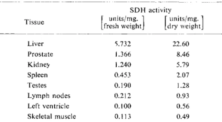
S D H activity
Tissue [" units/mg. 1 [ units/mg. ' Tissue
[fresh weight] [dry weight
Liver 5.732 22.60
Prostate 1.366 8.46
Kidney 1.240 5.79
Spleen 0.453 2.07
Testes 0.190 1.28
Lymph nodes 0.212 0.93
Left ventricle 0.100 0.56
Skeletal muscle 0.113 0.49
Physiologically the cytoplasmic S D H may be considered to be coupled with a TPN-specific aldose reductase
8 - 9
*
1
) according to the following reactions:
reductase
(1) D-Glucose + T P N H + H+ ; = = ± D-sorbitol + T P N + S D H
(2) D-Sorbitol + D P N + D-fructose + D P N H + H+
The determination of S D H activity is of clinical i m p o r t a n c e
5 - 7
. 1 0 - 1 3 ) because the sera of healthy individuals contain very little, but in liver cell damage (infectious, toxic or hypoxic in origin) it becomes demonstrable, therefore the S D H activity of serum can be of value as a fairly specific indicator of liver cell damage.
1) R. L. Blakley, Biochem. J. 49, 257 [1951].
2
) O. Hoffmann-Ostenhof: Enzymologie. Springer, Wien 1954, p. 485.
3) H. Holier, J. Haon and S. Schneider, Biochem. Z. 326, 451 [1955].
4
) H. G. Williams-Ashman and J. Banks, Arch. Biochem. Biophysics 50, 513 [1954].
s) U. Gerlach, Klin. Wschr. 35, 1144 [1957].
6) U. Gerlach in W. H. Hauss and H. Losse: Struktur und StofTwechsel des Herzmuskels. G. Thieme, Stuttgart 1959, p. 148.
7
) U. Gerlach, Klin. Wschr. 37, 93 [1959].
7
*) H. Holzer and H. W. Goedde, Biochim. biophysica Acta 40, 297 [i960].
8) H. G. Hers, Biochim. biophysica Acta 22, 202 [1956].
9) H. G. Hers: Proceedings of the International Symposium on Enzyme Chemistry (Tokyo and K y o t o 1957). Maruzen, T o k y o 1958, p. 109.
9a) S. Hollmann, Hoppe-Seylers Z. phvsiol. Chem. 317, 193 [1959].
io) U. Gerlach, unpublished.
n) U. Gerlach, Therapie des Monats 10, 211 [I960].
12) U. Gerlach and H. Kronsbein, Klin. Wschr. 37, 595 [1959].
13) U. Gerlach and E. Schiirmeyer, Z. ges. exp. Med. 132, 413 [I960].
Ulrich Gerlach
Sorbitol dehydrogenase ( S D H ) , which was first described by Blakley
l
\ has been found in the liver and the genital organs of many mammals
2
~
4 )
. In man the liver has the largest concentration of S D H
5 - 7
) ; the concentration of the enzyme in other organs is given in Table 1. The serum and haemo- lysed blood of healthy human beings contains hardly any S D H
1
\ The enzyme is also known as polyol dehydrogenase
? a
) .
Table 1. S D H activity in human tissues obtained at autopsy.
762 Section C: Measurement of Enzyme Activity
Principle
The principle of the measurements is given in equation (2). The S D H activity can be measured in either direction but the use of fructose as substrate is more favourable for measurements in serum.
The decrease in D P N H absorption with time at 340 or 366 mjx is a measure of the S D H activity.
Optimum Conditions for Measurements
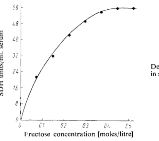
Figure 1 shows the dependence of S D H activity in serum on the concentration of D-fructose.
The optimum concentration is ca. 0.4 M (in triethanolamine buffer p H 7.4)
1 3
»
1 4
) .
The p H optimum of the reaction with fructose in triethanolamine or tris-hydroxymethyl-amino- methane (tris) buffer is p H 6.1. The reaction is slightly faster in tris than in triethanolamine buffer, but in tris buffer a higher concentration of fructose is required to saturate the enzyme.
In several buffers the S D H reaction is 1 0 - 2 0 times faster with D P N H than with T P N H . Raising the temperature by 10°C, increases the reaction rate (between 20 and 35° C) by 150 %
1 0 )
.
The best working conditions for the measurement of S D H activity in human sera in different diseases are given in the following procedure.
Reagents *>
1. Triethanolamine hydrochloride
2. Reduced diphosphopyridine nucleotide, DPNH
sodium salt, D P N H- N a 2 ; commercial preparation, see p. 1011.
3. D-Fructose, crystalline, pure 4. Sodium hydroxide, 2 N
5. Sodium hydrogen carbonate solution, 1 % (w/v) Preparations of Solutions
I. Triethanolamine buffer (0.2 M; pH 7.4):
Dissolve 3.72 g. triethanolamine hydrochloride in about 80 ml. distilled water, add 7.2 ml. 2 N NaOH, mix and dilute to 100 ml. with distilled water. Check pH with a glass
Fig. 1
Dependence of S D H activity in serum on the fructose con
centration.
0 01 02 03 OL Ob
Fructose concentration [moles/litre]
electrode.
*) Complete reagent kits are available commercially, see p. 1037.
1
4
) E. Schmidt and F. W. Schmidt, personal communication.
II.2.e Sorbitol Dehydrogenase 763
II. Reduced diphosphopyridine nucleotide (1.2 x 1 0 -2
M (3-DPNH):
Dissolve 15 mg. DPNH-Na 2 in 1.5 ml. 1 % N a H C 0 3 solution.
III. D-Fructose (72% w/v):
Dissolve 72 g. fructose in distilled water and make up to 100 ml.
Stability of the s o l u t i o n s
The D P N H solution should be prepared freshly each week and stored at 2°—4° C. The other solutions are stable for a considerable period, as long as they are not contaminated with bacteria.
Procedure
Spectrophotometric m e a s u r e m e n t s
Wavelength: 340 or 366 mu.; light path: 1cm.; final volume: 3 ml.; temperature 24°C.
Measure against air or water.
For each series of determinations a reagent blank is prepared containing water instead of serum.
Pipette successively into the cuvette:
1.6—2.4 ml. triethanolamine buffer (solution I) 0.1ml. DPNH solution (II)
0.2 — 1.0 ml. serum.
Allow to stand ca. 30 min. until the optical density is constant (metabolites in the serum oxidize DPNH with the aid of dehydrogenases also present in the serum). Start the SDH reaction by addition of
0.3 ml. fructose solution (III).
Read the optical density at 30 or 60-second intervals for 5 — 8 min.
Calculations A u n i t
3
.
7
) is the amount of enzyme which causes an optical density change of 0.001/min. at 366 mu, and 24° C, in a final volume of reaction mixture of 3 ml. and with a 1 cm. light path. Measurements, made at 340 mu. are divided by 1.89 to allow for the difference in the extinction coefficient of D P N H at 366 and 340 mu..
All optical density changes A E read at 30-second intervals are multiplied by 2 and these values or those read at 60-second intervals are averaged. From the definition of the activity unit it follows
that:
A E
3 6 6 m ' [ x
x 1 0 0 0
=
S D
H
units/test mixture.
The units per ml. of serum are obtained by dividing by the amount of serum used.
Under the test conditions the activity is proportional to the amount of serum added and the optical density changes are linear with time. The error with duplicate determinations, depending on the activity of the sample, is not more than 3 %.
Multiplication of the S D H units/ml. serum by the factor 0.0545 gives the S D H activity in ^moles substrate reduced per ml. serum per hour at 24° C. The S D H activity in the serum of healthy people is < 1 unit/ml.
764 Section C : Measurement of Enzyme Activity
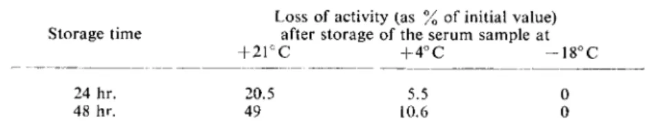
Stability of the Enzyme in the Serum Sample
The enzyme is relatively thermolabile. The decrease in activity
15
> at different temperatures is given in Table 2.
Table 2. Stability of S D H in serum at different temperatures. The initial value is the activity of freshly collected s e r u m
1 0
) .
Loss of activity (as % of initial value) Storage time after storage of the serum sample at
+ 2 1 ° C + 4 ° C - 1 8° C
24 hr. 20.5 5.5 0 48 hr. 49 10.6 0
Sources of Error
S D H activity is inhibited by ethylene-diamine-tetra-acetate and mercury ions; mercaptans, sodium borate, mono-iodoacetate and K C N reduce the activity of the rat liver enzyme; heparin does not substantially affect the a c t i v i t y
1 5
.
1 9
) .
Details for Measurements in Tissues
For the determination of S D H activity in t i s s u e
3
.
5 -
7
a
^
a
, i o , B , 15-17) the sample is homogenized and centrifuged at high speed in the cold. T h e clear supernatant can be used directly for the assay, but the optimum conditions used for human serum measurements are not always valid for tissue preparations;
other conditions apply in the case of purified S D H preparations
1
.
7 a
.
9 a
. 20, 21). With sorbitol as substrate, favourable conditions for measurements in rat liver extract are obtained by use of tris buffer (pH 9 )
1 0
.
1 8
> and 0.15 M sorbitol. Proportionality between activity and amounts of enzyme added, as well as a linear increase in optical density with time are obtained. In the opposite direction the reaction (with 0.4 M fructose) proceeds about 8 times faster in triethanolamine buffer (pH 7.4) and about 5 times faster in tris buffer (pH 7.4).
It is to be noted that the estimation of liver S D H usually includes cytoplasmic S D H and the mitochondrial xylitol dehydrogenase. Both enzymes are practically homospecific, but are not i d e n t i c a l
9 a
) .
1
5
) E. Schmidt, F. W. Schmidt and E. Wildhirt, Klin. Wschr. 37, 1221 [1959].
!6) H. Kalk, E. Schmidt, F. W. Schmidt and E. Wildhirt, Klin. Wschr. 38, 421 [I960].
17) E. Schmidt, F. W. Schmidt and E. Wildhirt, Klin. Wschr. 37, 1229 [1959].
18
) F. H. Bruns, personal communication.
1
9
) H. Schon and H. Wilst, Klin. Wschr. 38, 497 [I960].
20) s. K. Wolfson and H. G. Williams-Ashman, Proc. Soc. exp. Biol. Med. 99, 761 [1958].
2D J. B. Wolff in S. P. Colowick and TV. O. Kaplan: Methods in Enzymology. Academic Press, N e w York 1955, Vol. I, p. 348.