VIL 2. MULTIPLE FUNCTIONS OF SULFUR IN MITOSIS*
Herbert Stern
Chemistry Division, Science Service, Canada Department of Agriculture, Ottawa, Ontario, Canada
I. Introduction 391 II. The Intracellular Distribution of Thiol Compounds 392
1. Histochemical Studies 393 2. Cell Fractionation 393 3. Conclusions 394 III. Soluble Sulfhydryls in Relation to the Mitotic Cycle 394
1. Glutathione Accumulation and the Onset of Division 395
2. Functions of Glutathione in Cell Division 397
3. Conclusions 400 IV. Protein Thiols in Relation to Growth and Division 401
1. Methods 402 2. Thiol Changes During Microscope Development 402
3. The Seat of Protein Thiol Variation 404
V. General Conclusions 405
I. Introduction
In the history of cell biology there have been a number of theories designed to explain the phenomenon of cell division. Very few of these have survived. It is impressive, therefore, to consider that for over twenty- five years the one theory of cell division to retain a prominent place in the thinking of cell physiologists has been the sulfhydryl theory of mitosis as set forth by Rapkine (1). In a field so complex and untrodden as the physiology of cell division, such persistence of an idea is a major tribute to the creative influence of its originator.
Rapkine neither discovered the importance of thiols in metabolism nor first recognized their association with cell division. But he did what his contemporaries did not do: he proposed a simple and exciting scheme to explain the phenomenon. To do this, he combined Hopkin's equation for the reversible exchange of hydrogen between protein and glutathione with Warburg's evidence and argument for the glycolytic nature of mitotic metabolism (2). The scheme runs as follows: Events leading to cell divi- sion have their origin in a partial denaturation of cellular proteins. This
* Contribution No. 401, Chemistry Division, Science Service.
391
392 HERBERT STERN
denaturation exposes otherwise protected thiol groups which then reduce the oxidized glutathione present in the cell. The reduced glutathione acti- vates glycolysis and thus initiates the metabolic events leading to cell division.
The purpose of this discussion is to present evidence bearing on the validity of Rapkine's theory. Is it correct in its particulars? And if not in its particulars, does its presumption that thiol groups act as a motive force in cell division have general application? The importance of thiols and their disulfide counterparts in mitotic phenomena is not being ques- tioned. This has long been established in a general way. Moreover, both Mazia (3) and Nickerson (4) have recently provided important instances of specific thiol functions in cell division.
Before considering the evidence relating to thiols as a motive force in cell division, I want to clarify my general point of view. I do not believe in a single causal mitotic substance even though the existence of one has been assumed by many investigators (5). However common the chemical mechanisms underlying cell division may be, they operate under a divers- ity of conditions. Different species of cells and different developmental forms of the same species have correspondingly different internal me- tabolic environments. Given the complexity of metabolic pathways operat- ing within a cell, it is easy to see how any one of a number of reactions could limit a whole set of metabolic developments. Anything removing this limitation could be properly regarded as a causal agent of the phe- nomenon in question. Indeed, if one surveys the biological kingdom, it be- comes apparent that Nature has lavishly used this feature of susceptibility in cell metabolic organization to provide a variety of mechanisms by which multicellular organisms control the proliferation of their constituent cells.
For the above reason I do not interpret the sulfhydryl theory of mitosis to mean that glutathione acts as a causal agent of mitosis. It is worth re- membering, nevertheless, that in the early days of the theory, belief in the specific power of the thiol group was exceedingly strong. Hammett called glutathione the "mitotic hormone" (β), and Rapkine thus quoted Ham- mett without any note of criticism (1). However, causality aside, if the theory is valid it should describe a universal pattern of biochemical events which are indispensable to the performance of cell division.
II. The Intracellular Distribution of Thiol Compounds
It is apparent that the stimulus to cell division, whatever its nature, must ultimately act within the nucleus. If chromosomes and nuclei were particularly rich in thiols it would strengthen the view that these com- pounds have a singular and critical importance in the initiation of divi-
M U L T I P L E F U N C T I O N S OF S U L F U R I N M I T O S I S 393 sion. For many years the nitroprusside reaction was used histochemically to demonstrate the presence and distribution of —SH protein in proliferat- ing cells (3, 7). The unreliability of the chemical technique led, however, to few lasting conclusions. Recently, newer techniques in histochemistry and cell fractionation have been applied. The results are still of a pre- liminary nature, but superficially at least, histochemical and cell frac- tionation methods yield contradictory answers.
1. H I S T O C H E M I C A L S T U D I E S
Of the two approaches to thiol distribution, the histochemical one has yielded the most spectacular results. In France, Zagury (8) and Idelman (9) found lipoprotein thiols to be concentrated in the vicinity of inter- phase and mitotic chromosomes. The thiol groups were found to be par- ticularly evident in the nucleolar region of nondividing cells. In metaphase cells, thiol reagents stained chromosomes deeply against a weakly staining cytoplasmic background. Similar results were obtained in Japan by Shimamura and colleagues (10). Among the tissues studied by the Japa- nese workers were pollen mother-cells of the lily. In these, nuclei and chromosomes stained deeply with Bennett's sulfhydryl reagent, the sulfur in the chromosomes being largely in the disulfide linkage until the latter phases of the meiotic cycle.
2. C E L L F R A C T I O N A T I O N
Studies by this technique have revealed no selective concentration of thiols in nuclear structures. Glutathione has been found in all mammalian nuclei isolated in nonaqueous media, but at concentrations similar to those present in the cytoplasm (11). We have recently isolated nuclei from em-
T A B L E I
DISTRIBUTION OF PROTEIN THIOLS IN CELLS OF THE P E A EMBRYO0
Minoles —SH/mg. protein Ν
Cell fraction Untreated Treated
Homogenate 0.27 0.36
Homogenate less nuclei 0.29 0.32
Nuclear fraction 0.26 0.26
Nuclei 0.24 0.22
Nuclei less nucleoli 0.16 0.22
Nucleoli 0.19 0.22
* Data taken from unpublished work of Stern and co-workers (12). Measure- ments were made amperometrically according to the procedure of Benesch et al. (28).
"Treated" column indicates reduction of proteins with thioglycolate prior to titration.
394 H E R B E R T S T E R N
bryos of dried peas and prepared nucleoli from these nuclei (12). Differ- ent fractions of the embryo were analyzed for protein thiols before and after reduction with thioglycolate. The results reported in Table I are not spectacular. Reduced or readily reducible thiols are evenly distributed throughout the embryonic cells. Nucleoli are no exception, although the French workers found them to be strongly sulfhydryl positive.
3. C O N C L U S I O N S
It is not difficult to reconcile apparent contradictions between histo- chemical and cell fractionation data: what is difficult is to determine their respective significances. The spatial sensitivity of the histochemical method is beyond the reach of gross in vitro separations. Surface localizations would be missed by the in vitro method of analysis and this may have occurred in the distribution data reported for the pea embryo. On the other hand, it is difficult to attach a quantitative meaning to most histo- chemical staining. Not even a semiquantitative histochemical method has yet been applied to the localization of thiols in nuclear structures.
Assuming the histochemical results to be qualitatively correct, and the chemical analyses to be quantitatively so, the sole conclusion which can be drawn is that thiols are important elements in nuclear and chromosome organization, and that they are probably equally important in cytoplasmic functions. At present, data on intracellular distribution of thiols cannot throw much light on the validity of the sulfhydryl theory of mitosis, even though histochemical results provide excellent pointers on mitotic and cell division mechanisms.
III. Soluble Sulfhydryls in Relation to the Mitotic Cycle
The evidence to be presented in this and the following sections has been obtained from studies of developing anthers in two closely related plants—lily and trillium.* Reasons for choosing this material have been previously stated (18). Our principal interest is in the microsporocytes and microspores of the anther. They are suspended in a thick and some- times pasty fluid, the antheral sap, and probably constitute no more than 2 5 % of the dry matter of the anther. A layer of cells called the tapetum surrounds the sporogenous tissue in its early development; beyond the tapetal layer are sturdier cells of different types which comprise the wall of the anther. Although patterns of chemical change obtained by analysis of whole anthers usually reflect cyclic variations in the synchronously
*The "Croft" variety of Lilium longiflorum and the pink trillium, Trillium erectum were used throughout. Both plants are readily obtainable from commercial sources.
M U L T I P L E F U N C T I O N S O F S U L F U R I N M I T O S I S 395 developing sporogenous tissue, they are nevertheless damped patterns.
For this reason we have sought in many instances to analyze the sporo- genous tissue itself. The approach, however, is not helpful in the case of soluble components such as glutathione, which move readily between the antheral sap and the cell interior. Trillium microspores and microsporo- cytes have been shown to be unusually permeable to a variety of solutes
(14)7 and it is probably best to assume that these sporogenous cells are highly dependent upon and much influenced by the organic environment in which they bathe.
1. G L U T A T H I O N E A C C U M U L A T I O N A N D T H E O N S E T OF D I V I S I O N
The importance of the antheral sap is clearly indicated in studies of soluble sulfhydryl compounds. Microspores isolated in a sucrose medium contain virtually no thiols, yet at least half of the soluble thiol content of the anther is found in the medium in which the spores are suspended. It is unlikely that the wall tissue is the source of these thiols, as only a small fraction of the wall cells is destroyed in isolating the microspores and relatively little protein is found in the thiol-rich medium. We assume, therefore, that the varying concentration of soluble thiols in the anther reflects the composition of the antheral sap, an assumption which will be further justified in later experiments.
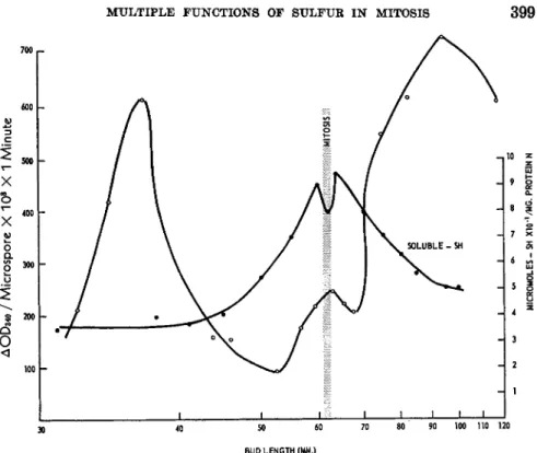
There is a striking parallel between the pattern of soluble thiol con- centration in the developing lily anther and the occurrence of division in the sporogenous tissue (15). In a superficial way at least, the near-trebling of concentration from interphase to pre-mitosis appears to be part of the pattern of chemical changes associated with and indispensable to nuclear division (Fig. 1). To broaden our results we turned to trillium, a closely related plant, but one which provided a contrasting set of physiological conditions. In trillium, meiotic and mitotic divisions of the immature pollen occur in late fall and early winter while the rest of the plant is dormant. We found the difference in physiological state of lily and trillium at the time of pollen development to be matched by a difference in their respective patterns of soluble thiol concentration. In trillium anthers, solu- ble thiols increase but once before meiosis during the autumn months.
With the onset of meiosis a variable but uninterrupted decline begins which extends through microspore mitosis (15). It therefore appears that soluble thiols need not increase before each division. The common re- quirement is probably the presence of an adequate concentration of solu- ble thiols to support divisional processes; in the case of trillium, sufficient glutathione must have been formed before the beginning of winter and so obviated the need for a rise prior to microspore mitosis. This speculation is supported by the fact that on a protein-nitrogen basis, the peak thiol
396 HERBERT STERN
concentration in trillium is four times that in lily. Thus our results are consistent with the substance of Rapkine's view on glutathione accumu- lation, but they do not support his scheme of a glutathione cycle.
Two other particulars of the theory, the identity and source of soluble thiols, have also been tested. Rapkine did not identify glutathione in his preparations; he held to the prevailing belief that the soluble thiols of a cell were principally, if not entirely, glutathione. That his assumption was correct may be seen from the data collected in Table II. Glutathione is the major component of the soluble thiol fraction of the anther; it increases more sharply than the other thiols as the microspores approach mitosis.
On the other hand, there is no evidence to support Rapkine's idea that
1.1 p
1.0 -
FIG. 1. Concentrations of soluble thiols in developing lily anthers. Copied from Stern (10).
glutathione accumulates from the reduction of its oxidized form by de- natured proteins. We have measured the changes in protein —SH of the anther, and these are of far too small an order of magnitude to account for the increases in soluble thiols (13). But, more important, at no stage in the development of the lily anther have we found a significant pool of oxidized glutathione. The amount of soluble disulfides alters little during anther development (Table II) and it is even probable that much of the disulfide found arises from oxidation in the course of extraction. In trillium anthers, oxidized glutathione does accumulate towards the end of meiosis, but this appears to be a consequence of the process, not a prelude to it (15). Un- fortunately we have insufficient data on trillium to know the full course of glutathione oxidation.
M U L T U R E F U N C T I O N S OF S U L F U R I N M I T O S I S 397 T A B L E I I
SOLUBLE THIOLS IN DEVELOPMENT OF LILY ANTHERS
Atmoles — S H / m g . protein Ν
Bud length Oxidized Total Total Per cent
(mm.)" thiols thiols glutathione glutathione
40-45 0.090 0.321 0.227 70
46-51 0.113 0.515 0.371 72
52-56 0.110 0.670 0.544 81
57-M 0.041 0.770 0.641 83
Mitotic interval (M) 0.097 0.778 0.619 80
M-70 0.063 0.854 0.773 91
74-85 0.068 0.630 0.476 75
85-100 0.072 0.552 0.333 60
° Bud length is used as an index of development. The "mitotic interval" includes anthers with microspores before, in, or just beyond mitosis. The methods of measure- ment have already been described (15).
Certain conclusions may be drawn from these results. First, the high concentration of glutathione surrounding the nuclear divisions of imma- ture pollen supports the long held belief that glutathione plays a neces- sary role in cell division. The old views of Voegtlin and Chalkley (16) that glutathione is associated with the nuclear phase of division are in line with the behaviour of the microspores which undergo a nuclear, but not a cytoplasmic division. Second, glutathione must be present above a certain level for division to occur but its formation need not precede each cycle of mitosis. Third, glutathione increases as a result of de novo syn- thesis, and not by a reduction of its oxidized counterpart. The last two conclusions contradict the principles of the sulfhydryl theory of mitosis.
2. F U N C T I O N S OF G L U T A T H I O N E I N C E L L D I V I S I O N
Thiols in general, and glutathione in particular, may play one or both of two roles in the phenomena accompanying cell division. They may par- ticipate in the mechanical and structural changes or they may function in the supporting metabolic pathways. The importance of thiols in the spin- dle apparatus was first considered extensively by Brächet (7). Recently, Mazia (3) has investigated the relationship between glutathione and pro- tein thiol bonding. To what extent glutathione is also related to the histo- chemical changes in chromosome thiols as described by Shimamura et al.
(10) remains to be investigated.
According to the sulfhydryl theory of mitosis, the critical role of gluta- thione is in its activation of glycolysis which Warburg (2) still regards as the key pathway to mitosis. The proliferation of tumor cells aside, it is
398 HERBERT STERN
clear from a variety of studies that the whole of the division cycle is not characteristically anaerobic. In both lily and trillium only part of the mitotic phase of division is associated with anaerobiosis (17, 18). The interval of chromosome reproduction is marked by a high rate of oxygen consumption, and so too is the interval immediately following telophase.
Since the high levels of glutathione in the microspores cover both of these respiratory peaks, there can be no singular relationship between gluta- thione concentration and fermentation as Rapkine conceived it.
A more restricted, though equally deep-seated application of the sulf- hydryl theory is nevertheless possible. Chromosome separation requires energy and since respiration is minimal during the interval of separation, it might be supposed that glycolysis then acts as the energy source. Some investigators have proposed that pre-mitotic respiration produces the nec- essary store of energy for mitosis (19, 20), but there is no evidence as to whether a particular phase of mitosis utilizes the store, if such a store is actually formed. We have analyzed developing lily anthers for their acid- labile phosphate content in the hope that if a phosphate energy store ex- isted it would show up in the same way as the soluble components previ- ously studied. The results however yielded a straight line plot with no evident inflection accompanying the mitotic interval. If a store does exist, it must therefore be different from the one sought or of an order of mag- nitude far smaller than the accumulations noted for glutathione and ascorbic acid.
We also sought a more direct demonstration of the relationship between mitosis and glycolysis. We followed the production of phosphoglyceric acid from hexose diphosphate in isolated microspores (21) (Fig. 2 ) . For tech- nical reasons only part of the glycolytic cycle was measured. Nevertheless, it is the —SH moiety of the dehydrogenase measured which has been con- sidered to be the site of glutathione action with respect to affecting the course of glycolysis (7). The results of these studies are contained in Fig.
3. The most striking feature of the curves for glutathione concentration and dehydrogenase activity is their nearly reciprocal behaviour. On either side of the mitotic cycle, glutathione concentration falls as dehydrogenase activity rises. If crystalline aldolase is not added to the test medium, the activities of the microspores are lower but the shape of the curve is the same except for the absence of a small temporary rise during the mitotic interval. Thus, in the sequence of steps involved in the primary phase of glycolysis, the activity of glycolysis runs counter to the concentration of glutathione. Furthermore, for the interval that oxygen consumption of the isolated microscopes has been measured (18), respiration has been found to follow a course nearly opposite to that of dehydrogenase activity. Apart from the brief mitotic interval during which oxygen consumption falls,
M U L T I P L E F U N C T I O N S O F S U L F U R I N M I T O S I S 399
I ι ι ι 1 1 1—ι—ι—I
30 40 50 60 70 80 90 100 110 120 BUD LENGTH (MM.)
FIG. 2 . Changes in rates of hexose diphosphate breakdown by isolated lily micro- spores. Cells were obtained in the same way as for protein-thiol analysis, and ho- mogenized prior to assay. Measurements were made in 1 ml. cuvettes at temperatures ranging from 2 2 . 5 - 2 4 ° . Activities are expressed as the increase in optical density per minute at 3 4 0 τημ for the indicated number of microspores in a 1 ml. volume and with light path of 1 cm. The incubation medium had the following composition (in Atmoles): hexose diphosphate, 1 0 ; D P N , 0 . 1 ; tris buffer (pH 8.6), 100; sodium arse- nate, 2 0 ; sucrose, 2 5 ; NaCN, 0.5; final volume = 1 ml. 5 μ\. of a solution of crystalline aldolase was also added in each run. The curve of glutathione concentration was ob- tained by methods described earlier (15).
high respiration is matched by high glutathione concentration. The oppo- site should have occurred if the postulated glutathione-glycolysis relation- ship were valid.
The section of the dehydrogenase curve in the region of mitosis indi- cates, nevertheless, that a comparatively small rise in glycolysis may coincide with the drop in oxygen consumption. Here again, the change cannot be related directly to glutathione concentration since the latter also drops slightly during that same interval. But the coincidence of high dehydrogenase activity with low respiration does point to glycolysis as a probable source of energy for at least part of the mitotic interval. To be sure, capacity and performance are not identical properties of a cell. We
HERBERT STERN
30 40 50 60 70 80 90 100 110 120 BUD LENGTH (MM.)
FIG. 3 . Changes in protein thiols of isolated lily microspores. Samples of micro- spore protein (generally about 0.2 ml.) were added to 1 0 ml. of the titration medium described by Benesch et al. (23). The medium also contained 0.25% sodium lauryl sulfate. To prevent precipitation of the detergent in course of titration 1 ml. of 25%, ethanol was added making the total volume 1 1 ml. The protein was titrated with 0.0005 Ν AgNOa and the current followed with a Rubicon galvanometer.
do not yet know whether glycolysis in the living microspore runs a course parallel to that of its enzymatic capacities. But, whatever the relation- ship between enzymatic potential and metabolic performance, it is most unlikely that the striking differences noted in the one are without bear- ing on the other.
3. C O N C L U S I O N S
It is apparent from the results reported above that the glycolytic ca- pacity of a cell can and does change sharply, but that glutathione level is not the major cause of change, nor mitosis the major result. The belief of Rapkine and of Warburg that glycolysis is intimately associated with cell division may indeed be correct insofar as it relates to the mechanical phases of mitosis, but the association cannot have the grand significance given to it by these investigators.
The above conclusions return us to the original question respecting the function of glutathione in mitosis. The striking rise in its concentration as lily microspores approach division is clearly not designed to stimulate
M U L T I P L E F U N C T I O N S O F S U L F U R I N M I T O S I S 401
Control Maleimide
Days Thiols Thiols
after Weight (/xmoles Weight (μΠ1θ1β8
excision (mg.) per anther) Cytology (mg.) per anther) Cytology
0 132 0.145 — 138 0.163 —
1 — — — 132 0.114 —
2 133 0.155 Prophase 142 0.123 Interphase 3 131 0.180 J Binucleate 140 0.163 Interphase 4 139 0.130 Binucleate 138 0.173 Prophase
binucleate
6 — — — 156 0.146 90% binucleate
° The data are taken from the unpublished work of Stern and Katznelson (22).
Buds of 49-50 mm. length were used as source of anthers for these experiments. The anthers were grown on a 1% agar medium with or without 0.002 M A7-ethylmaleimide.
irreversible bond with the soluble thiols, this experiment further illustrates the capacity of detached anthers to form glutathione. Mitosis does not begin until the glutathione level of the treated anthers rises to the appro- priate value (22). Glutathione concentration appears to have a critical influence on pre-mitotic metabolism not directly related to formation of the mitotic apparatus.
IV. Protein Thiols in Relation to Growth and Division
The role of protein thiols in formation of the mitotic apparatus seems to be well established ; so too does their role in cytoplasmic division. One glycolysis. It could be supposed that glutathione functions solely in the manner indicated by Mazia, namely, as a component of the mitotic ap- paratus. But the evidence, thin though it is, points also in other directions.
A metabolic role rooted in the reducing power of the thiol radical is sug- gested by the fact that ascorbic acid concentration follows a course simi- lar to that of glutathione (11). The peak concentrations of these com- pounds cover two intervals of D N A synthesis, one preceding and one following the mitotic cycle. It is conceivable that a relationship exists between these compounds and the reduction of ribosides to the deoxy- form. We have only begun to look into this possibility. We have found that an induced lowering of thiol concentration during the pre-mitotic period delays the onset of mitosis. The experiments indicating this are summarized in Table III. Since iV-ethylmaleimide probably forms an
T A B L E III
T H E EFFECT OF 2V-ETHYLMALEIMIDE ON MICROSPORE MITOSIS IN CULTURED LILY ANTHERS"
4 0 2 H E R B E R T S T E R N
proposition of the sulfhydryl theory of mitosis remains to be examined—
the denaturation of protein as an antecedent of cell division processes.
We have already discounted the possibility of such denaturation giving rise to soluble thiols, but this need not eliminate the imaginative concep- tion of a reversible unfolding of proteins as a basis for initiating the divi- sion cycle.
1. M E T H O D S
The studies to be described were all performed on microspores isolated from lily anthers at appropriate stages of development. The anthers were slit longitudinally with a sharp blade, cut transversely three or four times, and stirred gently with a rod in a 0 . 5 M solution of sucrose. The fluid por- tion was passed through two layers of cheesecloth and the residue treated once more with fresh sucrose solution. The microspores settled on standing or were sedimented by a brief centrifugation. A few tests were made on microspores isolated in salt-sucrose media, or in higher sucrose concentra- tions, but no appreciable differences were found in thiol titers.
Microspores thus isolated were either precipitated directly with sulfo- salicyclic acid or treated overnight with a mixture of 0 . 1 2 M sodium thio- glycolate and 0 . 2 5 M sucrose at a pH of approximately 6 . 5 to 7. All operations were carried out at 0 ° . Thioglycolate-treated microspores were washed four times with sulfosalicyclic acid before being titrated for thiol content.
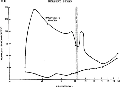
2 . T H I O L C H A N G E S D U R I N G M I C R O S P O R E D E V E L O P M E N T
It may be seen from the curves plotted in Fig. 3 that protein — S H undergoes little change in the course of microspore development. To this extent microspores and whole anthers behave in much the same way.
Since the titrations were performed in the presence of a strong denaturing agent we have no way of telling whether there were any changes in the proportion of easily titratable —SH. It is apparent, however, that the more dramatic changes occur in the protein fraction which yields —SH groups after reduction with thioglycolate. T o set the time of these changes in their proper perspective, it should be noted that the interphase between meiosis of the microsporocyte and mitosis of the microspore is about four weeks. The abrupt rise in disulfide protein occurs six to eight days before mitosis and the return to the original level takes place three days after mitosis. The nature of the change apart, it is clear that the major varia- tions in protein thiols are not immediately bound to cell division. Indeed, if a correlation exists, it must be with the over-all growth of the micro- spore which is rapid during the interval being considered. Protein thiols
M U L T I P L E F U N C T I O N S O F S U L F U R I N M I T O S I S 403
have a relationship to cellular development broader than that supposed in the sulfhydryl theory of mitosis.
The disulfide links reduced by thioglycolate probably existed as such in the intact microspores. We infer this from results obtained employing ethyl maleimide. Microspores obtained from buds about 38 mm. in length were isolated in the presence of 0.01 M ΛΓ-ethylmaleimide. About one- third to one-half of the original — S H groups remained uncombined, indi- cating at least a partial penetration of the reagent into the protein fabric.
There was no decrease in the disulfide titer. This eliminated the probabil- ity that exposed — S H groups were oxidized in the course of microspore isolation, and since the conditions of denaturation were the same for treated and untreated lots of protein, the increase in —SH titer is very likely due to the reduction of disulfide links originally present in the microspore protein.
It is possible that a change akin to denaturation underlies the pattern of protein disulfide concentration seen in Fig. 3. Two points bear on this conclusion. First the values are plotted on a per cell basis. The abrupt initial rise in — S — S — content must therefore be due either to a de novo synthesis of disulfide protein or to a change in physical state of the protein exposing more disulfide links to thioglycolate action. There is no com- pelling reason for choosing either alternative, although present evidence would appear to favor the second of these. One such piece of evidence is the observation of Nasatir (18) that protein begins to increase in the microspores about a day or two after the appearance of the high disulfide titer. It is clear that during this same interval the amount of reducible protein disulfide does not increase in the microspores. Premitotic protein synthesis cannot therefore include those proteins exhibiting the disulfide behavior noted in Fig. 3. A second point bearing on the possibility of some physical change occurring in the cellular protein is the fact that under the conditions of the experiment thioglycolate does not reduce all the disulfide linkages. Preparations treated with 0.1% deoxycholate give higher — S H titers after reduction with thioglycolate than those maintained in a sucrose-thioglycolate mixture. Thus, the — S — S — values plotted reflect to some extent the steric properties of the protein.
Taken together, the results show a striking relationship between pro- tein thiols and cellular development. The relationship may consist, in part at least, of a physical change in some of the cellular proteins. To the ex- tent that this is correct, Rapkine's postulate of a reversible denaturation of protein may prove to be valid. But the particular significance which Rapkine attached to such denaturation appears to lack substance. Mitosis is not the immediate and primary consequence of the change; if anything,
404 H E R B E R T S T E R N
it is growth of the cell which is tied to the thiol shift observed. There is a smaller change represented by a dip in disulfide concentration, associ- ated with mitosis, but this change is in line with Mazia's results on the formation of the mitotic apparatus (3). If the previous supposition, that disulfide titers vary with the physical state of the microspore protein, is correct, it follows that the dip reflects the tighter organization of disulfide linkages in the mitotic apparatus.
3. T H E S E A T OF P R O T E I N T H I O L V A R I A T I O N
It is of obvious interest to know whether the observed pattern of pro- tein — S — S — changes reflects the behavior of the cellular proteins in general or whether it originates in a specific protein fraction. Our initial studies, exploratory though they are, throw appreciable light on this ques- tion.
Different portions of the lily anther show markedly different responses to thioglycolate treatment (Table IV). The only fraction which yields a
T A B L E I V
RATIOS OF —S—S—/—SH PROTEIN IN FRACTIONS OF LILY ANTHER"
Fraction —SH Protein —S—S— Protein Ratio —S—S—/—SH
Whole anther 0.17 0.05 0.3
Sucrose medium 0.02 0.01 0.5
Unbroken residue 0.09 0.06 0.7
Microspores 0.007 0.033 5.0
a Values are expressed as μπιοΐββ —SH equivalents per anther. Determinations were made as described in text and under Fig. 3. The "sucrose medium" contained the components of the antheral sap plus whatever leaked out of the cells. The "unbroken residue" contained all the wall tissues and those microspores which were not freed in the course of separation. Buds of length 64.0 mm. were used in this experiment.
high — S H titer after reduction with thioglycolate is that containing the microspores. One may infer from this that the disulfide proteins under study are specifically associated with cell proliferation. This conclusion, attractive though it is, to be taken seriously only if a similar situation were shown to prevail in other cell types.
We have attempted to fractionate the proteins of the microspore by differential centrifugation. Although we have been unable to obtain micro- scopically homogeneous preparations, one property of the disulfide pro- teins has been made clear: they are consistently sedimented with the subcellular particles. Treatment with desoxycholate, lauryl sulfate, or thioglycolate does not bring them into solution. It appears, therefore, that the sulfur-containing proteins which vary most in relation to growth
M U L T I P L E F U N C T I O N S OF S U L F U R I N M I T O S I S 405
and division are those incorporated into the structural elements of the cell.
V. General Conclusions
The principal objective of this presentation was to examine the valid- ity of Rapkine's sulfhydryl theory of cell division. The main conclusion reached is that the theory is not generally valid. A number of mechanisms proposed in the theory are certainly operative in cell division processes;
but on the critical issue, the denaturation-glutathione-glycolysis sequence, the theory lacks basis in experimental fact. The importance of glutathione in the mitotic cycle is clearly confirmed, but not in the all-powerful role once assigned to it as the "mitotic hormone" (6). Glutathione appears rather as a store of thiol radicals and perhaps of reducing power. During cell division—and possibly during other phases of growth—there is a strong demand on the glutathione store. This demand is probably created by a number of metabolic channels. It do^s not seem reasonable to assume that the concentration of glutathione per se is sufficient to transfer thiol radicals or to effect a reduction of cellular components. More probably, the utilization of glutathione is governed by specific enzymatic and hor- monal mechanisms, certain of which are directed towards fulfillment of the mitotic cycle. It may be disturbing to the biochemist (and perhaps gratifying to the physiologist) to know that in attempting to hang the whole of a complex biological phenomenon on the hook of a single chemical substance, scheme and substance become ensnared in a network of meta- bolic circuits. Such failure is a testimony to the astuteness of evolution in adapting the smallest number of chemical substances to a maximum num- ber of physiological purposes.
R E F E R E N C E S
1. L. Rapkine, Ann. physiol. physicochim. biol. 7, 382 (1931).
2. 0 . Warburg, Science 124, 270 (1956).
8. D. Mazia, in this volume, see Section VII. 1.
4. W. J. Nickerson and G. Falcone, in this volume, see Section VII. 3.
5. H. Stern, Ann. Rev. Plant Physiol. 7, 91 (1956).
6. F. S. Hammett, Protoplasma 7, 535 (1929).
7. J. Brächet, "Chemical Embryology." Interscience, New York, 1950.
8. D . Zagury, Compt. rend. 244, 1825 (1957).
9. S. Idelman, Compt. rend. 244, 1827 (1957).
10. T. Shimamura, T. Ota, and T. Hishida, Symposia Soc. Cellular Chem. (Tokyo) 6, 21 (1957).
11. H. Stern and S. Timonen, J. Gen. Physiol. 38, 41 (1954).
12. H. Stern, F. B. Johnston, and G. Setterfield, submitted for publication.
13. H. Stern, Science 124, 1292 (1956).
406 H E R B E R T S T E R N
14· H . Stern, Trans. Roy. Soc. Can. 40, 1 4 1 ( 1 9 4 6 ) . 15. H . Stern, J. Biophys. Biochem. Cytol. 4, 1 5 7 ( 1 9 5 8 ) .
16. C. Voegtlin and H . W. Chalkley, U. S. Public Health Repts. 45, 3041 ( 1 9 3 0 ) . 17. H . Stern and P. L. Kirk, J. Gen. Physiol. 31, 2 4 3 ( 1 9 4 8 ) .
18. M. Nasatir, Ph. D . Thesis, University of Pennsylvania, Philadelphia, Pennsylvania,
1958.
19. W. S . Bullough, Biol. Revs. Cambridge Phil. Soc. 27, 133 ( 1 9 5 2 ) . 20. M. M. Swann, Quart. J. Microscop. Sei. 94, 3 6 9 ( 1 9 5 3 ) .
21. M. Nasatir and H. Stern, submitted for publication.
22. H . Stern and H . Katznelson, in preparation.
23. R . E. Benesch, H . A. Lardy, and R . Benesch, J. Biol. Chem. 216, 6 6 3 ( 1 9 5 5 ) .
Discussion
RACKER: I was wondering why you necessarily relate the activity of a single en- zyme to the over-all glycolytic activity? Is there any evidence that a single enzyme is limiting?
STERN : No, there is no such evidence. I tried to make myself clear on this point.
Technical factors prevented our studying total glycolysis. It is of interest, neverthe- less, that the sulfhydryl theory implicated glutathione as the stimulant of dehydro- genase. That relationship clearly does not hold. I tried, moreover, to put together the fact that at the time that dehydrogenase and aldolase are high, respiration is low, and vice versa. Thus, the evidence points to but does not prove an inverse relationship between glutathione concentration and glycolytic activity.
I can only add that we are trying to find out how the process as a whole is behav- ing.
BENESCH: There are a number of technical points I would like to ask. How did you remove the thioglycolic acid?
STERN : We precipitated the proteins with sulfosalicyclic acid and washed the pre- cipitate four times. We measured the SH content, after the third and fourth wash, and got the same value for both.
BENESCH: HOW did you determine the oxidized glutathione?
STERN : We reduced the extract in a mercury cell.
BENESCH: Electrolytic reduction?
STERN : Yes. We always found some, as I pointed out.
BENESCH: But not enough.
STERN : Hardly of the order required in supposing a formation of GSH from the oxidized form. The pattern, if anything points in the other direction. What we did find, but didn't emphasize, is consistent with Dan Mazia's scheme of GSH-protein interaction. At the mid-point of mitosis one generally finds the highest concentration of oxidized thiols. At the beginning when the oxidized form should have been present as a large store, if Rapkine's theory were correct, one finds little, if any. The lowest concentration of oxidized glutathione is found when that of the reduced form is lowest.
BENESCH : I see that on the slide in which you showed reduced and oxidized gluta- thione, values were expressed as a percentage of the total. How did you determine the total?
STERN : Amperometric titration. Your method.
BENESCH : I understand from Dan Mazia that the so-called soluble glutathione at least in sea urchin eggs has recently been shown by Japanese workers to be actually a soluble —SH protein. Would you care to comment on that?
MULTIPLE FUNCTIONS OF SULFUR IN MITOSIS 407 MAZIA : This matter is discussed in a little more detail in the published text of my paper than it was in my talk. I rather hesitate to take advantage of Professor Katsuma Dan's kindness in letting me see the data by Sakai and himself in advance of publica- tion, but perhaps a few remarks are permissible. We are all familiar with Rapkine's famous experiment, which Dr. Stern has discussed. The essence of this was that the trichloroacetic-acid-soluble SH in the dividing sea urchin egg first dropped rapidly during the period up to metaphase or thereabouts and then rose to its original value.
The period of decrease is identifiable with the preparations for mitosis, the period of restoration corresponds to that of growth and chromosome movement in the mitotic apparatus. It was supposed that the TCA-soluble SH was glutathione. As I mention in the text, Dr. Elizabeth Neufeld, in our laboratory, found that the curve could not be duplicated with methods of protein precipitation which should separate glutathione effectively from the proteins. Sakai and Dan in Tokyo (Exptl. Cell Research, in press) made a very detailed study, also using sea urchin eggs, and solved the paradox, I think. They found that the TCA precipitation did not bring down a constituent that was precipitable by other means. We can guess that it is a TCA-soluble protein or large polypeptide. When they used TCA, they obtained a time-concentration curve very much like that of Rapkine. When they removed the "TCA-soluble protein," as we may call it, by other précipitants, they obtained a result very much like that of Dr. Neufeld. In other words, we can say—and this is my conclusion, which Sakai and Dan may not agree with—that this component is cycling according to the pattern described by Rapkine. The mechanism we proposed for the formation of the mitotic apparatus, involving the transformation of intramolecular —S—S— to intermolecular
—S—S— by the mediation of a soluble SH compound is still possible, but that com- pound cannot be glutathione.
BENESCH: What protein precipitant did you use, Dr. Stern?
STERN : Sulfosalicylic acid.
BENESCH: D O you think this would extract this protein?
MAZIA : I have no idea.
STERN : If I may make this remark, Nature has more than one way of doing the same thing. Reducing groups, if necessary, need not be supplied by glutathione. One has to allow for that variability. There is a point I might make about comparing the sea urchin egg with the lily microspore. The sea urchin egg has the whole of its early developmental needs stored within a single cell. These spores have not. They are de- pendent both on the environment within the anther and on the activities of the whole plant. I don't know whether protein would behave as glutathione in the glyoxalase test. It is conceivable. A significant point, it seems to me, is that whether the peptide is glutathione or some other peptide, its pattern of change is at variance with that required by the sulfhydryl theory of mitosis.
RACKER: I don't believe that there is a single enzymatic assay which we could consider to be completely specific for glutathione. Glyoxalase has been known for many years to react with aspartathione and isoglutathione. More recently Th.
Wieland, G. Pfleiderer, and H. H. Lau (Biochem. Ζ. 327, 393, 1956) have shown that a number of SH-peptides and thiol esters, including some with 4 or 5 amino acids, act as substrates for the glyoxalases. This was shown to be the case with purified enzymes, and I would not be surprised if there are additional thiol esterases in crude enzyme preparations. Dr. Stern, did you use purified glyoxalases for your assay?
STERN : No, it was a crude preparation. On a molar basis, would these peptides be as active as glutathione? You may recall that most of the —SH present is accountable by the glyoxalase assay.
408 HERBERT STERN
RACKER: Some of Wieland 's —SH compounds are almost as active as GSH. With others the rate is 20-50% that of GSH.
MAZIA: May I re-emphasize a point that Herbert Stern was making earlier con- cerning differences among cells of various types. Our experiments deal with two extremes. He has been describing the events in cells which are synthesizing various compounds, going through division on a biochemical "pay as you go" basis. The cells of which I was speaking, egg cells, carry on relatively little synthesis; they are draw- ing upon a full biochemical bank account. The pictures of the biochemical changes in cells at these two extremes may be very different, but this does not necessarily mean that the fundamental design of the division process itself is different.