Molecular Human Reproduction Vol.12, No.1 pp. 31–34, 2006
Advance Access publication January 10, 2006 doi:10.1093/molehr/gal001
© The Author 2006. Published by Oxford University Press on behalf of the European Society of Human Reproduction and Embryology. All rights reserved. For
Permissions, please email: journals.permissions@oxfordjournals.org 31
Three mechanisms in the pathogenesis of pre-eclampsia suggested by over-represented transcription factor-binding sites detected with comparative promoter analysis
B.Vásárhelyi
1,5, Á.Cseh
1, I.Kocsis
1,2, A.Treszl
1,2, B.Györffy
3and J.Rigó Jr
41Research Laboratory of Pediatrics and Nephrology, Hungarian Academy of Sciences, 2First Department of Pediatrics,
3Szentágothai János Knowledge Centre and 4First Department of Obstetrics and Gynecology, Semmelweis University, Budapest, Hungary
5To whom correspondence should be addressed at: First Department of Pediatrics, Budapest, Bókay u. 53, H-1083 Hungary.
E-mail: vasbar@gyer1.sote.hu
Microarray studies generating lists of genes with altered expression in placentas from pregnancies complicated with pre-eclampsia (PE) have so far been published in several different studies. Working under the assumption that altered gene expression in PE may be the result of altered expression of regulatory transcription factors (TFs), we looked for over-represented TF-binding sites (TFBSs)—which indicate the involvement of TFs in gene regulatory networks—in lists of genes (n = 143) compiled in these stud- ies. We compared the prevalence of TFBSs in the promoter regions of 68 genes with the background prevalence of TFBSs in pro- moters of the human genome. The prevalence of the E47, sterol regulatory element binding protein (SREBP) and NFKB-p50 TFBSs was higher (P < 0.005) in the promoter sequences of the PE gene lists than in the background model. Each of these TFBSs could be implicated in the development of PE. The E47 protein is an E-protein or basic helix-loop-helix (bHLH) TF. Data support the role of bHLHs in the differentiation of placental tissue. SREBP-1, a lipid-sensing sterol regulatory element-binding protein, is a critical regulator of fatty acid homeostasis in the placenta. The target genes of NFKB-p50 determine inflammatory response, and aberrant cytokine homeostasis is a further sign of PE. These TFs may provide an insight into the pathogenesis of the disease.
Key words: microarray/pre-eclampsia/transcription factor/transcription factor-binding site
Introduction
Pre-eclampsia (PE) is a major cause of maternal mortality, affecting approximately 5% of all pregnancies (Anonymous, 2000). PE is the cause of 150,000 deaths worldwide each year. PE also affects fetal development, causing intrauterine growth retardation. Growth retarda- tion is even more pronounced when the disease starts before 32nd week of pregnancy (Ventura et al., 2001). Most cases of PE present with increased blood pressure, proteinuria and edema, but some cases may progress into a complex disorder that involves the central nerv- ous system, liver and kidneys and results in multiple organ failure (Gillham and Hayman, 2004). The etiology of PE remains unclear; it is aptly termed as ‘disease of theories’. Blood pressure usually nor- malizes and proteinuria subsides after termination of pregnancy, lend- ing support to the assumption that the placenta plays an important role in the pathogenesis and pathophysiology of PE (Redman and Sargent, 2003). Placental dysfunction may be caused by uncoordinated expres- sion of genes in the placenta. We used data from four recent microar- ray studies comparing gene expression in placental tissue of women with PE to gene expression in placental tissue of women without PE (Reimer et al., 2002; Tsoi et al., 2003; Pang and Xing, 2004a;
Soleymanlou et al., 2005). From these data, we generated a list of genes showing altered expression in the placentas of women with PE.
Altered gene expression in PE may be linked to altered expression of regulatory transcription factors (TFs). TFs may up-regulate or down- regulate gene expression. Transcriptional regulatory regions—so called
promoter sequences—located before the start codon of each gene con- tain binding sites (TFBS) for TFs. The TFBS is short–usually 4–10 bases long—and one TF may bind to several TFBSs (Aerts et al., 2003). Therefore, one TF may bind to the promoters of several genes or the promoter sequence of one gene may contain several TFBSs.
TFs with over-represented TFBSs in the promoter region may play a role in the regulation of expression of co-regulated genes. If they do, certain TFBSs should be over-represented in the promoter regions of genes with altered expression.
To test our hypothesis, we looked for over-representation of some TFBSs in the promoter regions of gene sets with altered expression in PE.
Methods
We searched the PubMed archive (http://www.ncbi.nlm.nih.gov/entrez/) for papers presenting lists of genes with altered expression in PE using the key- words PE, placenta, DNA-chip or microarray. We found four articles in which the altered expression of 143 genes in placental tissue was described (Reimer et al., 2002; Tsoi et al., 2003; Pang and Xing, 2004a; Soleymanlou et al., 2005).
For TFBS analysis, we applied the same method as described previously (Mayer et al., 2004). We used the Java program TOUCAN for comparative promoter analysis of the selected genes (Aerts et al., 2003). Proximal promoter sequences (1 kb upstream and 0.05 kb downstream of the transcriptional start sites) were extracted from the genomic databases using the EZ-Retrieve (Zhang et al., 2002). The TOUCAN tool MotifScanner, which searches the
Downloaded from https://academic.oup.com/molehr/article-abstract/12/1/31/1046984 by Hungary EISZ Consortium user
on 31 August 2018
B.Vásárhelyi et al.
32
TRANSFAC database (Wingender et al., 2001), was used to detect TF binding sites (TFBSs) in the sets of sequences. The prior (stringency level) was set to a value of 0.1, and the human promoter set of the Eukaryotic Promoter Database (EPD) was chosen as a third-order background model. We applied TOUCAN’s statistics tool to the data produced by MotifScanner to detect over-representation (showing positive significance values) in the sets of the selected genes. The level of significance was set at P = 0.005.
Results
The promoter sequences were available for 68 genes and we identified 226 different TFBSs. The prevalence of three TFBSs was signifi- cantly higher in the analyzed promoter sequences of PE gene lists than in the background model: E47 protein (TF accession number:
T00207) (prevalence: 0.147 versus 0.0000229, P = 0.0011), sterol regulatory element binding protein (SREBP-1) (TF accession number:
T01556) (prevalence: 0.221 versus 0.0000278, P = 0.0002) and nuc- lear factor-kappa-beta-p50 (NFKB-p50) (TF accession number:
T00593) (prevalence: 0.176 versus 0.0000513, P = 0.0005)
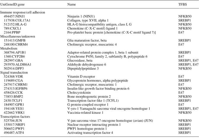
These TFBSs were present on 26 genes. Table I lists the genes with over-represented TFBSs in the analyzed promoter sequences. Most of the 26 genes contain TFBSs for one of the three identified TFBSs while both E47 and SREBP-1 had TFBSs on the promoter sequence of three genes.
Of the 26 genes with over-represented TFBS, five genes were impli- cated in metabolism and five in cell adhesion and immune response.
The proteins encoded by ten genes play a role in intracellular signaling while the proteins encoded by four genes are themselves TFs.
Discussion
In our study, we identified three over-represented TFBSs in the promoter regions of genes associated with PE. The results of our
analysis suggest that these TFBSs could be implicated in the develop- ment of PE.
E47 protein
An association between altered E47 function and PE could be implied by two different mechanisms. E47 protein belongs to E-proteins or basic helix-loop-helix (bHLH) TFs. Although the exact role of E47 in pregnancy has not been investigated, other structurally related E-pro- teins have been found to play a role in placental development (Hemberger and Cross, 2001; Liu et al., 2004). Data obtained in knock- out mice models show that trophoblast differentiation and placental morphogenesis are regulated at multiple checkpoints by bHLH factors and their regulators. In the placentas of women with PE, abnormalities of trophoblast differentiation could be caused by bHLH TFs, includ- ing E47. In normal pregnancy, cytotrophoblast invasion of the uterine spiral arteries is accompanied by the loss of musculo-elastic tissue in these vessels (Wang and Alexander, 2000). An important structural abnormality of the placenta in PE is the incomplete cytotrophoblast invasion of the uterine spiral arteries. As a result, spiral arteries fail to lose their elasticity and instead of becoming low resistance vascular channels as in normal pregnancy, the resistance of arteriolas in PE pregnancies remains high. E47 could contribute to altered musculo- genesis in the spiral arteries. Although the importance of heterodimers of E47 proteins with muscle-specific receptor (MSR) proteins has been tested only in the development of skeletal muscles (Carlsen and Gundersen, 2000), other bHLH/MSR heterodimers have been shown to play a role in smooth muscle differentiation (Dhulipala et al., 2001). Altered E47 protein expression could contribute to inadequate loss of musculo-elastic tissue in the vascular wall in PE.
Table I. List of genes associated with pre-eclampsia having binding sites for the over-represented transcription factor-binding sites (TFBS) in their promoter sequences
UniGeneID:gene Name TFBS
Immune response/cell adhesion
494457:NINJ1 Ninjurin 1 (NINJ1) NFKB50
117938:COL17A1 Collagen, type XVII, alpha 1 SREBP1
512152:HLA-G HLA-G histocompatibility antigen, class I, G NFKB50
789:CXCL1 Chemokine (C-X-C motif) ligand 1 NFKB50
2164:PPBP Pro-platelet basic protein [chemokine (C-X-C motif ligand 7)] E47
Miscellaneous/unknown
151413:GMFB Glia maturation factor, beta SREBP1
248100:CHRM4 Cholinergic receptor, muscarinic 4 E47
Metabolism
368794:AP1B1 Adaptor-related protein complex 1, beta 1 subunit SREBP1
1360:CYP2B6 Cytochrome P450, family 2, subfamily B, polypeptide 6 E47
282997:GBA Glucosidase, beta SREBP1, E47
293970:ALDH6A1 Aldehyde dehydrogenase 6 SREBP1, E47
502914:DPP3 Dipeptidylpeptidase 3 NFKB50
Signal transduction
524368:VDR Vitamin D receptor E47
119689:CGA Glycoprotein hormones, alpha polypeptide SREBP1
247917:CHRM1 Cholinergic receptor, muscarinic 1 E47
274313:IGFBP6 Insulin-like growth factor binding protein 6 NFKB50
458426:CCK Cholecystokinin E47
73853:BMP2 Bone morphogenetic protein 2 NFKB50
2430:TCLF1 Transcription factor-like 1 (TCFL1) SREBP1
184907:GPR1 G protein-coupled receptor 1 E47
194148:YES1 V-yes-1 Yamaguchi sarcoma viral oncogene homologue 1 SREBP1, E47
422662:VRK1 Vaccinia-related kinase 1 NFKB50
Transcription factors
525704:JUN V-jun sarcoma virus 17 oncogene homologue (avian) (JUN) NFKB50
155017:NRIP1 Nuclear receptor interacting protein 1 SREBP1
506652:PWP1 PWP1 homologue protein 1 SREBP1
496487:ATF4 Activating transcription factor 4 SREBP1
Downloaded from https://academic.oup.com/molehr/article-abstract/12/1/31/1046984 by Hungary EISZ Consortium user
on 31 August 2018
Promoter analysis of gene lists in pre-eclampsia
33 SREBP
Epidemiological data show that PE and cardiovascular disorders share several risk factors, including insulin resistance, obesity and diabetes (Wolf et al., 2004). PE also presents an increased maternal risk of car- diovascular disease later in life. SREBP has recently emerged as a possible link between these disorders. SREBPs are essential for the control of intracellular lipid accumulation. Intracellular lipid accumu- lation might be a link between insulin resistance, visceral obesity and increased lipid deposition in non-adipose tissue, including the arterial wall, therefore, SREBPs are being investigated as a possible target for atherosclerosis (Muller-Wieland and Kotzka, 2002). The role of SREBPs in lipid homeostasis of the human placenta has also been suggested. Recent studies have demonstrated that SREBPs regulate the transcription of ACC and FAS, important enzymes for the synthe- sis of fatty acids (Duttaroy, 2004). Insulin is known to act through SREBP-1c by augmenting the nuclear content of SREBP-1c while LCPUFA suppresses the nuclear content. SREBP may also play a role in insulin resistance syndrome. Studies have proposed that, along with other TFs for fatty acid regulation, such as peroxisome-proliferator activating receptor gamma (PPAR-g), alterations in the amount and activity of SREBPs could offer a key to understanding the association of insulin resistance with cardiovascular risk factors at the cellular or gene regulatory level. In PE, several mechanisms may contribute to the possible alteration of SREBP expression. Consumption of large amounts of polyunsaturated fatty acids could increase risk of PE (Clausen et al., 2001). As expression of SREBP has been shown to be regulated by cholesterol and polyunsaturated fatty acids, altered SREBP expression after ingestion of fatty acids could be an explanation for the link between nutrition and PE (Kim et al., 2002; Jump, 2002).
Another possible explanation for the alteration of the hypothesized alteration of SREBP expression is a change in HCG production (Richardson et al., 2005). In PE, HCG levels are abnormally high (Kharfi et al., 2005). Because HCG has been shown to cause a shift between SREBP isoforms as well as increase the expression of SREBP-1c, therefore, the link between HCG and SREBP could be a mechanism leading to altered expression of SREBP-regulated genes.
NFKB-p50
NFKB-p50, the third identified TF, belongs to the NFKB/Rel family, which plays an important role in the intracellular regulation of immune response, inflammation and cell cycle regulation (Liou and Hsia, 2003). The most prevalent activated form of NFKB is the het- erodimer of subunit NFKB-p65, associated with either subunit NFKB-p50 or -p52. p50 homodimers lack the transcription activation domain but still bind to B-consensus sites and therefore function as transcription repressors. A growing number of NFKB/Rel target genes have been identified in the past several years. These include cytokines, chemokines, cytokine/chemokine receptors, adhesion molecules, sur- vival factors, cell cycle regulators and inducible effector enzymes.
Inadequate functioning of the cytokine network has been implicated in the disturbance of trophoblast remodelling in PE pregnancies by induc- ing enhanced apoptosis (Allaire et al., 2000; Kharfi et al., 2003) as well as in the triggering and maintenance of the maternal systemic inflam- matory response. One study has postulated that NFKB-p50, as the conductor of cytokine regulation, plays a role in PE. This hypothesis was supported recently by an immunohistochemical study which showed increased expression of NFKB-p50 in PE (Aban et al., 2004). This finding was confirmed by the results of our TFBS analysis, which also indicate an over-representation of binding sites for NFKB-p50.
Although the results of our analysis provide attractive hypotheses for the pathogenesis of PE, one should take into account the limita- tions of our analysis.
In this study we analyzed promoter sequences of genes with altered expression published in four papers. These papers listed more than 350 genes that are up- or down-regulated in PE; however, only a minority of these genes were listed in the papers (n = 143) and only a fraction of them (n = 68) possessed well-defined promoter sequences in EZ- Retrieve (Zang et al., 2002). The length of promoter sequences used for the analysis was also arbitrary (1000 bases). (We postulated that TFs with TFBSs on these proximal promoter regions would have the greatest effect on gene transcription.) If longer promoter sequences and/or more genes had been involved in the analysis, it may have affected our results.
Furthermore, our computer-assisted analysis does not provide information about the functional impact of the identified TFs on gene activation. Our results are putative and the resulting hypothesis should be tested by experimental methods. One way to investigate the com- plex interaction of TFs and DNA is to use functional assays such as an electrophoretic mobility shift assay (EMSA). (Fried and Crothers, 1984). (EMSAs make use of the observation that protein : DNA com- plexes migrate more slowly than free DNA molecules when subjected to non-denaturing polyacrylamide or agarose gel electrophoresis used to investigate sequence-specific interactions.)
Interestingly, the TFs highlighted in our analysis are different from those identified by Pang and Xing (2004a,b) and Reimer et al. (2002).
A probable explanation for this apparent contradiction is the methods used. These authors drew their conclusions after the comparison of gene expression data while we used a computer-simulated statistical analysis. Our analysis, however, does not exclude the implication of TFs other than E47, SREBP-1 and NFKB-p50 in the pathogenesis of PE. (Indeed, 4 of the genes with TFBSs for these TFs are TFs them- selves or regulators of cell signalling). There may be a mutual interac- tion between E47, SREBP-1 and NFKB-p50 and the products of these genes, but the analysis of these connections is beyond the scope of our present study.
In summary, altered gene expression of the placenta may contribute to the development of PE. Based on the results of our computer-simu- lated analysis of the promoter regions of genes with altered gene expression, we hypothesize that the altered function of three TFs in the placenta may contribute to the disease. These TFs play a role in the regulation of tissue differentiation, lipid homeostasis, inflamma- tion and apoptosis.
Acknowledgements
This study was supported by grants OTKA T046086 and NKFP 1A/002/ 2004.
References
Aban M, Cinel L, Arslan M, Dilek U, Kaplanoglu M, Arpaci R and Dilek S (2004) Expression of nuclear factor-kappa B and placental apoptosis in pregnancies complicated with intrauterine growth restriction and preeclamp- sia: an immunohistochemical study. Tohoku J Exp Med 204,195–202.
Aerts S, Thijs G, Coessens B, Staes M, Moreau Y and De Moor B (2003) Toucan: deciphering the cis-regulatory logic of coregulated genes. Nucleic Acids Res 31,1753–1764.
Allaire AD, Ballenger KA, Wells SR, McMahon MJ and Lessey BA (2000) Placental apoptosis in preeclampsia. Obstet Gynecol 96,271–276.
Carlsen H and Gundersen K (2000) Helix-loop-helix transcription factors in electrically active and inactive skeletal muscles. Muscle Nerve 23,1374–1380.
Clausen T, Slott M, Solvoll K, Drevon CA, Vollset SE and Henriksen T (2001) High intake of energy, sucrose, and polyunsaturated fatty acids is associated with increased risk of preeclampsia. Am J Obstet Gynecol 185,451–458.
Dhulipala PD, Lianos EA and Kotlikoff MI (2001) Regulation of human P2X1 promoter activity by beta helix-loop-helix factors in smooth muscle cells.
Gene 269,167–175.
Duttaroy AK (2004) Fetal growth and development: roles of fatty acid trans- port proteins and nuclear transcription factors in human placenta. Indian J Exp Biol 42,747–757.
Downloaded from https://academic.oup.com/molehr/article-abstract/12/1/31/1046984 by Hungary EISZ Consortium user
on 31 August 2018
B.Vásárhelyi et al.
34
Fried MG and Crothers DM (1984) Kinetics and mechanism in the reaction of gene regulatory proteins with DNA. J Mol Biol 25,263–282.
Gillham J and Hayman R (2004) Maternal complications. In Baker PN and Kingdom JCP (eds), Pre-Eclampsia. Parthenon Publishing Group, Boca Raton, London, New York, Washington DC, pp. 145–154.
Hemberger M and Cross JC (2001) Genes governing placental development.
Trends Endocrinol Metab 12,162–168.
Jump DB (2002) Dietary polyunsaturated fatty acids and regulation of gene transcription. Curr Opin Lipidol 13,155–164.
Kharfi A, Giguere Y, Sapin V, Masse J, Dastugue B and Forest JC (2003) Tro- phoblastic remodeling in normal and preeclamptic pregnancies: implication of cytokines. Clin Biochem 36,323–331.
Kharfi A, Giguere Y, De Grandpre P, Moutquin JM and Forest JC (2005) Human chorionic gonadotropin (hCG) may be a marker of systemic oxida- tive stress in normotensive and preeclamptic term pregnancies, Clin Biochem,38,717–721.
Kim HJ, Miyazaki M, Man WC and Ntambi JM (2002) Sterol regulatory ele- ment-binding proteins (SREBPs) as regulators of lipid metabolism: polyun- saturated fatty acids oppose cholesterol-mediated induction of SREBP-1 maturation. Ann N Y Acad Sci 967,34–42.
Liou HC and Hsia CY (2003) Distinctions between c-Rel and other NF-kap- paB proteins in immunity and disease. Bioessays 25,767–780.
Liu YP, Burleigh D, Durning M, Hudson L, Chiu IM and Golos TG (2004) Id2 is a primary partner for the E2–2 basic helix-loop-helix transcription factor in the human placenta. Mol Cell Endocrinol 222,83–91.
Mayer H, Bilban M, Kurtev V, Gruber F, Wagner O, Binder BR and de Martin R (2004) Deciphering regulatory patterns of inflammatory gene expression from interleukin-1-stimulated human endothelial cells. Arterioscler Thromb Vasc Biol 24,1192–1198.
Muller-Wieland D and Kotzka J (2002) SREBP-1: gene regulatory key to syn- drome X? Ann N Y Acad Sci 967,19–27.
Pang ZJ and Xing FQ (2004a) Comparative profiling of metabolism-related gene expression in pre-eclamptic and normal pregnancies. Arch Gynecol Obstet 269,91–95.
Pang ZJ and Xing FQ (2004b) DNA microarrays detect the expression of apoptosis-related genes in preeclamptic placentas. J Perinat Med 32,25–30.
Redman CW and Sargent IL (2003) Pre-eclampsia, the placenta and the mater- nal systemic inflammatory response – a review. Placenta 24,S21–S27.
Reimer T, Koczan D, Gerber B, Richter D, Thiesen HJ and Friese K (2002) Micro- array analysis of differentially expressed genes in placental tissue of pre- eclampsia: up-regulation of obesity-related genes. Mol Hum Reprod 8,674–680.
Anonymous (2000). Report of the National High Blood Pressure Education Program Working Group on High Blood Pressure in Pregnancy. Am J Obstet Gynecol,183,S1–S22.
Richardson MC, Cameron IT, Simonis CD, Das MC, Hodge TE, Zhang J and Byrne CD (2005) Insulin and human chorionic gonadotropin cause a shift in the balance of sterol regulatory element-binding protein (SREBP) isoforms toward the SREBP-1c isoform in cultures of human granulosa cells. J Clin Endocrinol Metab 90,3738–3746.
Soleymanlou N, Jurisica I, Nevo O, Ietta F, Zhang X, Zamudio S, Post M and Caniggia I (2005) Molecular evidence of placental hypoxia in preeclampsia.
J Clin Endocrinol Metab 90,4299–4308.
Tsoi SC, Cale JM, Bird IM and Kay HH (2003) cDNA microarray analysis of gene expression profiles in human placenta: up-regulation of the transcript encoding muscle subunit of glycogen phosphorylase in preeclampsia. J Soc Gynecol Investig 10,496–502.
Ventura SJ, Martin JA, Curtin SC, Menacker F and Hamilton BE (2001) Births: final data for 1999. Natl Vital Stat Rep, 49,1–100.
Wang Y and Alexander JS (2000) Placental pathophysiology in preeclampsia.
Pathophysiology 261–270.
Wingender E, Chen X, Fricke E, Geffers R, Hehl R, Liebich I, Krull M, Matys V, Michael H, Ohnhauser R et al. (2001) The TRANSFAC system on gene expression regulation. Nucleic Acids Res 29,281–283.
Wolf M, Hubel CA, Lam C, Sampson M, Ecker JL, Ness RB, Rajakumar A, Daftary A, Shakir AS, Seely EW et al. (2004) Preeclampsia and future cardi- ovascular disease: potential role of altered angiogenesis and insulin resist- ance. J Clin Endocrinol Metab 89,6239–6243.
Zhang H, Ramanathan Y, Soteropoulos P, Recce ML and Tolias PP (2002) EZ- Retrieve: a web-server for batch retrieval of coordinate-specified human DNA sequences and underscoring putative transcription factor-binding sites.
Nucleic Acids Res, 30, e121.
Submitted on September 27, 2005; accepted on December 7, 2005
Downloaded from https://academic.oup.com/molehr/article-abstract/12/1/31/1046984 by Hungary EISZ Consortium user
on 31 August 2018