ACTA DERMATOVENEROLOGICA CROATICA 169
The First Reported Case of Trichothiodystrophy in Hungary: A Young Male Patient with Mutations in the ERCC2 Gene
Klara Veres1, Nikoletta Nagy2, Béla Háromszéki1, Ágnes Solymosi1, Viktoria Vass3, Márta Széll2, Zsuzsanna Zsófia Szalai1
1
Department of Pediatric Dermatology of Heim Pál Children’s Hospital, Budapest, Hungary;
2University of Szeged Department of Medical Genetics, Szeged, Hungary;
3Department of Pathology of Heim Pál Children’s Hospital, Budapest, Hungary
Corresponding author:
Klara Veres, MD
Department of Pediatric Dermatology of Heim Pál Children’s Hospital
Üllői u. 86 1089 Budapest Hungary
klariii@yahoo.com
Received: November 14, 2016 Accepted: May 15, 2018
Acta Dermatovenerol Croat 2018;26(2):169-172 CASE REPORT
ABSTRACT Trichothiodystrophy, also called sulphur-deficient brittle hair syndrome, is a rare autosomal recessive genetic disorder of DNA repair and transcription. Trichothiodysthrophy is characterised by dry, thin, easily broken hair, showing alternating light and dark pattern called
‘tiger tail’ banding under polarizing light microscopy. According to our knowledge, our report is the first one on this rare disorder from Hungary:
a case of a 9-year-old boy showing clinical features typical of trichotio- dystrophy. Sequence analysis of the ERCC2 gene identified two recur- rent trichothidodystrophy missense heterozygous mutations – c.934G/
A p.Asp312Asn (CM015299) and c.2251A/C p.Lys751Gln (CM004814) – suggesting compound heterozygous state of the patient and confirm- ing the clinically suspected diagnosis of trichothiodystrophy.
KEY WORDS: hair disorders, genetic diseases/mechanisms, photosen- sitivity
INTRODUCTION
Trichothiodystropy (TTD), also called sulphur-defi- cient brittle hair syndrome, is a rare autosomal reces- sive genetic disorder of DNA repair and transcription.
TTD develops as the consequence of mutations in the excision repair complementing defective in Chinese hamster 2 (ERCC2) gene. In the USA and in Western Europe, the prevalence of trichothiodystrophy is 1- 1.2 per million (1,2).
The first cases were reported by Tay in 1971 (3), described as nonbullous ichthyosiform erythroderma, premature aging, growth and mental retardation.
The hair of the patients was short, sparse, lustered, microscopically showing pili torti and trichorrhexis
nodosa (3). The term trichothiodystrophy for sulphur- deficient hair was introduced in 1980 by Pierce at al (4). The name trichothiodystrophy originates from the Greek language: tricho – hair, thio – sulfur, dys – faulty, and trophe – nourishment (1,5).
Trichothiodystrophyy is characterised by brittle, dry, sparse hair due to the lack of sulphur of the hair (5,6). Under light and electron microscopy trichoschi- sis, trichorrhexis nodosa-like fractures, irregular hair surface and diameter, absence of cuticle can be seen (5,7). Under polarizing microscopy, alternating light and dark banding pattern, called ‘tiger tail’ banding, is characteristic of this syndrome (7,8). Although in
170 ACTA DERMATOVENEROLOGICA CROATICA
Veres et al. Acta Dermatovenerol Croat
Trichothiodystrophy 2018;26(2):169-172
mild cases only the hair is involved, in more severe cases there is a wide variety of clinical features, in- cluding delayed development, significant intellectual disability, microcephaly, cranial dysplasia, abnormal facial structures, protruding ears, dental abnormali- ties, growth retardation with short stature, premature aging, infertility and recurrent respiratory infections (1,5,6,8,9). Already the intrauterine development can be complicated by preeclampsia or HELLP syndrome.
Premature birth, low birth weight and slow growth are common findings (10). Patients suffering from the disease are highly social – they have an outgo- ing, engaging personality (10). Cutaneous manifesta- tions can be ichythyosis, eczema, follicular keratosis, cheilitis, telangiectasia, hypohidrosis, photosensitiv- ity, freckles or atopic dermatitis (9). Collodion baby manifestation is also possible (1). Nails are often dys- trophic with koilonychia, ridging, lamellar splitting and spotted leukonychia. Ocular lesions like cataract, conjunctivitis, nystagmus, photophobia, retinal dys- trophy and ectropian can be also present. Severely affected individuals, depending especially on the neurological manifestations and immune deficiency, have reduced life expectancy (1).
CASE REPORT
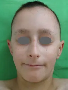
A 9-year-old boy was presented at the Depart- ment of Dermatology of the Heim Pál Children’s Hos- pital, with sparse, brittle hair and extremely dry skin (Figure 1-2.). He was born at term as the first child of non-consanguineous parents with the birth weight
of 2000 grams and with vacuum extraction because of poor maternal pushing. His APGAR scores were 3/5/7, his status was stabilized after manual ventila- tion, but he was put in incubator and oxygen cabin for a short time.
His motoric development was appropriate, but he had mild mental retardation and minimal micro- cephaly.
His medical history included varicella, inguinal hernia, penicillin and milk allergies. His family history was positive for diabetes, hypertonia, and rheuma- toid arthritis. The appearance and consistency of his mother’s and grandmother’s hair were similar to his hair.
Repeated ultrasound examination of the head and neurological examination did not find any additional alterations. Cognitive delays were observed; his IQ level was between 78 and 82. The diagnosis of hyper- kinetic conduct disorder was established by psycho- logical examination. He was controlled by cardiolo- gists because he had ASD II and mild pulmonary valve stenosis. He was under orthopaedic care because of scoliosis. X-ray examinations demonstrated normal bone age. No evidence of other bone alterations was observed. Ophthalmologic examination revealed severe hypermetropy, amblyopia. He complained of photosensitivity. His sweating was normal.
Physical examination revealed a prematurely aged appearance, with asthenic body type. His per- centile for weight was between 3rd and 10th and un- der the 3rd percentile for height. He had a long face, frontal bossing, high arched palate and carious teeth.
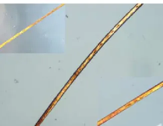
His skin was extremely dry and lichenification was present over the knees. The hair was sparse and brit- tle, with sparse eyebrows (Figure 1, Figure 2). Under polarizing light microscopy, the hair shaft exhibited dark and light banding in a ‘tiger tail’ pattern (Figure 3). The nails were not affected.
Figure 1. Long face, sparse, brittle, dry hair, sparse eye-
brows, and dry skin. Figure 2. Easily broken, dry, sparse hair.
ACTA DERMATOVENEROLOGICA CROATICA 171
Full blood count, electrolytes, renal and liver func- tion, TSH, rheumatoid factor and serum immuno- globulin were within the normal ranges.
The clinical features and hair microscopy findings were consistent with the diagnosis of TTD, therefore molecular genetic investigations were performed.
Direct sequencing of the coding regions and the flanking introns of the ERCC2 gene revealed two re- current missense heterozygous mutations – c.934G/A p.Asp312Asn (CM015299) and c.2251A/C p.Lys751Gln (CM004814) – suggesting compound heterozygous state of the patient and confirming the clinically sus- pected diagnosis of trichothiodystrophy (Figure 4, Figure 5).
He is followed up closely by a multidisciplinary team including dermatologists, neurologists, psy- chologists, cardiologists, orthopaedists and ophthal- mologist.
DISCUSSION
TTD is characterized by sparse, dry and easily broken, brittle hair due to lack of sulphur of the hair (1,5,11). Sulphur is an element that normally gives the strength of the hair (9). Light microscopy of hair shaft might reveal trichoschisis, trichorrhexis nodosa or absence of cuticle. Under polarizing microscopy, the typical appearance of alternating light and dark bands called “tiger tail” pattern is seen, which is char- acteristic for the syndrome. Amino acid analysis is a useful diagnostic method of TTD due to quantifying sulfur (specifically cystein), which correlates inversely with the percentage of affected hairshafts (2).
According to the presence of variant clinical symp- toms like photosensitivity, icthyosis, brittle hair and nails, intellectual impairment, decreased fertility and short stature, PIBIDS, IBIDS, BIDS are distinguished from each other.
Depending on the presence or absence of clinical and cellular photosensitivity, TTD is divided into two major groups: photosensitive (TTD1-3) and nonpho- tosensitive (TTD4 or TTDN1) forms (5,9).
In the photosensitive form, in most cases the xeroderma pigmentosum complementation group D (XPD) gene, rarely the xeroderma pigmentosum complementation group B (XPB) and TTD comple- mentation group A (TTD-A) genes are affected (2).
The encoded proteins of XPD, XPB and TTD-A are parts of the TFIIH transcription factor, a multi-subunit protein complex, playing important role in transcrip- tion initiation, cell cycle regulation and DNA repair (1,5,6,8,9,12). In DNA repair, a part of the DNA nu- cleotide excision repair (NER), a multi-step pathway, Figure 3. Polarizing microscopy showed alternating light
and dark regions, called ‘tiger tail’ banding.
Figure 4. Sequence analysis of the ERCC2 gene: novel missense heterozygous mutation (c.934G/A p.Asp312Asn (CM015299)) in the tenth exon (see arrow).
Figure 5. Sequence analysis of the ERCC2 gene: novel missense heterozygous mutation (c.2251A/C p.Lys751Gln (CM004814)) in the twenty-third exon (see arrow).
Veres et al. Acta Dermatovenerol Croat
Trichothiodystrophy 2018;26(2):169-172
172 ACTA DERMATOVENEROLOGICA CROATICA
TFIIH has at least two major functions: transcription- coupled DNA repair, which removes lesions only in the actively transcribed DNA, and the global genome repair, which removes lesions in any sequence of the genome (2,11,12). The TFIIH complex consists of two modules: core and CAK (CDK-activating kinase).
The core consists of XPB, which has predominantly ATPase activity, TTD-A and 4 other subunits (2,11,12).
The core and CAK sub-complexes are bridged by XPD, which has predominantly helicase activity (1,12).
The TFIIH subunit XPD is encoded by ERCC2, XPB is encoded by ERCC3 and TTDA is encoded by GTF2H5 (general transcription factor IIH polypeptide 5). ERCC2 variants cause TTD1, cerebrofacioskeletal syndrome syndrome, and xeroderma pigmentosum group D (6,9). ERCC3 variants cause TTD2 and xeroderma pig- mentosum group B (6,9). Mutations in the GTF2H5 gene can result in TTD3 (9). Although the same genes are affected in Xeroderma pigmentosum (XP) and TTD, in TTD no predisposition to malignant lesions has been reported (6,10). In XP, mutations primarily affect DNA repair, whereas in TTD transcriptional al- terations, in addition to impaired nucleotide excision repair (NER), is dominant (2,6).
The nonphotosensitive TTD form is said to be caused by mutations in the MPLKIP (C7orf11) gene, which is also called TTDN1. Products of these genes can play a role in cell cycle events like mitotic entry, centrosome maturation and separation, spindle pole integrity, kinetochore attachment, and cytokinesis, but the exact pathogenesis still remains unknown (6,9).
The diagnosis of TTD is established by typical clin- ical features, the ‘tiger tail’ pattern of the hair shaft un- der polarizing light microscopy and molecular inves- tigation. The syndrome should be differentiated from ectodermal dysplasias, Sjögren Larsson, Menkes and Netherton syndromes, based on the sulphur content,
‘tiger tail’ pattern of the hair and trichoschisis.
In the presented case, molecular investigation identified two novel missense mutations in the tenth (c.934G/A p.Asp312Asn; CM015299) and twenty-third (c.2251A/C p.Lys751Gln; CM004814) exons of the ERCC2 gene, which supported the clinical diagnosis of photosensitive TTD.
CONCLUSION
Until now there is no known curative treatment for the syndrome. Photo protection, prevention of pulmonary infections, skin rehydration, physiother- apy and other symptomatic treatment methods are important to improve the patient’s lifetime and qual- ity of life.
References:
1. Stefanini M, Botta E, Lanzafame M, Orioli D. Tricho- thiodystrophy: from basic mechanisms to clinical implications. DNA Repair (Amst). 2010;9:2-10.
2. Hashimoto S Egly JM. Trichothiodystrophy view from the molecular basis of DNA repair/transcrip- tion factor TFIIH. Hum Mol Genet. 2009;18:224- 30.
3. Tay CH. Ichthyosiform erythroderma, hair shaft ab- normalities, and mental and growth retardation:
a new recessive disorder. Arch Derm. 1971;104:4- 13.
4. Price VH, Odom RB, Ward WH, Jones FT. Trichothio- dystrophy: sulfur-deficient brittle hair as a marker for a neuroectodermal symptom complex. Arch Derm. 1980;116:1375-84.
5. Itin PH, Sarasin A, Pittelkow MR. Trichothiodystrop- hy: update on the sulfur-deficient brittle hair syn- dromes. J Am Acad Dermatol. 2001;44:891-920.
6. Moriwaki S, Saruwatari H, Kanzaki T. Trichothio- dystrophy group A: a first Japanese patient with a novel homozygous nonsense mutation in the GTF2H5 gene. J Dermatol. 2014;41:705-8.
7. Shao L, Newell B. Light Microscopic Hair Abnor- malities in Children: Retrospective Review of 119 Cases in a 10-Year Period. Ped and Develop Path.
2014;17:36-43.
8. Oon HH, Sze Chan AW, Lee JS, Leow YH, Giam YC.
Leow YH, et al. Trichothiodystrophy in a Child with Occult Learning Disorder. Int J Trichol. 2013;5:35- 7.
9. Shah K, Ali RH, Ansar M. Mitral regurgitation as a phenotypic manifestation of nonphotosensi- tive trichothiodystrophy due to a splice variant in MPLKIP. BMC Med Genet. 2016;17:13.
10. Kuschal C, Botta E, Orioli D. GTF2E2 Mutations De- stabilize the General Transcription Factor Complex TFIIE in Individuals with DNA Repair-Proficient Tri- chothiodystrophy. Am J Hum Genet. 2016;98:627- 42.
11. Egly JM, Coin F. A history of TFIIH: two decades of molecular biology on a pivotal transcription/re- pair factor. DNA Repair (Amst). 2011;11:714-21.
12. Singh A, Compe E, Le May N, Egly JM. TFIIH subu- nit alterations causing xeroderma pigmentosum and trichothiodystrophy specifically disturb seve- ral steps during transcription. Am J Hum Genet.
2015;96:194.
Veres et al. Acta Dermatovenerol Croat
Trichothiodystrophy 2018;26(2):169-172