COMMENTARY
Factor XIII: What does it look like?
Z S U Z S A B A G O L Y *† and LAS Z L O M U S Z B E K *
*Division of Clinical Laboratory Science, Department of Laboratory Medicine, Faculty of Medicine, University of Debrecen; and†MTA-DE Cerebrovascular and Neurodegenerative Research Group, Debrecen, Hungary
To cite this article:Bagoly Z, Muszbek L. Factor XIII: What does it look like?.J Thromb Haemost2019;17: 714–6.
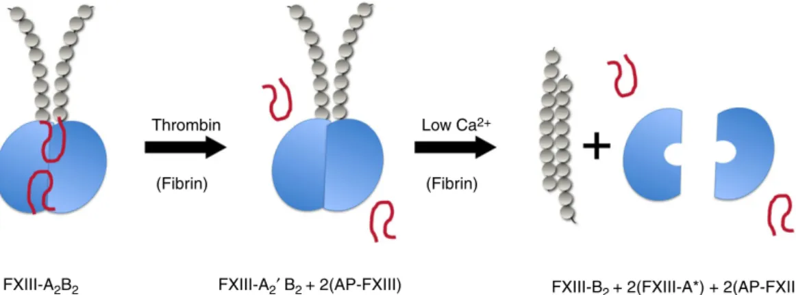
Coagulation factor XIII (FXIII) is a stepchild among clotting factors. As opposed to all other zymogenic clot- ting factors, it is not the precursor of a proteolytic enzyme but of a transglutaminase. It is of tetrameric structure consisting of two types of subunits (FXIII- A2B2). In the final step of the coagulation cascade the A subunit (FXIII-A) is transformed into an active enzyme by the concerted action of thrombin and Ca2+. Thrombin cleaves off an activation peptide (AP-FXIII) of 37 amino acids from the N-terminal end of FXIII-A. Subsequently, in the presence of Ca2+, the protective/inhibitory B sub- units (FXIII-B) dissociate from the cleaved A´ subunits, which then assume an active configuration (FXIII-A*).
Activated FXIII is a transglutaminase, the main function of which is the cross-linking of fibrinaand fibrincchains and the attachment of a2-plasmin inhibitor to fibrin through e(c-glutamyl) lysyl isopeptide bonds. This way FXIII is involved in mechanically strengthening the fibrin clot and protecting it from fibrinolytic degradation. A dimer of FXIII-A lacking the B subunits is also expressed in several cell types, including platelets, monocytes, macrophages, osteoblasts, chondrocytes, and preadipo- cytes in which it exerts diverse functions. The intracellular activation of FXIII-A2 does not need limited proteolytic involvement. The elevation of the Ca2+ level in the cyto- plasm is sufficient to convert FXIII-A2 to an active con- figuration (FXIII-A°). (For comprehensive reviews see references [1–5].)
X-ray crystallography revealed the atomic resolution structure of FXIII-A2 and demonstrated that the FXIII- A monomer contains four domains (b-sandwich, cat- alytic core, b-barrel 1, and b-barrel 2) and AP-FXIII
[6,7]. The crystallographic structure of FXIII-B is not available. This protein is of mosaic structure, it consists of 10 short tandem repeats (sushi domains) each con- taining about 60 amino acids and held together by a pair of disulfide bonds [8]. Our knowledge on the struc- ture of FXIII and on structural changes during FXIII activation was enriched by important studies using elec- tron microscopy, size exclusion chromatography, analyti- cal ultracentrifugation, H-D exchange, mass spectrometry, and molecular modeling [2,9–13]. The use of high-resolution single-molecule atomic force micro- scopy (AFM) in the study of Protopopova et al., pub- lished in this issue of JTH, provides a new dimension to this extensive line of research [14]. This technique visual- izes the surface topology/contour of the molecule rather than the protein backbone, and this way an additional aspect of structural information is revealed. The authors investigated the morphology of the tetrameric FXIII- A2B2 complex and the individual subunits. The struc- tural changes induced by thrombin and Ca2+ were also demonstrated.
The structure of FXIII and its subunits
Based on early electronmicroscopic studies it was sug- gested that two FXIII-B subunits are wrapped around the FXIII-A dimers [9]. This hypothesis was supported by the finding that the presence of FXIII-B protects the A sub- units from proteolytic inactivation [10,14]. The AFM pic- tures produced by Protopopova et al. provide a remarkably novel insight into the structure of FXIII- A2B2. In the published study it is clearly demonstrated that within the tetrameric structure, FXIII-A2 is repre- sented by a globular part of moderate ovality. In the majority of cases two strands corresponding to mono- meric FXIII-B extend from the globular part; however, there were also minor structural variants with only one or no thin flexible extensions. This structural diversity very likely reflects the equilibrium between association and dis- sociation of FXIII-B subunits from the A2 dimer.
According to the AFM data, 26% of the B subunits dis- sociate from the globular core, while 74% are connected to the globules, which is in excellent agreement with the values calculated from the actual FXIII concentration
Correspondence: Laszlo Muszbek, Division of Clinical Laboratory Science, Department of Laboratory Medicine, Faculty of Medicine, University of Debrecen, Nagyerdei Krt. 98, 4032 Debrecen, Hungary Tel: +36 52 43 1956
Email: muszbek@med.unideb.hu
Received: 3 March 2019
Manuscript handled by: Ton Lisman Final decision: 11 March 2019
©2019 The Authors.Journal of Thrombosis and Haemostasispublished by Wiley Periodicals, Inc. on behalf of International Society on Thrombosis and Haemostasis.
This is an open access article under the terms of the Creative Commons Attribution-NonCommercial License, which permits use, distribution and reproduction in any medium, provided the original work is properly cited and is not used for commercial purposes.
Journal of Thrombosis and Haemostasis,17: 714–716 DOI: 10.1111/jth.14431
(~6.3 nM) and the Kd (4.179 10 10M) [15]. Earlier reports suggested that the first two N-terminal sushi domains of FXIII-B are involved in the association to FXIII-A2 [10,15], which was confirmed by the AFM pic- tures. As expected, the individual FXIII-A monomers in the complex are not resolved by AFM. The location of FXIII-B in the complex is somewhat surprising. As dis- cussed later, the individual free FXIII-B molecules form dimers, and it is not clear how the FXIII-B dimer falls apart into monomers to be able to combine with an indi- vidual FXIII-A molecule. The binding site for FXIII-B on FXIII-A also needs to be specified to explain how the two FXIII-B monomers enter the compact/closed struc- ture of FXIII-A2. Such investigations might be relevant for FXIII-A-deficient patients receiving recombinant FXIII-A2 for prophylaxis: in these patients the binding of recombinant FXIII-A2 to the patient’s FXIII-B is essen- tial for the formation of heterotetramers, which in con- trast to uncomplexed FXIII-A2 have a long half-life in the plasma.
Earlier reports concerning the dimeric or monomeric state of free FXIII-B were controversial. Sedimentation analysis and low-resolution electronmicroscopic images suggested free FXIII-B to be a flexible and kinked mono- meric structure [9], while gel filtration experiments sug- gested the protein to be a dimeric structure [10,16]. In the present study the dimeric nature of free FXIII-B was clearly confirmed. It has been shown that the measured length of FXIII-B dimer corresponded to a previously sug- gested model, in which the two FXIII-B subunits have an antiparallel orientation, which is ensured by the interaction of sushi domains 4 and 9 [10]. Accepting this model, further experiments are needed to explain the mechanism of com- plex formation considering that the two N-terminal sushi domains are positioned at the opposite ends of the dimeric strand. One possibility is that the two opposite ends of FXIII-B2 bind to FXIII-A molecules present in distinct FXIII-A dimers. The other possibility is that the binding of
a FXIII-B subunit to FXIII-A2induces the dissociation of the FXIII-B2and the released FXIII-B monomer becomes available for combining with FXIII-A2. Obviously such speculations and others call for experimental support.
Structural changes upon FXIII activation
Earlier reports suggested that after activation of FXIII- A2B2and FXIII-A2by thrombin and Ca2+the two active subunits stick together (FXIII-A2*) and express transglu- taminase activity. Most recently this paradigm has been challenged. The dissociation of FXIII-A2 into FXIII-A* monomers upon both proteolytic and non-proteolytic activation was demonstrated by analytical ultracentrifuga- tion, and FXIII-A*also remained monomeric upon bind- ing of substrate analogs [11]. Protopopova et al.provided direct evidence on the monomeric form of FXIII-A* by AFM [14]. Using volume calculations the FXIII-A2dimer was found to be 2.3-fold larger than FXIII-A*. Molecular modeling also supported a monomeric form for FXIII-A* [13,17]. The monomeric structure of all other active trans- glutaminases is in line with these findings [2,18]. Based on the results of Protopopovaet al.and on earlier findings a schematic representation of FXIII activation is proposed in Fig. 1. The transformation of FXIII-A does not induce drastic conformational changes within a particular domain. However, the changes in the position of individ- ual domains indicate considerable structural transitions, usually termed as transition from closed/compact to open/extended structure (reviewed in references [2,19]).
The resolution of single-molecule AFM does not seem sufficient to capture such a difference.
The introduction of high-resolution single-molecule AFM opens new perspectives in FXIII structural research. It helps in evaluating controversial issues and might confirm or contradict existing paradigms. New findings by this technique contribute to our better under- standing of the structural-functional relationship of this
Thrombin
FXIII-A2B2 FXIII-A2′ B2 + 2(AP-FXIII) FXIII-B2 + 2(FXIII-A*) + 2(AP-FXIII) Low Ca2+
(Fibrin) (Fibrin)
FIGURE 1.Schematic representation of proteolytic FXIII activation. The A subunit of (FXIII-A) is depicted in blue. Elongated strands con- sisting of gray pearls represent the FXIII B subunit (FXIII-B) consisting of sushi domains. Activation peptides (AP-FXIII) are shown as red loops. FXIII-A´, FXIII-A cleaved by thrombin; FXIII-A*, FXIII activated by thrombin; Ca2+(active transglutaminase).
© 2019 The Authors. Journal of Thrombosis and Haemostasis published by Wiley Periodicals, Inc. on behalf of International Society on Thrombosis and Haemostasis.
Commentary715
unique coagulation factor and its role in the hemostatic mechanism.
Addendum
The authors were equally involved in writing and con- structing the commentary.
Acknowledgments
The authors were supported by the National Office for Research and Technology (FK128582, K129287) and the GINOP 2.3.2-15-2016-00050 project co-financed by the European Union and the European Regional Develop- ment Fund.
Disclosure of Conflict of Interests
Neither author had any real or potential conflict of interest.
References
1 Lorand L. Factor XIII: structure, activation, and interactions with fibrinogen and fibrin.Ann NY Acad Sci2001;936: 291–311.
2 Komaromi I, Bagoly Z, Muszbek L. Factor XIII: novel struc- tural and functional aspects.J Thromb Haemost2011;9: 9–20.
3 Muszbek L, Bereczky Z, Bagoly Z, Komaromi I, Katona E.
Factor XIII: a coagulation factor with multiple plasmatic and cellular functions.Physiol Rev2011;91: 931–72.
4 Schroeder V, Kohler HP. Factor XIII: structure and function.
Semin Thromb Hemost2016;42: 422–8.
5 Mitchell JL, Mutch NJ. Let’s cross-link: diverse functions of the promiscuous cellular transglutaminase factor XIII-A.J Thromb Haemost2019;17: 19–30.
6 Yee VC, Pedersen LC, Le Trong I, Bishop PD, Stenkamp RE, Teller DC. Three-dimensional structure of a transglutaminase:
human blood coagulation factor XIII.Proc Natl Acad Sci USA 1994;91: 7296–300.
7 Weiss MS, Metzner HJ, Hilgenfeld R. Two non-proline cis pep- tide bonds may be important for factor XIII function. FEBS Lett1998;423: 291–6.
8 Ichinose A, McMullen BA, Fujikawa K, Davie EW. Amino acid sequence of the b subunit of human factor XIII, a protein com- posed of ten repetitive segments.Biochemistry1986;25: 4633–8.
9 Carrell NA, Erickson HP, McDonagh J. Electron microscopy and hydrodynamic properties of factor XIII subunits. J Biol Chem1989;264: 551–6.
10 Souri M, Kaetsu H, Ichinose A. Sushi domains in the B subunit of factor XIII responsible for oligomer assembly. Biochemistry 2008;47: 8656–64.
11 Anokhin BA, Stribinskis V, Dean WL, Maurer MC. Activation of factor XIII is accompanied by a change in oligomerization state.FEBS J2017;284: 3849–61.
12 Woofter RT, Maurer MC. Role of calcium in the conforma- tional dynamics of factor XIII activation examined by hydrogen-deuterium exchange coupled with MALDI-TOF MS.
Arch Biochem Biophys2011;512: 87–95.
13 Gupta S, Biswas A, Akhter MS, Krettler C, Reinhart C, Dodt J, Reuter A, Philippou H, Ivaskevicius V, Oldenburg J. Revisiting the mechanism of coagulation factor XIII activation and regula- tion from a structure/functional perspective. Sci Rep 2016; 6: 30105.
14 Protopopova AD, Ramirez A, Klinov DV, Litvinov RI, Weisel JW. Factor XIII topology: organization of B subunits and changes with activation studied with single-molecule atomic force microscopy.J Thromb Haemost2019; https://doi.org/10.1111/jth.
14412 [Epub ahead of print].
15 Mary A, Achyuthan KE, Greenberg CS. b-chains prevent the proteolytic inactivation of the a-chains of plasma factor XIII.
Biochim Biophys Acta1988;966: 328–35.
16 Katona E, Penzes K, Csapo A, Fazakas F, Udvardy ML, Bagoly Z, Orosz ZZ, Muszbek L. Interaction of factor XIII sub- units.Blood2014;123: 1757–63.
17 Seelig GF, Folk JE. Noncatalytic subunits of human blood plasma coagulation factor XIII. Preparation and partial charac- terization of modified forms.J Biol Chem1980;255: 8881–6.
18 Stieler M, Weber J, Hils M, Kolb P, Heine A, Buchold C, Pasternack R, Klebe G. Structure of active coagulation factor XIII triggered by calcium binding: basis for the design of next- generation anticoagulants. Angew Chem Int Ed Engl 2013; 52: 11930–4.
19 Lorand L, Iismaa SE. Transglutaminase diseases: from biochem- istry to the bedside.FASEB J2019;33: 3–12.
20 Iismaa SE, Mearns BM, Lorand L, Graham RM. Transglutami- nases and disease: lessons from genetically engineered mouse models and inherited disorders.Physiol Rev2009;89: 991–1023.
©2019 The Authors.Journal of Thrombosis and Haemostasispublished by Wiley Periodicals, Inc. on behalf of International Society on Thrombosis and Haemostasis.
716 Commentary