APPLICABILITY OF ROTARY SPUN
HYDROXYPROPYL CELLULOSE MICROFIBERS FOR THE FORMULATION OF ORODISPERSIBLE
TABLETS OF POORLY SOLUBLE DRUGS
Ph.D. thesis
Péter Szabó
Doctoral School of Pharmaceutical Sciences Semmelweis University
Supervisor: Dr. Romána Zelkó, D.Sc., professor Official reviewers:
Dr. Ildikó Kovácsné Bácskay, Ph.D., associate professor Dr. Krisztina Ludányi, Ph.D., associate professor
Head of the Final Examination Committee:
Dr. Krisztina Takácsné Novák, D.Sc., professor
Members of the Final Examination Committee:
Dr. Piroska Révész, D.Sc., professor
Dr. Lívia Budai, Ph.D., assistant professor
Budapest
2016
1
1. TABLE OF CONTENTS
1. TABLE OF CONTENTS ... 1
2. LIST OF ABBREVIATIONS ... 4
3. INTRODUCTION ... 7
3.1. Solubility and the rate of dissolution ... 9
3.1.1. The biopharmaceutical aspects of solubility ... 11
3.2. Pharmaceutical strategies to overcome poor solubility in solid dosage forms ... 13
3.2.1. Particle size reduction ... 14
3.2.2. Amorphous solid dispersions ... 16
3.2.3. Solid solutions ... 19
3.3. Polymeric micro- and nanofibers in the pharmaceutical research ... 20
3.3.1. Structure and physicochemical features of polymeric fibers ... 20
3.3.2. Polymers for fiber formation ... 22
3.3.3. Fiber formation techniques ... 24
3.3.3.1. Electrospinning ... 25
3.3.3.2. High speed rotary spinning ... 27
3.3.4. Preparation of drug loaded micro- and nanofibers ... 29
4. RESEARCH OBJECTIVES ... 31
5. MATERIALS AND METHODS ... 32
5.1. Materials ... 32
5.2.1. Hydroxypropyl cellulose ... 33
5.2.2. Carvedilol ... 33
5.2. Sample preparations ... 34
5.2.1. Preparation of aqueous HPC gels ... 34
5.2.2. Preparation of drug containing HPC gels ... 34
5.2.3. Fiber formation ... 35
2
5.2.4. Preparation of physical mixtures ... 35
5.2.5. Preparation of orodispersible tablets ... 36
5.3. Measurements ... 37
5.3.1. Texture analysis ... 37
5.3.2. Percentage yield ... 38
5.3.3. Morphological evaluation ... 39
5.3.4. Milling process ... 39
5.3.5. Particle size characteristics ... 39
5.3.6. UV-Vis spectroscopy ... 40
5.3.7. Powder X-ray diffraction (XRD) ... 40
5.3.8. Positron lifetime measurements ... 40
5.3.9. Differential scanning calorimetry ... 41
5.3.10. Attenuated total reflectance - Fourier transform infrared (ATR-FTIR) spectroscopy examinations ... 41
5.3.11. Tablet parameters ... 41
5.3.12. Dissolution test ... 42
5.3.13. Comparison of the dissolution curves ... 42
5.3.14. Accelerated stability study ... 43
6. RESULTS ... 44
6.1. Preformulation study ... 44
6.2. Preparation and investigation of drug loaded microfibers ... 48
6.2.1. MD loaded fibers ... 48
6.2.2. CD loaded microfibers ... 50
6.3. Formulation and examination of orodispersible tablets ... 54
6.3.1. MD containing orodispersible tablets ... 54
6.3.2. CD containing orodispersible tablets ... 57
6.4. Accelerated stability test ... 60
7. DISCUSSION ... 64
7.1. Preformulation study ... 64
7.2. Preparation and investigation of drug loaded microfibers ... 65
3
7.3. Formulation and examination of orodispersible tablets ... 66
7.4. Accelerated stability test ... 67
8. CONCLUSIONS ... 69
9. SUMMARY ... 71
9. ÖSSZEFOGLALÁS ... 72
10. REFERENCES ... 73
11. LIST OF PUBLICATIONS ... 93
11.1. Publications relevant to the dissertation ... 93
11.2. Other publications ... 93
12. ACKNOWLEDGEMENTS ... 95
4
2. LIST OF ABBREVIATIONS
A - surface area
Å - ångström
Adiss,n - dissolved amount of a solute at the n-th segment of the gastrointestinal tract
ADMET - absorption, distribution, metabolism, excretion and toxicity ASD - amorphous solid dispersion
ATR-FTIR - attenuated total reflectance - Fourier transform infrared BCS - Biopharmaceutical Classification System
Bq - becquerel
C - concentration
°C - degree Celsius
Ca - capillary number
CD - carvedilol
CDER - Center for Drug Evaluation and Research Cs - saturation concentration
Cs,n - solubility of the compound at the particle surface at the n-th segment of the gastrointestinal tract
Cu - copper
D - diffusion coefficient
D10% - the particle size at the intercept of the cumulative distribution curve and 10%
D50% - median diameter, or the particle size at the intercept of the cumulative distribution curve and 50%
D90% - the particle size at the intercept of the cumulative distribution curve and 90%
Δr - electron layer thickness, equals to 1.66 Å DSC - differential scanning calorimetry
η - dynamic viscosity
ECavitation - cavitation energy
ECM - extracellular matric ECrystal Packing - crystal packing energy
5
ESolvation - solvation energy
f1 - difference factor f2 - similarity factor FCent - centrifugal force
FDA - U.S. Food and Drug Administration FTIR - Fourier transform infrared spectroscopy 𝛾𝛾𝑠𝑠l - solid-liquid interfacial tension
g - Gram
GI - gastro-intestinal GIT - gastro-intestinal tract
h - thickness of the diffusion layer happ - apparent diffusion layer thickness HPC - hydroxypropyl cellulose
kB - Boltzmann constant, equals to 1.38x10⁻²³ JK-1
kV - kilovolt
λ - wavelength
logP - logarithm of partition coefficient
m - mass
M - molar (molar concentration)
Mw - molecular weight
mA - milliamper
MD - model drug
MPa - megapascal
µg - microgram
mg - milligram
min - minute
mJ - millijoule
ml - millilitre
mm - millimetre
N - newton
nA - nanoamper
ω - angular velocity
6 o-Ps - ortho-positronium
𝜋𝜋 - pi, mathematical constant, approximately 3.14159 PALS - positron annihilation lifetime spectroscopy
pH - pondus hydrogenii
pKa - acid dissociation constant
ps - picosecond
r - radius
R - universal gas constant, equals to 8.314 JK-1mol-1
𝜌𝜌 - volumetric mass density
RH - relative humidity
rpm - revolutions per minute
Rt - dissolution value of reference at t time
s - second
S - solubility
S∞ - normal solubility of a material with a plane surface SEM - scanning electron microscope
Sr - solubility of a particle of r radius
SS - solid solution
t - time
𝜏𝜏3 - ortho-positronium lifetime
T - temperature
Tt - dissolution value of test at t time U - polymer jet exit speed
v - number of ions dissociating from the solute V - volume of dissolution medium
Vlumen,n - volume of the liquid presented in the n-th segment of the gastrointestinal tract
% v/v - volume concentration
% w/w - mass fraction, percentage by mass XRD - powder X-ray diffraction
7
3. INTRODUCTION
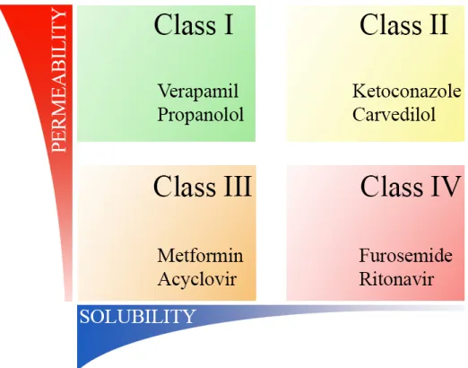
Recent trends in pharmaceutical industry enforce the consideration of novel dosage forms in course of the development of pharmaceutically active ingredients. The phenomenon of the increasing demands for special therapeutic purposes and the expanding proportion of actives of undesirable physicochemical properties have been of the major concerns for pharmaceutical researchers. A widely accepted approach for classifying pharmaceuticals is the biopharmaceutical drug classification system (BCS) in which actives are categorized based on their solubility and permeability is represented in Fig. 1 (Amidon et al., 1995).
Figure 1 Illustration of Biopharmaceutical Classification System including typical representatives of each class
As can be seen from Fig. 1, members of class I possess the most advantageous physicochemical features enabling their rapid and complete dissolution and absorption.
The general view is that formulation is an effective way to improve the gastrointestinal behavior of drugs belonging to class II, while chemical modification should be considered for actives of classes III and IV (Pouton, 2006). A remarkable part of these formulations
8
focuses on the dissolution and solubility enhancement of the incorporated ingredients.
However, it can be noted that several research papers were published turning the spotlight on the permeation and bioavailability improvement of drugs of class III and IV (Kaukonen et al., 2007; Özdemir et al., 2000; Sinha et al., 2010; Yu et al., 1999).
Moreover, the assessment of class II drugs is further complicated since it has been demonstrated that an enhanced in vitro dissolution does not result necessarily in an increased oral bioavailability (Sarnes et al., 2014). Another crucial obstacle is that formulations usually cannot address such phenomenon as high rate of metabolism or extensive efflux transport associated with class II-IV drugs (Wu and Benet, 2005).
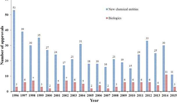
The real problem lies in the ever-decreasing number of approvals of new chemical entities, which is partly believed as a consequence of the extensive proportion of drugs of unfavorable physicochemical features (Fig. 2).
Figure 2 Number of original drug approvals per year by FDA (FDA, 2016)
The root of this tendency lies in the regnant approaches in modern drug development: the rational drug discovery and the high throughput screening lead to poorer permeability and poorer solubility, respectively (Lipinski, 2000). Hann et al. have identified this issue as the aftermath of the upset balance between potency and ADMET features resulting in molecular obesity. This term refers to the large molecular size and lipophilicity, as well
9
as the poor aqueous solubility of the actives (Hann, 2011; Hann and Keseru, 2012).
Against this background, it is not surprising that approximately 70% of new chemical entities and 30% of marketed drugs belong to the BCS class II (Wu and Kesisoglou, 2010). The reason why the proportion of BCS class II drugs is smaller among marketed drugs is that such molecules had been neglected during the early development stages.
3.1. Solubility and the rate of dissolution
In physical chemistry, two kinds of solubilities can be distinguished: the thermodynamic solubility and the kinetic solubility. The thermodynamic solubility refers to a physically long-term stable condition, and can be described as the concentration of a solute in a solution in equilibrium with the normal sized powder of the most stable crystalline state.
The kinetic solubility is not a physically long-term stable condition and is defined as the concentration of a solute in a solution in equilibrium with a metastable crystalline state.
The kinetic solubility is usually higher than thermodynamic solubility (Janssens and Van den Mooter, 2009; Mauludin et al., 2009).
Generally, three quantities have been recognized, which affect the solubility (S) of a drug, and their relationship with solubility is described in the following equation:
𝑆𝑆 = 𝑓𝑓(𝐸𝐸
𝐶𝐶𝐶𝐶𝐶𝐶𝐶𝐶𝐶𝐶𝐶𝐶𝐶𝐶 𝑃𝑃𝐶𝐶𝑃𝑃𝑃𝑃𝑃𝑃𝑃𝑃𝑃𝑃+ 𝐸𝐸
𝐶𝐶𝐶𝐶𝐶𝐶𝑃𝑃𝐶𝐶𝐶𝐶𝐶𝐶𝑃𝑃𝐶𝐶𝑃𝑃+ 𝐸𝐸
𝑆𝑆𝐶𝐶𝐶𝐶𝐶𝐶𝐶𝐶𝐶𝐶𝑃𝑃𝐶𝐶𝑃𝑃)
(1)where ECrystal Packing refers to the endoergic energy responsible for the disruption of the crystalline lattice in order to remove molecules from. ECavitation means the endoergic energy related to the disruption of the water molecules creating a cavity suitable to host the solute, and ESolvation represents the resultant energy deriving from the interactions of the solute and the solvent (Lipinski et al., 2012).
The applied solvent, solvent mixture, the temperature, pH, solid state, ionic strength are the main parameters affecting the solubility of a given compound (Stegemann et al., 2007).
Another important characteristic of the drug liberation is the rate of dissolution, which is equivalent to the amount of drug dissolved per unit time. Noyes and Whitney described
10
the proportionality between the dissolution rate and the difference of the concentration of the saturated solution and the solution in question (Noyes and Whitney, 1897).
Later on, Nernst and Brunner published an improved equation (Eq. (2)) on the basis of the diffusion layer approach:
𝑑𝑑𝐶𝐶
𝑑𝑑𝐶𝐶
=
𝐷𝐷𝐷𝐷𝑉𝑉ℎ(𝐶𝐶
𝑆𝑆− 𝐶𝐶)
(2)where D is the diffusion coefficient, A the surface area of the interface between the compound to be solved and the solution, V the volume of the dissolution medium, h the thickness of the diffusion layer, CS the saturation solubility and C the concentration of the solute in the bulk phase at t time (Brunner, 1904; Dokoumetzidis and Macheras, 2006;
Nernst, 1904).
Regarding the huge number of research papers focusing on particle size reduction and the formulation of nanoscale drug delivery systems it is essential to discuss the impact of particle size and interfacial tension on the solubility. The dependence of the saturation solubility on the particle size was given by Ostwald and Freundlich (Eq. (3)):
𝜌𝜌𝜌𝜌
𝑀𝑀𝑅𝑅𝑅𝑅𝑤𝑤
𝑙𝑙𝑙𝑙
𝑆𝑆𝑆𝑆𝑟𝑟∞
=
2𝛾𝛾𝐶𝐶𝑠𝑠𝑠𝑠(3)
where ρ is the volumetric mass density, r the radius of the particles, v represents the number of ions dissociating from the solute, M the molecular weight of the solute, γsl the solid-liquid interfacial tension, R the ideal gas constant, T the temperature Sr and S∞ the solubility of the particle of r radius and the normal solubility of a plane surface, respectively (Wu and Nancollas, 1998). In other words, the solubility of a compound of a given particle size changes exponentially with the reciprocal radius. However, it must be noted that the particle size reduction related solubility enhancement is significant only below a particle size of 2 µm and even more below 1 µm (Müller et al., 2000).
Since Eqs. 2 and 3 are applicable only in the existence of a steady state condition suggesting a linear concentration gradient along with the diffusion layer, Wang and Flanagan introduced a new equation taking into account that the dissolution rate is influenced by the surface curvature and that the width of apparent diffusion layer alters
11
with change of the particle size. The authors proposed that in case of monodispersed spherical particles, h in Eq. (2) should be replaced with happ, the apparent diffusion layer thickness, which was given as
1
ℎ𝑎𝑎𝑎𝑎𝑎𝑎
=
ℎ1+
1𝐶𝐶 (4)where h is the diffusion layer thickness, and r the particle radius (Wang and Flanagan, 1999, 2002).
Based on these considerations, to describe in vivo conditions, the following equation (Eq.
(5)) can be defined:
𝑑𝑑𝐷𝐷𝑑𝑑𝑑𝑑𝑠𝑠𝑠𝑠,𝑛𝑛
𝑑𝑑𝐶𝐶
= −4𝜋𝜋𝑟𝑟
2(𝑡𝑡) 𝐷𝐷 �
𝐶𝐶(𝐶𝐶)1+
ℎ1� �𝐶𝐶
𝐶𝐶,𝑃𝑃−
𝐷𝐷𝑑𝑑𝑑𝑑𝑠𝑠𝑠𝑠,𝑛𝑛𝑉𝑉𝑠𝑠𝑙𝑙𝑙𝑙𝑙𝑙𝑛𝑛,𝑛𝑛(𝐶𝐶)
�
(5)where, dAdiss,n/dt is the dissolution rate of the particle of r(t) radius at t time, D the diffusion coefficient, h the diffusion layer thickness, Cs,n the compound solubility at the partice surface, Adiss,n the dissolved amount of the solute, and Vlumen the volume of the liquid presented in the n-th segment of the gastrointestinal tract (GIT) (Jamei et al., 2009).
3.1.1. The biopharmaceutical aspects of solubility
Though there are some exceptions (active or the paracellular transport), the vast majority of the active ingredients is absorbed from the GIT via passive diffusion (Bravo-Osuna et al., 2008). The generally accepted model for the gastrointestinal absorption of these drugs requires for dissolved active compound at the absorption sites (Jamei et al., 2009; Rao et al., 2009; van De Waterbeemd et al., 2001). Two main scenario can be classified on this basis. One is that when the drug remains undissolved at the window of absorption because of its poor solubility. The other is that when the rate of dissolution is too low, therefore the transit time is not sufficient for the complete dissolution and solid drug particles pass through the absorption site (Hörter and Dressman, 2001).
12
Figure 3 The relationship of poor solubility and the ADME scheme (Leach et al., 2006;
Merisko-Liversidge and Liversidge, 2011; Wu and Benet, 2005)
The most pronounced effect of inadequate solubility features is the low bioavailability.
Alongside with the absorption issue, difficulties of the lead optimization and dosing are not negligible, as well. Furthermore, such active compounds often call for innovative dosage forms comprising excipients which are not indifferent to the human body.
Additionally, these approaches often try to ameliorate poor solubility properties by the kinetic solubility using a dosage form containing drugs in a metastable state (e.g.
amorphous state). Since the kinetic solubility is not a long-term stable condition and uncontrollable precipitation can occur, large variabilities of bioavailability can be expected. Parenteral and other dosage forms associated with discomfort and complication at administration can result in a diminished patients’ compliance (Kayser et al., 2003;
Merisko-Liversidge and Liversidge, 2011). With respect to acidic compounds, the formulation of an immediate release dosage forms can be arduous because of the dissolution hampering and precipitation facilitating effect of the acidic media in the upper tracts of the GIT (Maggi et al., 2015).
Fig. 3 illustrates how poor aqueous solubility influences the fate of drugs in the human body (Leach et al., 2006; Merisko-Liversidge and Liversidge, 2011; Wu and Benet, 2005).
13
Finally, it is important to emphasize the significance of the proper physicochemical and biological characterization of the actives. This is especially interesting for drugs, where low oral exposure is attributed to the poor aqueous solubility, whilst intensive efflux or presystemic metabolic activity are behind the phenomenon, thus low solubility veils poor permeability feature (Stella and Nti-Addae, 2007). Cyclosporine represents a typical example, where formerly the low oral performance was assigned to its poor aqueous solubility and high lipophilicity. Further investigations revealed that the real extent of absorption exceeds much more the presumed value and indicated that the observed low oral bioavailability is a consequence of the intestinal metabolism (Benet et al., 1996;
Hebert et al., 1992; Wu et al., 1995).
3.2. Pharmaceutical strategies to overcome poor solubility in solid dosage forms
Figure 4 Structural differences of the main approaches for the dissolution enhancement of poorly soluble drugs: a) particle size reduction, b) amorphous drug dispersion c) solid solutions
Apart from chemical approaches, e.g. pH adjustment, salt formation, complexation or the prodrug strategy, the most important means of overcoming the solubility issue are still resting on the basis of physical principles. Therefore this chapter focuses on approaches based on physical methods for improving solubility.
14
As can be seen in Fig. 4, the three major approaches of such efforts are the particle size reduction, the formation of amorphous dispersions and solid solutions.
3.2.1. Particle size reduction
Particle size reduction is a long-standing way of handling poorly soluble drugs. Lowering particle radius influences diffusion coefficient, lessens diffusion layer thickness (Eq. (4)) and increases concentration gradient, thus manifesting as an increment of dissolution rate and as the improvement of solubility, as well. The lower the particle radius the larger the diffusion coefficient according to Eq. (6) known as the Stokes-Einstein relationship:
𝐷𝐷 =
6𝜋𝜋𝜋𝜋𝐶𝐶𝑃𝑃𝐵𝐵𝑅𝑅 (6)where r is the radius of the spherical particle, kB is Boltzmann’s constant, T is the temperature and η is the dynamic viscosity of the solution (Edward, 1970). Based on Eq.
(2) it is easy to see that dissolution rate is directly proportional to the particle size dependent diffusion coefficient.
In case of an appropriate extent of size reduction, i.e. particle radius below 1 µm, the solubility surplus can reach such degree, which allows the formulation of poorly soluble compounds (Eq. (3)).
There are a great number of particle size reducing methods, which can be classified in two groups, the bottom up and the top down techniques. In the instance of bottom up approaches, fine particles are obtained from a homogenous system (usually solutions) exposed to a physical or chemical trigger. The latter refers to techniques, which scale down particle radius by means of mechanical or other physical energy. The conventional methods for particle size reduction are listed in Table 1.
15
Table 1 Overview of the particle size reducing techniques
Benefits Drawbacks Reference Top Down Methods
Milling basic and easy
technology, time-saving, solvent free
physical and thermal stress can induce drug degradation, erosion from milling pearls
(Keck and Müller, 2006;
Savjani et al., 2012)
High pressure homogenization
simple, time- saving, solvent free
multiple cycles required for desired particle size, small sample particles needed
(Chen et al., 2011; Hecq et al., 2005; Keck and Müller, 2006; Patravale et al., 2004) Bottom up techniques
Precipitation/Crystallization simple, low cost uncontrollable crystal growth during
precipitation, drug must be soluble at least in one solvent, rapid crystal growth can result in crystal
imperfection and inclusion of impurities
(Müller et al., 2000; Rabinow, 2004)
Spray drying narrow size
distribution,
thermal stress, drug should be
(Broadhead et al., 1992; Liu et
16 yields spherical particles,
one-step method
soluble at least in one solvent, costs of the method
al., 2015b; Patel et al., 2015)
The term micronization refers to procedures in which particle size is reduced to a few micrometers, usually to 1-10 µm (Bansal et al., 2011). Micronization itself facilitates drug dissolution, but it is not considered as a suitable way for solubility enhancement.
Therefore much more interest has been given to the preparation of nanosized particles or nanosuspensions.
One of the major drawbacks of particle size reduction arises from the increase of surface area and of surface energy resulting in a greater tendency to agglomerate. Thus the control of surface particles can be vital. Moreover, crystal imperfections, and formation of amorphous regions can decrease stability, as well (Shoyele and Cawthorne, 2006). On the other hand, declined flow properties and wettability may constitute barriers to further processing, e.g. tableting (Karavas et al., 2006).
3.2.2. Amorphous solid dispersions
Preparation of amorphous solid drug dispersions (ASDs) offers a promising means of increasing solubility. ASDs are multicomponent systems comprising an amorphous active ingredient in a carrier, that is usually a polymer (Newman et al., 2015). In ASDs, the active ingredient forms amorphous clusters in the carrier (Fig. 4). The principle of this method is the circumvention of low thermodynamic aqueous solubility by means of development of a solid system containing poorly soluble drug in a metastable state possessing an apparent solubility greater than that of the initial drug. ASDs are often referred to as spring-parachute systems. By virtue of their high chemical potential, drugs incorporated in ASDs are spouted in the dissolution fluid resembling a compressed spring. However, this higher energy form of the drug tends to precipitate, but the applied excipients act like a parachutes by hindering precipitation (Guzmán et al., 2007) . It is considered that the decisive energy among three quantities given in Eq. (1) is ECrystal Packing, that is greater than the other two values. For this reason any efforts done to decrease ECrystal Packing engenders in a notable solubility improvement (Brough and Williams Iii,
17
2013; Lipinski et al., 2012). Basically, in the course of getting contact with gastrointestinal fluids, ASD releases its active ingredient forming a thermodynamically unstable supersaturated solution (a solution in which a solute has a concentration greater than its equilibrium concentration) (Brouwers et al., 2009).
Because of their numerous benefits, such as enhanced wettability, higher porosity and increased specific surface area, ASDs have gained particular interest in new formulation strategies (Karavas et al., 2006). As a result of the presence of the polymeric matrix, ASDs exhibit better physicochemical stability than neat amorphous actives themselves.
This can be attributed to the decreased molecular mobility originating from drug- excipient interactions (Taylor and Zografi, 1997; Yoshioka et al., 1994; Zhou et al., 2007). Molecular mobility has an impact on phase separation and drug recrystallization, therefore it is crucial from the point of stability (Janssens and Van den Mooter, 2009).
Formation of strong drug-polymer interactions (ionic interactions and hydrogen bonds) and polymer-drug miscibility are of special impact for the physical stability (Li et al., 2014; Tian et al., 2014). In addition, polymers and surfactants hamper drug recrystallization during dissolution through the solubilization of the active. Another important aspect of ASDs relates to the wide range of polymers enabling the achievement of modified release.
On the contrary, the miscibility of the active and polymer is limited, therefore high weight fraction of the drug can lead to phase separation (Qi et al., 2010; Six et al., 2002; Six et al., 2004). Thus along with poor physical stability, the limited drug loading capacity can be considered one of the major disadvantages of this approach.
Preparation of ASDs can be carried out using a solvent, a melting or a solvent-melting method, as well (Brough and Williams Iii, 2013; Squillante and Sethia, 2003). Solvent methods are based on the evaporation of a solvent from the solution of the drug and the polymer. Melting techniques require for the heating of the drug-excipient mixture above its glass transition temperature, and then for a cooling step with due regard to avoid recrystallization. The most frequently applied methods are listed in Table 2.
18
Table 2 Overview of the solvent and melting techniques
Method Advantages Disadvantages References Melting methods
Hot-melt extrusion
modified release, reduced number of processing steps,
uniform drug distribution, continuous process,
high throughput rate, solvent-free
thermals stress, recrystallization upon cooling, high energy input, shear forces
(Breitenbach, 2002;
Crowley et al., 2007; Repka et al., 2007)
Injection molding
solvent-free, continuous process, rapid technique,
scalability and patentability, possibility of
autosterilization
high pressure, high temperature
(Konig et al., 1997;
Zema et al., 2012)
Solvent methods
Spray drying narrow size distribution, yields spherical particles, one-step method,
thermal stress, drug should be soluble at least in one solvent, costly, toxicity of organic solvents
(Broadhead et al., 1992; Liu et al., 2015b; Patel et al., 2015; Weuts et al., 2004)
Lyophilisation aqueous and organic solutions, high porosity,
costly, requires a great amount of water to dissolve poorly soluble drug, toxicity of organic solvents
(Ahmed et al., 2006; El-Badry and Fathy, 2006; Oetjen and Haseley, 2007)
19 3.2.3. Solid solutions
Solid solutions (SSs) are solutions comprising a solid solute in a solid solvent, hence the solute is molecularly dispersed resulting in the formation of a homogenous amorphous phase (Fig. 4). Glass solutions should be also noted here, where a solid is dissolved in glassy system, but in the relevant literature there is no consensus for the differentiation of these terms, they are often referred as synonyms (Chiou and Riegelman, 1971; Patterson et al., 2007). In general, the formulation of SSs result in a transparent system, while ASDs are usually opaque. It is not an effortless attempt to uncover the differences by using traditional physicochemical methods, e.g. differential scanning calorimetry (DSC), powder X-ray diffraction, Raman spectroscopy or Fourier transform infrared spectroscopy (FTIR), since the results of such measurements usually do not indicate the nature of the system. Nevertheless, modern techniques, such as solid-state nuclear magnetic resonance provide opportunity to gain information about the nature of the dispersion characteristics of the drug (Djuric et al., 2010; Ito et al., 2010; Pham et al., 2010; Stejskal et al., 1981).
The theoretical background of the solubility and dissolution enhancement effect of SSs is the same as expounded in the previous section above. The main difference lies in the distribution of the incorporated drug. By reason of the molecular dispersion, SSs represent the pinnacle of the amorphous formulations, since this approach takes advantage of particle size reduction and amorphous conversion as much as possible. In view of reduced molecular mobility and molecular dispersity, the statistical probability of encounter of drug molecules to form crystals is much lower than in other systems.
Basically, all of the techniques listed in Table 2 are capable for the formulation of SSs suggesting that the formation of SSs or ASDs depends on the applied excipients and process parameters rather than the chosen method. It has been found that the development of strong drug-excipient interaction plays a pivotal role from the point of the formation of SSs (Aldén et al., 1993). On the other hand ensuring good miscibility is also essential (Andrews et al., 2010).
Polymeric micro- and nanofibers represent unique carrier systems, that are also capable for the formulation of SSs and they are discussed in details in the next section.
20
3.3. Polymeric micro- and nanofibers in the pharmaceutical research
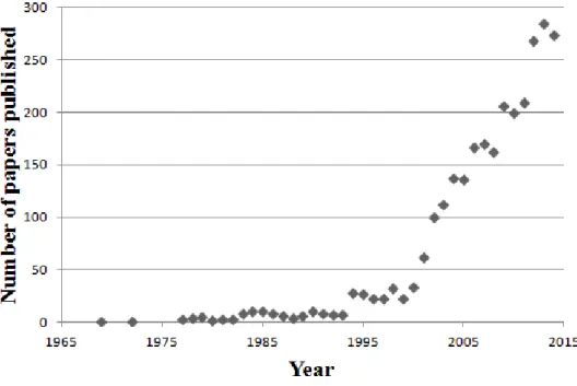
Even though, naturally occurring fibrous systems have been long present in the human history (such as cobweb, plant fibers or musculoskeletal tissues) the consideration of these structures as potential drug delivery systems is quite a new concept. Nowadays, the role of polymeric fibers in the formulation development of special dosage forms is unquestionable, which is well supported by the growing interest for these systems. Fig. 5 strikingly demonstrating that over the past 20 years the number of documents published concerning this topic have been sharply increased.
Figure 5 Number of published documents per year based extracted from Scopus search results analysis using the following keywords: fiber, drug delivery system
3.3.1. Structure and physicochemical features of polymeric fibers
Polymeric fibers owe their popularity to their unique physicochemical characteristics, which are well exploitable for the formulation of innovative drug delivery systems.
Because of their fibrous nature and small diameter, they can be characterized with high specific area-to-volume ratio (Tennent et al., 2000; Zhang et al., 2005b). High specific area-to-volume ratio is beneficial from the point of dissolution, since larger the surface area, the faster the drug dissolution, according to Eq. (2). High porosity and the possibility
21
to keep drugs in amorphous state are also a favorable properties for dissolution enhancement and for biological applications (Frenot and Chronakis, 2003; Szabó and Zelkó, 2015).
Since the structure of these fibers resembles to the extracellular matrix (ECM), these are potential candidates for tissue engineering applications. ECM has an important role in the regulation and maintenance of biologic function of living tissues through providing mechanical support and through the mediation of biological signals (Hay, 2013; Reddi, 2000). Fibrous scaffolds serve as an interim ECM until the regeneration and the formation of a native ECM.
Figure 6 Basic types of polymeric microfibers, a: blend type, b-d: core-shell type, e:
immobilized, f: hollow fibers
Furthermore, the availability of natural polymers, such as collagen makes these systems even more attractive for biological applications, since the more perfectly is the mimicking, the better repairing efficacy can be achieved (Pham et al., 2006).
As can be seen from Fig. 6, different types of fibers can be classified based on the distribution of the drug and the applied polymers.
Blend or matrix type fibers represent a simple structure consisting a drug uniformly dispersed in a carrier matrix. Immobilized fibers comprise a neat carrier and a drug attached to its surface by chemical or intermolecular bonds. Core-shell fibers are built up of two coaxial matrices, the drug can be incorporated in various arrangements as
22
illustrated in Fig. 6. Furthermore, the inner part of the fiber can consist of drug only. For the time being, hollow fibers have meager pharmaceutical importance (Li et al., 2005).
The fiber structure offers an opportunity to tailor release properties. While in case of blend type fibers, the drug release depends on the hydrophilicity of the chosen polymer, in case of core-shell type fibers sustained release, and biphasic release can be also achieved (Jiang et al., 2012; Szabó and Zelkó, 2015; Yu et al., 2013).
Surface properties play a critical role in drug delivery and biological applications.
Biocompatibility is usually a desired property, while biodegradability can be a supportive feature in certain cases (Pelipenko et al., 2015). Beyond hydrophilicity or hydrophobicity, more broadly, surface properties cover the surface functionalization of fibers, too. Surface functionalization is the chemical modification or the coating of fibers (Fang et al., 2008).
Changing the chemical environment on the surface of fibers offers a tool for adjusting release properties, drug loading and surface hydrophobicity (Jacobs et al., 2011; Xie et al., 2012). For instance, developing ionic groups allows the formation of ionic interactions (Jiang et al., 2014). Coating of fibers can aim at the preparation of stimuli- responsive drug delivery systems, such as pH-dependent drug dissolution or other release modifying purposes, e.g. reducing burst release (Jiang et al., 2014; Risdian et al., 2015;
Zeng et al., 2005a).
It has been demonstrated, that during the fiber formation, a supramolecular ordered structure can be developed (Cui et al., 2006; Sebe et al., 2013). The exact role of this kind of ordering in the function of fiber based formulations have been not revealed yet. But it can be proposed that this has an impact on molecular mobility and drug-excipient interactions, thus the stability of such formulations.
3.3.2. Polymers for fiber formation
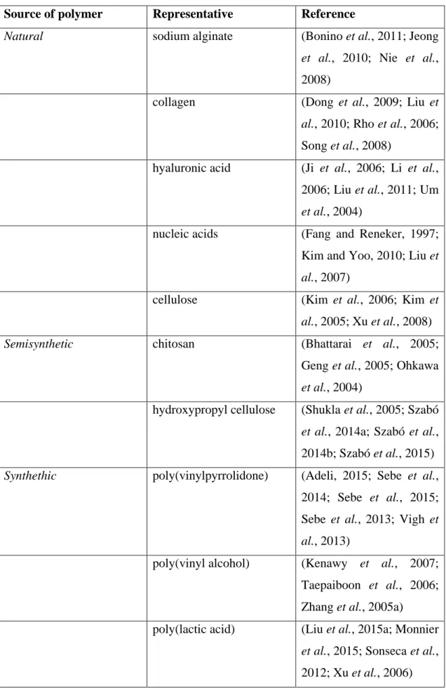
Since the dawn of modern pharmaceutical development and manufacturing, polymers have received a particular attention, and this is no different for both micro- and nanofibers, where most of the relevant papers focuses on the formulation of polymeric based fibers. Accordingly, scientific publications cover the whole spectrum of polymers available. Basically, natural, semisynthetic and synthetic polymers are equally frequently used (Table 3).
23
Table 3 Polymers frequently applied in fiber formation classified by their origin Source of polymer Representative Reference
Natural sodium alginate (Bonino et al., 2011; Jeong
et al., 2010; Nie et al., 2008)
collagen (Dong et al., 2009; Liu et al., 2010; Rho et al., 2006;
Song et al., 2008)
hyaluronic acid (Ji et al., 2006; Li et al., 2006; Liu et al., 2011; Um et al., 2004)
nucleic acids (Fang and Reneker, 1997;
Kim and Yoo, 2010; Liu et al., 2007)
cellulose (Kim et al., 2006; Kim et al., 2005; Xu et al., 2008)
Semisynthetic chitosan (Bhattarai et al., 2005;
Geng et al., 2005; Ohkawa et al., 2004)
hydroxypropyl cellulose (Shukla et al., 2005; Szabó et al., 2014a; Szabó et al., 2014b; Szabó et al., 2015) Synthethic poly(vinylpyrrolidone) (Adeli, 2015; Sebe et al.,
2014; Sebe et al., 2015;
Sebe et al., 2013; Vigh et al., 2013)
poly(vinyl alcohol) (Kenawy et al., 2007;
Taepaiboon et al., 2006;
Zhang et al., 2005a)
poly(lactic acid) (Liu et al., 2015a; Monnier et al., 2015; Sonseca et al., 2012; Xu et al., 2006)
24
poly(glycolic acid) (Boland et al., 2001; Dong et al., 2008; Hajiali et al., 2011)
poly(lactic-co-glycolic acid)
(Xie and Wang, 2006; You et al., 2006)
The paramount functionality-related characteristics of the polymers are the solubility, wetting properties, biodegradability and biocompatibility.
Solubility of the polymer plays a decisive role during the formulation, because the solubility of the chosen polymer has a great impact on the release kinetics of the active substance. Generally, the use of water soluble, hydrophilic polymers results in a rapid, immediate drug release, while hydrophobic or swelling polymers could be exploited in controlled release formulations (Szabó and Zelkó, 2015). So-called release modifying agents are low molecular weight hydrophilic polymers or surfactants (e.g. poly(ethylene glycol)), which can enhance surface hydrophilicity, thus facilitating the proper drug dissolution (Maretschek et al., 2008; Puhl et al., 2014). Biodegradability, i.e. the hydrolytic or enzymatic degradation of the carrier is relevant in terms of erosion controlled drug delivery systems, as well as biological applications, where the role of fabricated fibrous scaffold will be replaced by the continuous generation of the ECM. The relevance of biocompatibility is mainly pronounced in biological applications, where the interaction of the living tissues and their constitutive cells is a key element of the biological effect.
3.3.3. Fiber formation techniques
In the scientific literature several methods are known for fiber formation, however the vast majority of the publications focuses on only a couple of techniques. Classification of these methods can be performed considering various aspects, such as the nature of the sample or the driving force of the fiber formation. Thus we can distinguish physical and chemical methods, but it should be noted that the former is much more dominant. The driving force of the physical fiber formation can be solvent evaporation or cooling supported by electrostatic, pneumatic or centrifugal forces. Table 4 summarizes the available methods for fiber preparation. The table also highlights the versatility of
25
electrospinning, forcespinning and blowspinning, since these techniques can handle either solutions or melts; hence these are the most favorable choices for pharmaceutical purposes. Furthermore, electrospinning process can be facilitated with air blowing (Nayak et al., 2011).
Table 4 Fiber formation techniques (Nayak et al., 2011) Fiber formation method
Chemical methods Physical methods
Solvent based methods Melt methods interfacial polymerization electrospinning electrospinning template synthesis forcespinning forcespinning self-assembly solution-blow spinning melt-blow spinning
drawing template melt extrusion
phase separation 3.3.3.1. Electrospinning
Because of its easy configuration, versatility and scalability, electrospinning has emerged as the most popular technique for fiber formation. This technique provides a great control over product characteristics and enables the preparation of almost every kind of fiber structures. The principle of electrospinning is epitomized in Figure 7. This method requires for a viscoelastic sample; a solution or melt, which is put in a syringe with a needle on it and then it is exposed to a high voltage. When the applied voltage is large enough to overcome the surface tension, polymeric jets will be ejected. In more detail, the high voltage upsets the equilibrium between surface tension and electric field potential, which stabilizes the shape of a droplet. This disturbance leads to the conical deformation of the droplet developing the so-called Taylor cone (Fig. 7). The unstable state results in the fission of Taylor cone, thus jet formation can take place (Yarin et al., 2001). The formed electrostatic field also expedite fiber production by accelerating and stretching jets towards the collector. The solidification of the ejected jets by the evaporation of solvent or cooling enables the formation of solid fibers (Pham et al., 2006;
Teo and Ramakrishna, 2006). Finally, fibers are caught on the surface of the grounded collector. Electrospinning produces continuous fibers of a diameter ranging from a few
26
nanometers to a few micrometers. Hitherto, more than 200 polymers have been reported to be suitable for electrospinning remarkably indicating its significance (Bhardwaj and Kundu, 2010).
Figure 7 A schematic representation of a typical electrospinning arrangement, a: syringe equipped with a needle and filled with polymer solution or melt, b: collector, c: high voltage power supply, d: jet ejected from the needle, e: Taylor cone
Table 5 summarizes the critical parameters influencing fiber morphology including diameter and bead formation. These parameters can be classified as process, solution and ambient parameters.
27
Table 5 The most important factors influencing electrospinning (Pelipenko et al., 2013) Critical parameter
Process parameter Solution parameter Ambient parameter applied voltage polymer molecular weight temperature
flow rate viscosity humidity
needle-collector distance surface tension
needle construction solvent, solvent mixture
collector conductivity
dielectric constant
Beyond the basic arrangement displayed in Fig. 7, more complex apparatuses are available aiming at the preparation of special fiber structures or oriented fibers. The manufacturing of blend fibers is the more effortless, where drug and polymer are mixed together prior to the fiber formation. For the preparation of core-shell type fibers emulsion of two polymers or a specific coaxial electrospinning apparatus (equipped with a core- shell nozzle) is required (Yarin, 2011).
3.3.3.2. High speed rotary spinning
Forcespinning or high speed rotary spinning has been receiving an increasing attention for pharmaceutical applications. The fundamental of the method is depicted in Fig. 8.
In the course of this process, a viscoelastic polymeric solution or melt is put into a rotating reservoir, which has small wall orifices on its wall and is driven by a controlled engine.
A revolution speed is achieved that is large enough to develop a centrifugal force capable to overcome capillary forces, thus the polymeric sample is pressed through the orifices.
Finally, the lengthening jet will solidify upon solvent evaporation and rapid cooling. This method also produces continuous fibers. Beyond the driving force, one of the most important difference from electrospinning, that this method typically employs more concentrated polymeric solutions (Sarkar et al., 2010; Szabó and Zelkó, 2015).
28
Figure 8 Schematic and cutaway drawing of a spinneret applied for high speed rotary spinning, a: wall orifices, b: rotating reservoir, c: ejected polymeric jet or fiber, ω: angular velocity
Angular velocity and radius of the rotating reservoir determines centrifugal force, according to
𝐹𝐹
𝑃𝑃𝑐𝑐𝑃𝑃𝐶𝐶= 𝑚𝑚𝜔𝜔
2𝑟𝑟
(7)where Fcent is the centrifugal force, m, ω and r represent the weight, angular velocity and radius of the rotating mass, respectively (Eq. (7)).
Table 5 The most important factors influencing high speed rotary spinning (Badrossamay et al., 2010)
Critical parameter
Process parameter Solution parameter Ambient parameter angular velocity polymer molecular weight temperature
radius of reservoir viscosity humidity
diameter of orifice surface tension
solvent, solvent mixture volatility
29
Fiber formation is in strong connection with the capillary number which is defined as
𝐶𝐶𝐶𝐶 =
𝑈𝑈𝜋𝜋𝛾𝛾 (8)where Ca is the capillary number, η is the dynamic viscosity, γ is the surface tension and U is the polymer jet exit speed (Eq. (8)). It was found that above critical solution concentration (the concentration above which fiber formation take place) the higher the Ca, the better the quality of the prepared fibers and the smaller the tendency for bead formation (Badrossamay et al., 2010).
3.3.4. Preparation of drug loaded micro- and nanofibers
Preparation of drug loaded micro- and nanofibers can be carried out in several ways. The loading of polymeric fibers can be direct and indirect. In case of direct loading drug containing polymeric solutions, suspensions or melts are applied during the fiber formation process. In respect of indirect or active loading, neat fibers are prepared prior to the introduction of the active ingredient.
The use of drug containing gels or solutions can be considered as the most convenient approach. However, this way of loading has some limitations:
• the applied drug must be soluble at least in one solvent;
• the applied solvent should be a good solvent for each component in order to avoid phase separation;
• blend type fibers usually suffers from burst release;
• potential harm of solvent residues and
• the physicochemical stability of amorphous drug (Thakur et al., 2008; Zeng et al., 2005b).
The possibility to use melts is also advantageous, because of the solvent-free nature of the process. Nonetheless, the method is limited to thermoplastic polymers and to heat stable drugs.
30
Drug suspensions can be also subjected to spinning processes, but it is not a mainstream way of preparation drug loaded fibers. The ability of tailoring drug release through the modification of crystal characteristics, the thermodynamically stable nature of crystals are the advantages of the use of crystalline drug suspensions (Müller and Ulrich, 2012;
Puhl et al., 2014). It must be noted, that in case of high speed rotary spinning, the formed centrifugal force is incompatible with drug suspensions, because of its sedimentation effect.
Active loading was invented in order to address the issue of low drug loading capacity.
In the course of this method, neat fibers prepared prior to the loading step are immersed in a solution of the drug, of which solvent does not dissolve the fiber forming polymer.
Finally, the solvent will be evaporated. This proves also known as the non-solvent evaporation method (Kataria et al., 2014).
31
4. RESEARCH OBJECTIVES
The general aim of the thesis was to demonstrate the importance of fiber-based approaches in the formulation of poorly soluble drugs and at the same time to highlight the prosperous possibilities hidden in high speed rotary spinning. My intention was to provide a comprehensive overview of the progress of a fiber-based formulation. Thus the thesis and its underlying experiments cover a broad spectrum of pharmaceutical development from the preformulation to the stability testing, therefore my aspirations were as follows.
• Enrichment of the range of polymers capable for high speed rotary spinning, by selecting a pharmaceutical polymer, namely hydroxypropyl cellulose (HPC) for high speed rotary spinning that have not been reported to be rotary spun so far.
• Conducting preformulation studies with HPCs of different average molecular weights in order to establish critical and optimal concentrations for fiber formation. The investigation of the impact of textural-rheological properties on fiber characteristics.
• By means of the exploitation of HPC’s versatile solubility features (soluble both in water and ethanol), the preparation of drug loaded microfibers incorporating actives selected from BCS class II.
• Physicochemical and supramolecular characterization of drug loaded microfibers with a particular attention to the crystalline-amorphous transition.
• Formulation of a solid oral dosage form, i.e. orodispersible tablets from processed drug loaded microfibers for the dissolution enhancement of incorporated actives and for highlighting the inherent possibilities of oral administration of fibers.
• To obtain information on physicochemical stability of drug loaded fibers using accelerated stability test.
32
5. MATERIALS AND METHODS 5.1. Materials
Klucel® EXF Pharm and ELF Pharm type HPCs were selected for fiber formation (Ashland, USA). The average molecular weights are 80.000 and 40.000 (based on size exclusion chromatography), respectively, and the moles of substitution is 3.8 for each polymer.
Purified water (Ph. Eur. 8.) and the hydroalcoholic mixture of ethanol (96% v/v, Reanal, Hungary) and purified water (mixed in 3:1 volume ratio) were applied as solvents for the preparation of gels.
Pharmaceutical grade model drug (MD) for fiber preparation was selected from BCS class II, and can be defined with the following physicochemical properties: Mw < 500, pKa = 7.1 (with one basic centre), water solubility < 5.1 µg/ml, logP: 4–5.
The other pharmaceutically active ingredient was carvedilol (CD) (EGIS Pharmaceuticals Plc, Hungary, Ph. Eur.)
Citric acid monohydrate (Hungaropharma) was used for the preparation of drug stock solutions.
Concentrated hydrochloric acid (Molar Chemicals, Hungary), potassium dihydrogen phosphate (Molar Chemicals, Hungary), sodium hydroxide (Molar Chemicals, Hungary), and distilled water were applied in the preparation of the dissolution media.
The excipients of the orally disintegrating tablets were as follows: microcrystalline cellulose (Vivapur® 102 MCC) as filler and disintegrant, mannitol (Mannogem® EZ, SPI Pharma) or spray-dried lactose monohydrate (Flowlac®100, Meggle GmbH, Wasserburg, Germany) as filler, milled poly(ethylene glycol) 1500 (Macrogol 1500, Hungaropharma) or magnesium stearate (Ph.Eur. Hungaropharma, Hungary) as lubricant, equimolar mixture of milled citric acid anhydrate (Molar Chemicals, Hungary), and sodium bicarbonate (Hungaropharma) as effervescent agent and croscarmellose sodium (Vivasol®, JRS Pharma) as superdisintegrant agent.
33 5.2.1. Hydroxypropyl cellulose
Hydroxypropyl cellulose is a semisynthetic derivative of cellulose, a naturally occurring polymer. The polymer is official in different pharmacopoeias (e.g. European Pharmacopoeia, United States Pharmacopoeia) and widely applied for pharmaceutical purposes in oral and in topical dosage forms, as well. Chemical structure of HPC is illustrated in Fig. 9.
Figure 9 Chemical structure of hydroxypropyl cellulose
In pharmaceutical technology, HPC is mainly used as tablet binder, film coating agent and extended release matrix former (Rowe et al., 2009). The polymer is freely soluble in water (below 38 °C) and in polar organic solvents, such as ethanol, dioxane, propanol.
5.2.2. Carvedilol
CD is an arylethanolamine type nonselective beta and alpha-1 adrenoreceptor blocker, which is used in the treatment of hypertension, coronary artery disease and congestive heart failure (Frishman, 1998; Morgan, 1994; Packer et al., 1996; Ruffolo and Feuerstein, 1997). Chemical structure of CD is represented in Figure 10.
34 Figure 10 Molecular structure of carvedilol
CD exhibits poor aqueous solubility (27.11 ± 1.14 µg/mL in pH 6.8 buffer) and has a low oral bioavailability (below 25%), therefore it is a typical representative of BCS class II (v. Möllendorff et al., 1987; Zhao et al., 2011).
5.2. Sample preparations
5.2.1. Preparation of aqueous HPC gels
Aqueous gels were prepared in a beaker by the addition of water to HPC with due regard to their careful homogenization. Klucel® EXF gels were prepared in the concentration range of 38-52 % w/w, and Klucel® ELF gels were made in the concentration range of 42-60 % w/w with an increment of 2% w/w. In case of the former, gels were stored for 2 hours, in case of the latter, gels were stored for 1 hour at room temperature (T=25 °C) in order to ensure the complete dissolution of the polymers.
5.2.2. Preparation of drug containing HPC gels
For MD containing gels, the drug stock solution was prepared as follows. In a 50.00 ml volumetric flask, 5.000 g of drug was suspended with about the half of the necessary amount of hydroalcoholic mixture, until a lactescent dispersion was formed. Then 3.000 g of citric acid monohydrate (equimolar to drug) was added, and the dispersion was
35
shaken until a clear solution was formed. Finally, the solution was diluted to 50.00 ml with the solvent mixture.
For CD containing gels, the drug stock solution was prepared by the following way. In a 50.00 ml volumetric flask, 5.000 g of drug was suspended with about the half of the necessary amount of hydroalcoholic mixture, until a lactescent dispersion was formed.
Then 2.590 g of citric acid monohydrate (equimolar to drug) was added, and the dispersion was shaken until a clear solution was formed. Finally, the solution was diluted to 50.00 ml with the solvent mixture.
Drug loaded gels of 50 % w/w Klucel® ELF were prepared by the addition of the equal amount of drug stock solution to the amount of polymer in a beaker. After careful homogenization and covering with paraffin tape, the gels were let to rest at room temperature (T=25 °C) for 1 hour.
5.2.3. Fiber formation
HPC fibers were prepared via high speed rotary spinning technique. At first, the samples were put in a perforated aluminum rotating reservoir, which was powered by a WSE 602 M engine (AEG, Germany). The fiber formation was carried out at 10,500 rpm. The rotational speed was controlled with a toroidal transformer and determined with laser revolution counter (DT-10L, Voltcraft, Germany). The internal diameter of the two wall orifices were 0.5 mm.
5.2.4. Preparation of physical mixtures
Following analytical assay of the drug loaded microfibers, physical mixtures of the same composition were made by the combination of plain drug, Klucel® ELF and citric acid.
After sieving all components through a mesh sieve (nominal wire diameter 320 µm), the substances were homogenized in a Turbula (T2F model; Willey A Bachofen AG, Maschinenfabrik, Basel, Switzerland) at 23 rpm for 30 min in a cylindrical container. The mixture was used as control for the performed examinations, and for the preparation of control tablets.
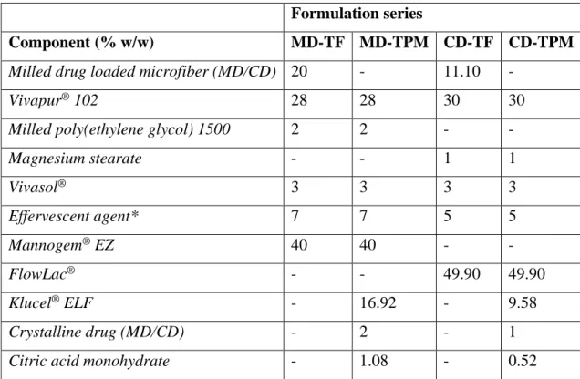
36 5.2.5. Preparation of orodispersible tablets
Orodispersible tablets containing 10 mg drug were prepared by direct compression technique using a single-punch tableting machine (Diaf TM20, Copenhagen, Denmark), with a shallow concave round punch of 13.5 mm. Formulas are given in Table 6, where microfiber based tablets were assigned as TF, and control tablets consisting physical mixture of hydroxypropyl cellulose, anhydrous citric acid and drug were assigned as TPM. After sieving all components through a mesh sieve (nominal wire diameter 320 µm), the substances were homogenized in a Turbula (T2F model; Willey A Bachofen AG, Maschinenfabrik, Basel, Switzerland) at 23 rpm for 30 min in a cylindrical container.
The batch size of 100 tablets was prepared for each composition and the tableting machine was powered manually.
To ensure specified hardness (30-35 N for MD and 40-45 N for CD containing tablets) the applied pressures were adjusted for each formula. Final weight of MD and CD containing tablets was adjusted to 500 and 600 mg, respectively.
Table 6 Composition of the prepared orodispersible tablets *Equimolar mixture of milled citric acid anhydrate and sodium bicarbonate
Formulation series
Component (% w/w) MD-TF MD-TPM CD-TF CD-TPM
Milled drug loaded microfiber (MD/CD) 20 - 11.10 -
Vivapur® 102 28 28 30 30
Milled poly(ethylene glycol) 1500 2 2 - -
Magnesium stearate - - 1 1
Vivasol® 3 3 3 3
Effervescent agent* 7 7 5 5
Mannogem® EZ 40 40 - -
FlowLac® - - 49.90 49.90
Klucel® ELF - 16.92 - 9.58
Crystalline drug (MD/CD) - 2 - 1
Citric acid monohydrate - 1.08 - 0.52
37 5.3. Measurements
5.3.1. Texture analysis
Figure 11 Calculation of adhesiveness from the load-displacement curve
Textural measurements were carried out using Brookfield CT3 Texture Analyzer with 4500 g load cell (Brookfield Engineering Laboratories, INC., USA) equipped with a cylindrical probe (TA-5, black delrin, diameter: 12.7 mm, length: 35 mm).
The compression test comprised two cycles applying the following parameters: pretest speed, test speed, and return speed of 2, 1 and 1 mm/s, respectively. Trigger load was set to 2 g. The probe compressed the gels through a depth of 10 mm as a target distance upon reaching the trigger load. Brookfield Texture Pro CT software was employed for evaluation. The adhesiveness is given by the total negative area of the load-distance curve of the first cycle (Figure 11).
38
Figure 12 Measuring arrangement for adhesiveness determination with the motions of the probe
Prior to the measurement, 10.00 g of each sample was transferred to a cylindrical glass container (internal diameter: 23 mm, height: 30 mm), which was fixed to the platform of the instrument. Three parallels of each concentration were measured. The principles of the analysis is depicted in Figure 12.
5.3.2. Percentage yield
Percentage yield on dry polymer of the fiber formation process was calculated as follows (Eq. (9)):
39
𝑌𝑌𝑌𝑌𝑌𝑌𝑙𝑙𝑌𝑌 % =
𝑚𝑚 𝑚𝑚𝑓𝑓𝑑𝑑𝑓𝑓𝑙𝑙𝑟𝑟𝑔𝑔𝑙𝑙𝑠𝑠𝑃𝑃𝑎𝑎𝑝𝑝𝑠𝑠𝑝𝑝𝑙𝑙𝑙𝑙𝑟𝑟
100
(9)where mfiber is the weight of the fibers prepared from a gel of a weight of mgel and a concenctration of cpolymer.
5.3.3. Morphological evaluation
Fiber morphology was characterized using both optical microscopes (LCD Micro type;
Bresser; Germany and Nikon SMZ 1000 type; Nikon, Tokyo, Japan) and scanning electron microscope (SEM) (Amray 1830-D4,equipped with a tungsten electron gun and EDAX-PV 9800 energy-dispersive spectrometer). In the course of light microscopy magnifications were: 40x, 100x and a standard micrometer scale was used for the calibration. For SEM pictures, the parameters were as follows: acceleration voltage of 15 kV, beam current of 0.1–0.5 nA, and samples were gold coated with JEOL JEE-4B vacuum evaporator. Images were analyzed employing Image Pro Plus 4.5 software (Media Cybernetics, Bethesda, U.S.).
5.3.4. Milling process
Rotary knife grinder (Gorenje SMK 150 B) was applied for the particle size reduction of citric acid anhydrate, sodium bicarbonate, poly(ethylene glycol) 1500 and drug loaded microfibers. Operating parameters were as follows: milling time of 6 min, frequency of rotation was 24000 rpm (determined with DT-10L laser revolution counter, Voltcraft, Germany). All of the milled materials were sieved through a mesh sieve (nominal wire diameter: 320 µm).
5.3.5. Particle size characteristics
Information on particle size distribution was obtained by laser scattering particle size distribution measurement. The instrument (LA-950V2 Horiba Co., Kyoto, Japan) was equipped with a dry feeder unit to determine the particle diameter of the excipients and the milled drug loaded fibers in the range of 0.011-3000 µm. The operating parameters were as follows: air pressure of 0.1 MPa; feeder intensity (0-200) was 80; and relative refractive index was 1.60.
40
Distribution span values were calculated to characterize the width of the distributions based on Eq. (10):
𝑆𝑆𝑆𝑆𝐶𝐶𝑙𝑙 =
𝐷𝐷90%𝐷𝐷−𝐷𝐷10%50% (10)
where D10%, D50% and D90% are the particle diameters at 10, 50 and 90% of the cumulative particles undersize plot. The results are the averages of five parallel measurements.
5.3.6. UV-Vis spectroscopy
Drug content of the milled fibers was measured by UV-vis spectroscopic assay (Agilent 8453 UV-Vis Diode Array System, USA for MD; and Jasco 530 UV–Vis spectrophotometer, Japan for CD) applying 0.1 M hydrochloric acid as solvent. The drug content of the samples was measured on the basis of the calibration curve recorded earlier.
Five parallel measurements were performed.
5.3.7. Powder X-ray diffraction (XRD)
X’Pert Pro diffractometer (PANAnalytical, Almelo, The Netherlands) system with CuKαI
radiation (λ = 1.5406 Å) over the interval of 2.0000 - 40.0014° was used to obtain X-ray diffraction patterns. The following conditions were applied: target of Cu; filter of Ni (thickness was 0.02 mm); voltage of 40 kV; current of 40 mA; angular step of 0.0334°;
counting time of 40.005 s.
5.3.8. Positron lifetime measurements
Supramolecular characterization of samples was carried out using positron annihilation lifetime spectroscopy (PALS). The method exploits the relatively long lifetime of the unstable exotic atom, ortho-positronium (o-Ps), which is formed after a positive beta decay upon bounding together with an electron. o-Ps atoms tend to be trapped in the free volume holes of polymeric excipients, and their annihilation is not influenced by their intrinsic lifetime but by the electron density in the holes. Deng et al. defined how o-Ps lifetime is associated with the size of the free volume around them (Eq. (11))
41
𝜏𝜏
3=
12�1 −
𝐶𝐶+∆𝐶𝐶𝐶𝐶+
2𝜋𝜋1sin �
𝐶𝐶+∆𝐶𝐶2𝜋𝜋𝐶𝐶��
−1 (11)where τ3 is the ortho-positronium lifetime, r the radius of the free volume hole, and Δr is the electron layer thickness (Deng and Jean, 1993). According to Eq. 11, the longer the lifetime, the larger the hole.
For o-Ps determination carrier-free 22NaCl positron source of an activity of 105-106 Bq was used with a fast-fast coincidence system based on BaF2 /XP2020Q detectors and Ortec® electronics. Three parallel spectra were measured at each composition to increase reliability. After summarizing the parallels, spectra were evaluated by the RESOLUTION computer code (Kirkegaard et al., 1981); the indicated errors are the deviations of the lifetime parameters obtained. Three lifetime components were found in all the samples.
5.3.9. Differential scanning calorimetry
Thermograms of CD loaded microfibers were obtained using Seiko Exstar 6000/6200 (Seiko Instruments, Japan) differential scanning calorimeter with an open aluminum pan.
The temperature range was 6°C - 200°C and the scanning rate was set to 5°C /min under air atmosphere. 0.0050 g of sample was used for the measurements.
5.3.10. Attenuated total reflectance - Fourier transform infrared (ATR-FTIR) spectroscopy examinations
ATR-FTIR spectra were collected on Jasco FT/IR-4200 spectrophotometer between 4000 and 2000 cm−1 with an ATRPRO470-H single reflection accessory (Jasco) equipped with flat pressure tip. The spectral measurements were performed in absorbance mode. The 200 scans at a resolution of 2 cm−1 were co-added by the FT-IR software (Spectra Manager-II, Jasco).
5.3.11. Tablet parameters
Hardness, friability and in vitro disintegration time were determined.
42
5 tablets of each composition were measured by tablet hardness tester (8M, Dr.
Schleuniger Pharmatron, Switzerland).
For friability testing, ca. 6.5 g of dedusted tablets was put in an Erweka friability tester (TAP, Offenbach/Main, Germany). The instrument was moved for 4 min with a revolution speed of 25 rpm. Percentage friability was calculated by the reweighting of the dedusted tablets.
In vitro disintegration times were determined applying Erweka Disintegration Tester (ZT 4, Germany). 900 ml of demineralized water was used as the media; the measurement was carried out at 37±2 °C by visual observation (disk was not applied). Six tablets from each composition were evaluated for their disintegration times. The observed minimum and maximum values are reported later.
5.3.12. Dissolution test
Orodispersible tablets were examined in a Hanson SR8-Plus (Hanson Research, Chatsworth, USA) type dissolution tester equipped with rotating paddles at 37±1 °C, with a rotation speed of 50 rpm. Solution of hydrochloric acid of pH 1.0 (Ph. Eur. 8.), phosphate buffer of pH 4.5 (Ph. Eur. 8.) and phosphate buffer of pH 6.8 (Ph. Eur. 8.) were applied as dissolution media (the volume was 500 ml). 3.00 ml of samples were taken at predetermined time points using a Biohit Proline 5.00 ml pipette without refilling. The samples were filtered through a 10µm UHMW polyethylene cannula dissolution filter.
The drug content of the samples was determined by UV-Vis spectroscopy (Agilent 8453 UV-Vis Diode Array System, USA for MD, and Jasco 530 UV–Vis spectrophotometer, Japan for CD) at the characteristic wavelength of the drug on the basis of the calibration curve recorded earlier. Three parallel measurements were carried out for each sample.
5.3.13. Comparison of the dissolution curves
Difference (f1) and similarity (f2) factors were calculated for the mathematical comparison of drug release profiles. Eqs. (12) and (13) were given by Moore and Flanner (Moore and Flanner, 1996) and implemented by FDA CDER (Center for Drug Evaluation and Research):