Preclinical investigation of potential new therapeutic targets in experimental cerebral large vessel occlusion
Doctoral thesis
Dr. Sándor Nardai Semmelweis University
János Szentágothai Doctoral School of Neurosciences
Consultant supervisor: Dr. Zoltán Nagy, D.Sc. Professor emeritus Dr. Judit Skopál, Ph.D. Senior research associate Official reviewers: Dr. Eszter Farkas, Ph.D. Senior research associate
Dr. Tibor Kovács, Ph.D. Associate professor of neurology Chairman of comprehensive examination committee:
Dr. Dániel Bereczki, D.Sc. Professor of neurology Members of comprehensive examination committee:
Dr. János Martos, Ph.D., Department chief Dr. Róbert Debreczeni, Ph.D., Adjunct lecturer
Budapest
2016
1 TABLE OF CONTENTS
ABBREVIATIONS ... 3
INTRODUCTION ... 6
Definitions ... 6
Epidemiology of stroke and ELVO ... 6
The natural history of ELVO... 9
Reperfusion therapy in ELVO ... 10
Safety issues of thrombectomy: prevention of bleeding complications and re- infarction ... 13
Potential adjunctive strategies to improve the functional outcomes of ELVO ... 15
Overview of the key regulatory genes influencing post-stroke recovery ... 16
Investigation of the molecular background of impaired recovery associated with gelatinase inhibition in the subacute phase of ELVO ... 25
Investigation of the neuro-restorative action of Selegine in ELVO ... 26
METHODS ... 27
The transient Middle Cerebral Artery Occlusion Model ... 27
Stereotactic delivery of gelatinase inhibitor ... 29
Administration of selegiline treatment... 29
Quantification of brain edema ... 30
Tissue sampling and mRNA extraction for the TaqMan® array analysis ... 31
TaqMan® array analysis... 32
In situ gelatin zymography ... 33
Antibody selection and tissue preparation for immunolabeling... 33
Immunolabeling for Rtn4r ... 34
Double immunolabeling of Rtn4r with cell markers ... 34
Immunolabeling of Notch 1 intracellular domain (NICD), and Jagged 1 and RECA ... 35
Double immunolabeling of NICD and Jagged 1 with cell markers ... 35
Microscopy, photography and image processing and quantification ... 36
Western blot for Rtn4r ... 37
2
Statistical analysis ... 37
RESULTS ... 38
Changes of mRNA expression in the peri-infarct cortex following delayed gelatinase inhibition ... 38
Rtn4r protein expression following MCAO and its alteration by gelatinase inhibition ... 40
Confirmation of the increased Rtn4r protein abundancy by Western Blot ... 41
Double immunolabeling of Rtn4r with cell markers ... 41
Selegiline induces Notch-Jagged signaling, anti-apoptosis markers, and the expression of glia associated genes ... 43
Double immunolabeling of NICD and Jagged 1 with cell markers ... 47
Measurement of microvascular density by RECA immunolabeling ... 48
MRI measurement of edema ... 49
DISCUSSION ... 51
The complex role of astrocytes in the neurovascular unit... 51
Induced Rtn4r expression in the astrocytes of the peri-infarct region following gelatinase inhibition ... 52
Notch signaling following MCAO... 53
The effect of selegiline following focal ischemia ... 54
HIGHLIGHTS OF NEW FINDINGS ... 56
CONCLUSIONS ... 57
SUMMARY ... 58
ÖSSZEFOGLALÁS ... 59
ACKNOWLEDGEMENTS ... 76
APPENDIX... 77
3 ABBREVIATIONS
ACA Anterior cerebral artery AIF Apoptosis-inducing factor
Ampa D,L-α-amino-3-hydroxy-5-methyl-isoxazolpropionic acid ASICs Acid-sensing ion channels
BA Basilary artery
BBB Blood-brain barrier
Bcan Brevican
Bdnf Brain derived neurotrophic factor CAP23 Membrane attached signal protein 1
Cat Catalase
CBF Cerebral blood flow
CI Confidence intervall
C-jun Jun proto-oncogene
CT Computed tomography
CTA Computed tomography angiography
Cuzd1 CUB and zona pellucida-like domains 1 Cxcl 12 Chemokine (C-X-C motif) ligand 12 CXCL8 C-X-C motif ligand 8
Cybb Cytochrom b-245, beta polypeptide Csnk2b Casein kinase 2 beta
Cspg4 Chondroitin sulfate proteoglycan 4
DAB 3,3-diaminobenzidine
DAPT Dual anti-platelet therapy DCC Deleted in colorectal carcinoma
DHC Decompressive hemicraniectomy
DISC Death inducing signaling complex DQ gelatin Dye-quenched gelatin
Egfr Epidermal growth factor receptor ELVO Emergent large vessel occlusion EphB1 Eph receptor B1
EU European union
FADD Fas associated death domain Flt1 FMS-related tyrosine kinase GAP 43 Growth associated protein 43
GAPDH Glyceraldehid 3 phosphate dehydrogenase Gpx1 Glutathion peroxidase 1
Hif1a Hypoxia inducible factor 1, alpha subunit IAT Intra-arterial therapy
Iba1 Ionized calcium-binding adapter molecule 1 ICA Internal carotid artery
4
Icam1 Intercellular adhesion molecule 1 ICH Intra-cerebral hemorrhage
Igf1 Insulin-like growth factor 1
Igfbp6 Insulin-like growth factor binding protein 6 IL1ᵦ Interleukin 1 beta
Infg Interferon gamma
IS Ischemic stroke
Jag1 Jagged 1
Kdr Kinase insert domain receptor L1 cam L1 cell adhesion molecule
LDF Laser doppler flowmetry
Lingo 1 Leucine rich repeat and Ig domain containing 1 LOC Level of consciousness
Lrp1 LDL receptor related protein 1
LVO Large vessel occlusion
Mag Myelin associated glycoprotein MAO B Monoamine oxidase B
Marcks Myristoylated alanine rich protein kinase MCA-M1 Middle Cerebral Artery proximal segment MCA-M2 Middle Cerebral Artery first division side branch Mmp12 Matrix metalloproteinase 12
Mmp2 Matrix metalloproteinase 2 Mmp9 Matrix metalloproteinase 9 Mog Oligodendrocyte glycoprotein
MRI Magnetic resonance imaging
mRNA Messenger ribonucleic acid
mRS Modified rankin scale
MTP Mitochondrial transition pores
MWW Mann-whitney-wilcoxon
Ncan Neurocan
NCBI National Center fo Biotechnology Information nDNA Nuclear deoxiribonucleic acid
NFM Neurofilament N
Ngf Nerve growth factor (beta polypeptide) Ngfr (P75NTR) Nerve growth factor receptor
NICD Intracellular domain of Notch1
NIHSS National Institute of Health Stroke Scale NMDA N-methyl-D-aspartate
Notch 1 Notch 1
Nogo receptor Alternative name of the Reticulon 4 receptor
Nrp1 Neuropilin
NSA Number of signal averages
Ntn 1 Netrin1
5
NVU Neurovascular unit
P21 Cyclin dependent kinase inhibitor1a PARP Poly (ADP-ribose) polymerase PCA Posterior cerebral artery PCR Polimerase chain reaction PirB Paired Ig-like-receptor B
Plat Plasminogen activator, tissue type Ptprz1 Protein tyrosine phosphatase receptor Z1 Rac1 Ras-related botulinum toxin substrate 1 RCT Randomized controlled trial
RECA Rat endothelial cell antigen
ROS Reactive oxigen species
Rtn4 Reticulon 4 (nogo a)
Rtn4r Reticulon 4 receptor (Nogo receptor)
SAH Subarachnoid hemorrhage
Sele Selecin E
Sema3a Semaphorin 3A
Sod1 Superoxide dismutase 1 Sod2 Superoxide dismutase 2 SPRR1 Small proline-rich protein 1
Stat3 Signal transducer and activator of transcription TBST Tris-buffer saline tween
TE Echo time
TGFᵦ 1 Transforming growth factor, beta 1 TIA Transient ischemic attack
TICI Thrombolysis in Cerebral Infarction TIMP1 Tissue inhibitor of metalloproteinase 1 TIMP2 Tissue inhibitor of metalloproteinase 2 TIMP3 Tissue inhibitor of metalloproteinase 3 TIMP4 Tissue inhibitor of metalloproteinase 4 tMCAO Transient Middle Cerebral Artery Occlusion
Tnf Tumor necrosis factor
TR Repetition time
TTC Tetraphyenil tetrazolium chloride
UV Ultra violet
VA Vertebral artery
Vcam1 Vascular cell adhesion molecule
Vcan Versican
Vegfa Vascular endothelial growth factor a
VISTA Volume isotropic Turbo spin echo Acquisition
vWF Von Willebrand factor
6
EXPERIMENTAL INVESTIGATION OF POTENTIAL NEW THERAPEUTIC TARGETS IN CEREBRAL LARGE VESSEL OCCLUSION
INTRODUCTION Definitions
A stroke is the acute neurologic injury occurring as a result of brain ischemia or brain hemorrhage.
Brain ischemia is either due to thrombosis, embolism, or systemic hypoperfusion, while brain hemorrhages can be classified into intra-cerebral hemorrhage (ICH) and subarachnoid hemorrhage (SAH) (Caplan (2009).
Our current work focuses on the cerebral infarcts caused by emergent large vessel occlusions (ELVO). These ischemic strokes (IS) develop as a consequence of a proximal thrombotic blockage of the intra-cranial arteries, and their natural history is most often characterized by poor functional outcomes and high mortality rates (Lima et al., 2014).
While the findings of our preclinical investigations may have implications for other stroke subtypes, the transient middle cerebral artery occlusion (tMCAO) model used in our experiments most accurately simulates the pathophysiology of the ELVO, therefore we will limit our conclusions to this well-defined clinical entity. Chronic causes of intra-cranial large vessel occlusion, including Moya-Moya disease are beyond the scope of our dissertation due to their fundamentally different pathobiology.
Epidemiology of stroke and ELVO
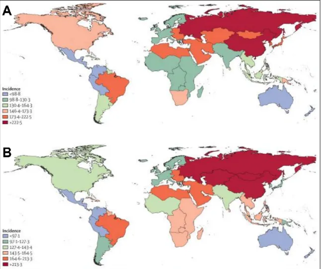
Stroke is the second most common cause of mortality and the third most common cause of disability worldwide. The incidence rate of stroke is changing inversely in the high- and low- income countries, with an increasing incidence in the low-income group (Krishnamurthi et al., 2013) (Fig. 1.). While the overall rate of stroke-related mortality is decreasing worldwide, the absolute number of people with stroke, stroke survivors, stroke-related deaths, and the global burden of stroke-related disability is high and increasing (Feigin et al., 2014).
7
Figure 1. Age-standardized incidence of ischemic stroke per 100 000 person-years for 1990 (A) and 2010 (B). During the last 20 years incidence rates were changing in the opposite directions in the high- and low- income countries (Krishnamurthi et al., 2013).
Globally, the incidence of stroke due to ischemia is 68 percent, while the incidence of hemorrhagic stroke (ICH and SAH combined) is 32 percent, reflecting a higher incidence of hemorrhagic stroke in low- and middle-income countries (Krishnamurthi et al., 2013).
Following multiple imaging studies reporting a broad range of incidence rates, the first prospective registry evaluating the frequency of ELVO among all-comer patients admitted for thrombolysis work up was conducted in the Capital Region of Denmark between July 2009 and December 2011 (Hansen et al., 2015). Among the 885 patients evaluated for the suspicion of hyper-acute stroke within 4.5 h after symptom onset, computed tomography
8
angiogram (CTA) was performed in 637 subjects, including 475 patients with a final diagnosis of IS (74.6%) and 162 (25.4%) diagnosed with transient ischemic attack (TIA).
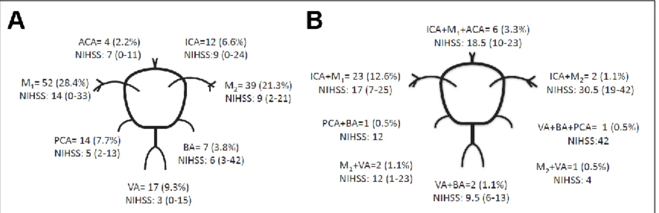
The rest of the patients were excluded from analysis due to ICH (n=99), stroke mimics (n=147) or technical reasons (n=2). Altogether 28.7% (n=183) of the patient undergoing CTA had large vessel occlusion with 15 different anatomical distributions (Fig. 2). Based on these results more than one out of five patients evaluated for thrombolysis harbored ELVO.
Important to note, that the majority of the large vessel occlusions involve the middle cerebral artery territory, confirming the adequacy of the tMCAO experiments for modelling this pathology.
Figure 2. Anatomical distribution of large vessel occlusions. Number and % of patients with intra-cranial occlusions and their median National Institute of Health Stroke Scale (NIHSS) score in single vessel (A) and multiple vessel (B) disease. (Hansen et al., 2015)
Currently no epidemiologic data is available on the frequency of ELVO in Hungary. The country-specific figures published recently in the framework of the EuroHope Register however suggest a particularly high incidence of hospitalization (407/100000) and a high case fatality rate (31% at 1 year) of stroke among the Hungarian population compared to other studied European Union countries (Malmivaara et al., 2015). While the authors have raised some queries about the reliability of the administrative data derived from hospital discharges, a more severe initial presentation of the stroke cases together with a potentially higher frequency of ELVOs due to the lack of effective cardiovascular prevention may also be responsible for the documented excess mortality.
9 The natural history of ELVO
A proximal occlusions in the main intra-cranial arteries affect large brain territories by severely decreasing their perfusion. In a prospective registry of patients with untreated, unilateral ELVO of the anterior circulation, less than half of the observed subjects (44%) could achieve functional independence, and almost one-fourth (22%) of them were dead at 6 months. The only predictors for good functional outcome at 6 months were less severe neurological status (NIHSS<11) at presentation (see Appendix), younger age, and better collateral circulation (Lima et al., 2014).
The adverse outcomes associated with the ELVOs are not only the consequence of the extensive loss of functional neuronal circuits, but the larger infarct size itself is also an independent predictor of symptomatic hemorrhagic transformation, which leads to further neurological worsening and potentially excess mortality (Tan et al., 2014).
Another dreadful complication of the ELVOs is the malignant middle cerebral artery syndrome, which is characterized by a complete infarction of the MCA territory accompanied by the mass effect of a space occupying edema. The malignant evolution of the infarction develops within 5 days of symptoms onset, and it is associated with about 80% mortality if left untreated (Hacke et al., 1996). The early identification of patients who are most likely to develop malignant edema after MCA infarction is of paramount importance to plan the necessary interventions, including a timely performed decompressive hemi-craniectomy (DHC) if necessary.
Many clinical variables were proposed as surrogate markers for adverse evolution. A fairly low positive predictive value can be attributed to NIHSS >20, thrombus at the carotid terminus location, and early involvement of >50% of the MCA territory on native CT.
Specificity is raised by the more precise volumetric analysis of the infarct size using MRI diffusion-weighted imaging: an infarct volume of >145cm3 measured within 14 hours of symptom onset detected patients at high risk with 100% sensitivity and 94% specificity.
Based on the results of randomized trials, early DHC significantly reduces mortality after malignant MCA infarction; however, it also increases the probability of survival with moderately severe disability. The early timing of the surgery is crucial in achieving favorable outcomes, and it appears to benefit younger patients the most (Staykov and Gupta, 2011).
10 Reperfusion therapy in ELVO
Until very recently, intravenous alteplase administered within 4.5 hours after symptom onset was the only reperfusion therapy with proven efficacy in patients with acute IS (Emberson et al., 2014). Besides the broad range of contra-indications of this therapeutic modality, including coagulopathy, recent surgery and previous intra-cranial bleeding, systemic thrombolysis was proven to be much less effective in opening proximal occlusions. In the ELVO cases involving the ICA terminus the early recanalization rate was only about 30%
using thrombolysis (Christou et al., 2001).
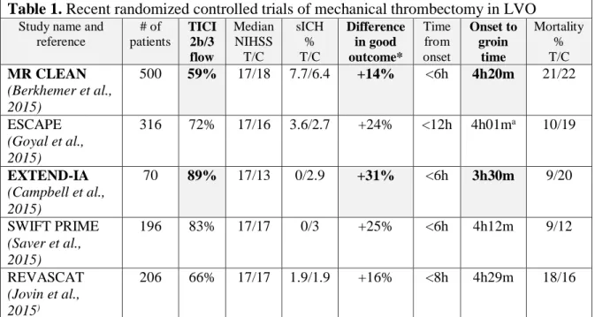
In 2015, five randomized clinical trials (Table 1) proved the higher efficacy of mechanical thrombectomy in the treatment of emergent LVOs compared to systemic thrombolysis, significantly changing the therapeutic recommendations for IS (Wahlgren et al., 2016). While the thorough presentation of these fundamental trials is beyond the scope of the thesis, some details are important to support the foundations of our experimental work.
Table 1. Recent randomized controlled trials of mechanical thrombectomy in LVO
Study name and reference
# of patients
TICI 2b/3 flow
Median NIHSS T/C
sICH
% T/C
Difference in good outcome*
Time from onset
Onset to groin
time
Mortality
% T/C MR CLEAN
(Berkhemer et al., 2015)
500 59% 17/18 7.7/6.4 +14% <6h 4h20m 21/22
ESCAPE (Goyal et al., 2015)
316 72% 17/16 3.6/2.7 +24% <12h 4h01ma 10/19
EXTEND-IA (Campbell et al., 2015)
70 89% 17/13 0/2.9 +31% <6h 3h30m 9/20
SWIFT PRIME (Saver et al., 2015)
196 83% 17/17 0/3 +25% <6h 4h12m 9/12
REVASCAT (Jovin et al., 2015)
206 66% 17/17 1.9/1.9 +16% <8h 4h29m 18/16
Table 1. Selected date from the five recent RCTs on mechanical thrombectomy show that faster and more effective recanalization leads to better outcome. *Functionally independent patients (mRS 0-2 at three months) a Time to reperfusion.
11
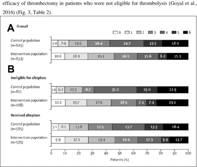
In contrast with the previous unsuccessful studies (Broderick et al., 2013; Kidwell et al., 2013), the new trials showing the improved functional outcomes associated with mechanical thrombectomy had some important common features. Only patients with confirmed large vessel occlusions were included, and the operators were able to rapidly achieve complete recanalization (TICI2b/3 flow) rate in a high proportion of the enrolled patients using the new generation stent retriever devices (Table 1). A recently published meta-analysis of the five trials showed similar positive results as the individual reports, and it also confirmed the efficacy of thrombectomy in patients who were not eligible for thrombolysis (Goyal et al., 2016) (Fig. 3, Table 2).
Figure 3. Functional outcomes of mechanical thrombectomy. Distribution of modified Rankin Scale scores at 90 days in the intervention and control groups in the overall trial population (A) and for patients treated with, or ineligible for intravenous alteplase (B). Data from the metaanalysis of the five RCTs (Goyal et al., 2016).
12
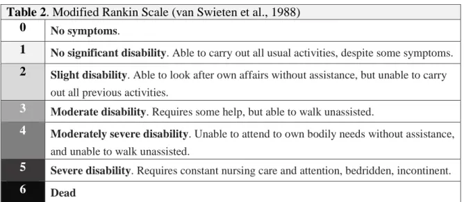
Table 2. Modified Rankin Scale (van Swieten et al., 1988)
0 No symptoms.
1 No significant disability. Able to carry out all usual activities, despite some symptoms.
2 Slight disability. Able to look after own affairs without assistance, but unable to carry out all previous activities.
3 Moderate disability. Requires some help, but able to walk unassisted.
4 Moderately severe disability. Unable to attend to own bodily needs without assistance, and unable to walk unassisted.
5 Severe disability. Requires constant nursing care and attention, bedridden, incontinent.
6 Dead
Table 2. Standardized classification of functional outcomes following neurological injury according to Modified Rankin Scale (van Swieten et al., 1988).
The speed of recanalization is probably the most important factor determining the efficacy of thrombectomy (Fig. 3), as the amount of salvageable brain tissue and the chances of achieving good perfusion both steadily decrease with time. For every hour of reperfusion- delay, the initially large benefit of IAT decreases; the absolute risk difference for a good outcome is reduced by 6% per hour of delay (Fransen et al., 2016).
While the importance of optimizing the time window cannot be overemphasized, we have to keep in mind that some optimally chosen patients with good collaterals may also benefit from thrombectomy beyond 6 hours from the onset of symptoms (Goyal et al., 2015).
There is an ongoing debate over the optimal use of imaging modalities to select patients for mechanical reperfusion therapy, especially to identify potential candidates among the late presenters. CT based perfusion imaging (Campbell et al., 2015) and the imaging of collaterals (Goyal et al., 2015) were both used in the recent clinical trials, and with certain limitations, they both proved to be fairly accurate in guiding patient selection. Yet other authors claim that MRI may be the ideal modality (Wouters et al., 2016), as the DWI/perfusion mismatch clearly defines the unviable territory as well as the salvageable brain tissue. The optimal selections criteria for such patients is yet to be determined by further clinical research.
13
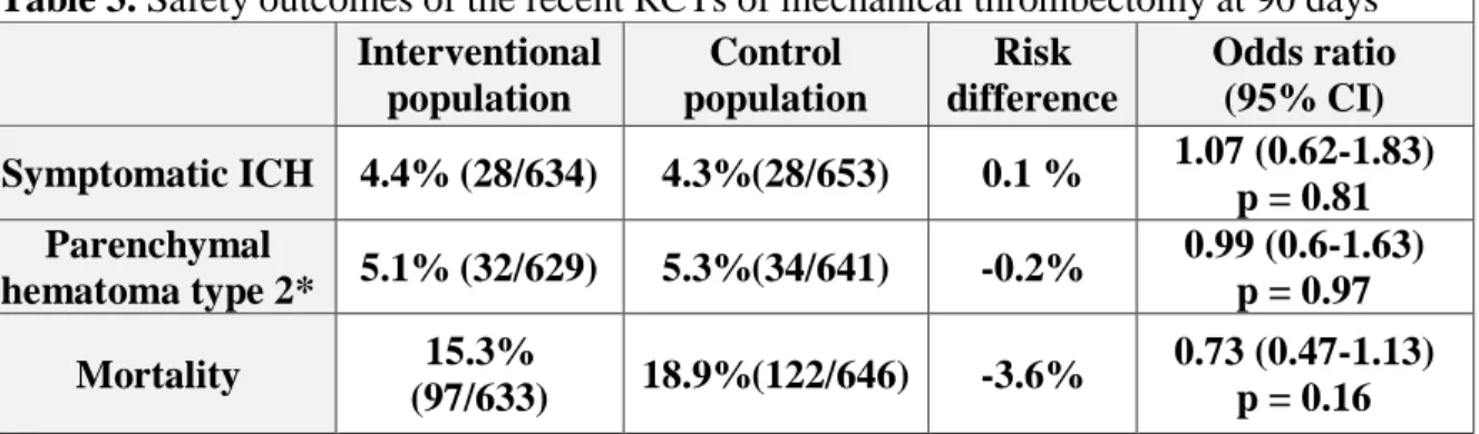
Safety issues of thrombectomy: prevention of bleeding complications and re-infarction A very important aspect of the interventional treatment of ELVOs is the safety of the procedures. The analysis of the procedural complications did not show any excess bleeding or mortality associated with the more invasive interventional treatment (Table 3). We have to remember however, that the systemic thrombolysis alone already increases bleeding risk compared to the conservative management without reperfusion therapy, causing an average absolute increased risk of early death from ICH of about 2% (Emberson et al., 2014).
Therefore further development is needed to prevent the reperfusion associated ICH, and the efficacy of thrombectomy alone in thrombolysis eligible patient should be also tested in new randomized controlled trails.
Table 3. Safety outcomes of the recent RCTs of mechanical thrombectomy at 90 days Interventional
population
Control population
Risk difference
Odds ratio (95% CI) Symptomatic ICH 4.4% (28/634) 4.3%(28/653) 0.1 % 1.07 (0.62-1.83)
p = 0.81 Parenchymal
hematoma type 2* 5.1% (32/629) 5.3%(34/641) -0.2% 0.99 (0.6-1.63) p = 0.97 Mortality 15.3%
(97/633) 18.9%(122/646) -3.6% 0.73 (0.47-1.13) p = 0.16 Table 3. Meta-analysis of the safety outcomes of the five recent thrombectomy trials showed no risk difference between the treatment groups (Goyal et al., 2016) *Blood clot occupying
>30% of the infarcted territory with substantial mass effect.
The optimal estimation of the hemorrhagic and thrombotic risk of each individual patient is crucial for optimizing treatment outcomes. The actual stroke etiology as well as the extent of the brain damage has to be taken into account while determining the timing and composition of the antithrombotic regimen following cerebral large vessel recanalization. Evidence from a randomized trial confirms, that in the first 24 hours following treatment with intravenous alteplase, aspirin or other antithrombotic agent should not be given (Zinkstok and Roos, 2012). Afterwards the use of early aspirin(160 to 325 mg/day) rather than no aspirin therapy or early anticoagulation is recommended for most patients with acute ischemic stroke or TIA (Jauch et al., 2013; Lansberg et al., 2012). Early parenteral
14
anticoagulation rather than aspirin is only suggested for select patients with acute cardio- embolic ischemic stroke, who have a high suspicion for intracardiac thrombus, as these patients are at high risk for recurrent ischemic stroke. In atrial fibrillation, early therapeutic anticoagulation following acute stroke should be reserved to those harboring only minor intracerebral lesions based on control imaging, since in unselected patients the clinical benefit from the prevention of recurrent strokes was offset by the excess occurrence of intra-cranial bleeds (Sandercock et al., 2015).
Stroke patients presenting with significant intracranial large vessel stenosis represent a special subgroup, and according to recent evidence, they require early double anti-platelet therapy (DAPT) with the combination of aspirin and clopidogrel (Derdeyn et al., 2014).
Patients receiving carotid stents as part of their acute stroke treatment also require DAPT after the exclusion of hemorrhagic transformation.
Important to note, that current antithrombotic strategies are mainly based on data from the old randomized trials of thrombolysis, therefore their validity following thrombectomy is not proven. Due to the fundamentally different recanalization efficacy and hemodynamic effect of the two reperfusion modalities, there is an urgent need for new RCTs in order to gather further evidence guiding the antithrombotic therapy following thrombectomy.
Another important, but currently less emphasized source of major complications in interventional stroke treatment is the access site bleeding. Due to the common co-occurrence of peripheral arterial disease and stroke, and the current therapeutic guidelines supporting the combined use of thrombolysis and thrombectomy, femoral artery punctures carry a non- negligible risk. Data from a recent metaanalysis comparing the safety of access sites in acute percutaneous coronary intervention show, that the use of radial artery can reduce vascular complication’s rate and even mortality compared to femoral access (Ando and Capodanno, 2015). Our group has published a randomized trial comparing radial and femoral puncture during carotid artery stenting, and our results confirmed, that radial puncture allows efficient access for the treatment of vascular lesions of the neck in most cases (Ruzsa et al., 2014).
Our findings also implicate, that radial approach may be used for thrombectomy in the future to avoid the complications associated with femoral puncture.
15
Potential adjunctive strategies to improve the functional outcomes of ELVO
The percutaneous revascularization of cerebral large vessel occlusions by mechanical thrombectomy is obviously a huge step forward in the optimal treatment of IS, and based on the above cited evidences the new therapeutic guidelines have given the highest level of recommendation for this treatment modality in eligible patients (Powers et al., 2015;
Wahlgren et al., 2016). Ongoing clinical trials will help to further improve the patient selection in order to avoid the futile, and in some cases even harmful revascularizations.
However there remains a large demand for the development of adjunctive therapies to offer alternative solutions to help the patients, who are non-eligible for reperfusion.
The infarcted brain tissue represents the common final stage of various different cascade mechanisms of cellular apoptosis and necrosis. The closely interrelated events occurring in the affected brain show a wide regional variety and diverse temporal pattern, making their investigation very challenging by conventional research methods. The system biology approach may provide a new insight into the pathophysiology of the brain vasculature (Clegg and Mac Gabhann, 2015), however the great amount of new information available at present time is not yet ready to formulate a new unifying concept for paving the foundations for new strategies of future stroke therapy.
Besides improving the outcomes with early reperfusion therapy, neuroprotective strategies or the effective augmentation of post-ischemic neuro-restorative mechanisms could be an alternative method to improve the prognosis of patients suffering from ELVO, however these approaches are still very far from being established treatment modalities. In order to find new therapeutic targets in stroke therapy, a fundamental understanding of the pathophysiology of ischemic brain lesions is crucial.
In the subsequent sections we will present the results of our recent investigations of two different pharmacological interventions, subacute gelatinase inhibition and selegiline treatment on a standard experimental model of ELVO in rodents. While the two treatments have been shown to inversely influence the functional outcomes after focal ischemia, their mechanism of action has not been fully explored before. We used an mRNA expression array analysis to study the effect of the interventions on the genes influencing post-stroke recovery.
16
Overview of the key regulatory genes influencing post-stroke recovery
The concept of the neurovascular unit (NVU) emphasizes the fact that neurons, astrocytes, smooth muscle cells, endothelial cells, pericytes, basement membranes, and the extracellular matrix all dynamically interact in the pathobiology of stroke (del Zoppo, 2009). The extent of recovery following brain injury is determined by the complex interplay of parallel processes involving the NVU, including synaptic and microvascular remodeling, inflammation, apoptosis, oxidative stress and structural alterations of the extracellular matrix.
We sought to investigate the simultaneous changes induced by the two tested pharmacological interventions on the mRNA expression profile of the most important regulatory genes driving the remodeling processes of the penumbra. Despite the presentation of the key genes under distinct categories, it is important to keep in mind their redundancy, and their potential parallel regulatory roles.
Stroke induces a process of axonal sprouting in the peri-infarct tissue that results in a substantial re-mapping of the connections of the cortical areas adjacent to the infarct. This post-stroke axonal sprouting response is correlated in location and magnitude with functional recovery (Calautti and Baron, 2003). Nervous system injury induces expression of both growth-promoting and growth-inhibitory genes that together determine the location and degree of axonal sprouting.
The growth-promoting gene products (Table 4A) mediate growth cone membrane signaling events, transcriptional control in the regenerating neuron, cytoskeletal reorganization and axonal extension, and include GAP43, CAP23, MARCKS, c-jun, members of the stathmin family, Tα1 tubulin, L1, p21/waf1, and SPRR1. These proteins are specifically upregulated during sprouting, reduce axonal sprouting when knocked down or out, and mediate enhanced axonal sprouting when overexpressed (Benowitz et al., 2002; Bomze et al., 2001;
Carmichael, 2003; Laux et al., 2000).
Stroke induces sequential waves of neuronal growth-promoting genes during the sprouting response: an early expression peak (SPRR1), a mid expression peak (p21, Ta1 tubulin, L1, MARCKS), a late peak (SCG10, SCLIP), and an early/sustained pattern (GAP43, c-jun). The expression of the growth inhibiting genes (Table 4B) show biphasic pattern. The growth-
17
inhibiting chondroitin sulfate proteoglycans aggrecan, brevican, versican, and phosphacan are induced late in the sprouting process; while the developmentally associated growth inhibitors ephrin-A5, ephB1, semaphorin IIIa, and neuropilin 1 are induced in the early phases of the sprouting response. At the cellular level, chondroitin sulfate proteoglycans are reduced in the region of axonal sprouting, during the peak of growth-promoting gene expression (Carmichael et al., 2005).
Table 4A Genes promoting axonal sprouting
Abbreviation Name NCBI Reference
Sequence
GAP 43 Growth associated protein 43 NM_017195.3
C-jun Jun proto-oncogene NM_021835.3
CAP23 Membrane attached signal protein 1 NM_022300.1
SPRR1 Small proline-rich protein 1 XM_574984.2
P21 Cyclin dependent kinase inhibitor1A NM_080782.3
L1 cam L1 cell adhesion molecule NM_017345.1
Marcks Myristoylated alanine rich protein kinase XM_002728965.1
DCC Deleted in colorectal carcinoma NM_012841.1
Cuzd1 CUB and zona pellucida-like domains 1 NM_054005.1
NFM Neurofilament N NM_017029.1
Ntn 1 Netrin1 NM_053731.1
Table 4B Sprouting inhibitor genes
Ncan Neurocan NM_031653.1
Vcan Versican NM_001170558.1
Ptprz1 Protein tyrosine phosphatase receptor Z1 NM_001170685.1
Cspg4 Chondroitin sulfate proteoglycan 4 NM_031022.1
Bcan Brevican NM_001033665.1
Sema3a Semaphorin 3A NM_017310.1
Nrp1 Neuropilin NM_145098.2
EphB1 Eph receptor B1 NM_001104528.1
Rtn4 Reticulon 4 (Nogo A) NM_031831.1
Rtn4r Reticulon 4 receptor (Nogo receptor) NM_053613.1 Ngfr (P75NTR) Nerve growth factor receptor NM_012610.2 Lingo 1 Leucine rich repeat and Ig domain containing 1 NM_001100722.1
Mag Myelin associated glycoprotein NM_017190.4
Mog Oligodendrocyte glycoprotein NM_022668.2
PirB Paired Ig-like-receptor B XM_001076302.2
Table 4. Genes promoting axonal sprouting (A) and sprouting inhibitors (B) included in the TaqMan® array analysis presented by the NCBI reference sequence, name and abbreviation.
18
The spatial and temporal dynamics of cerebral micro-vascular rearrangement after stroke are also closely related to neurological recovery. In a human post-mortem study sprouting angiogenesis was documented 5-92 days after stroke in the penumbra region (Krupinski et al., 1994). Scanning electron microscopic study of animal stroke models demonstrated, that radially arranged neocortical arterioles and small veins lost their regular patterns within one day of occlusion, and soon afterwards started to form a very dense network of anastomosing micro-vessels (Krupinski et al., 2003).
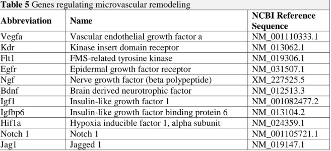
Compromised brain circulation activates complex processes that could facilitate the restoration of blood flow in the penumbra region. The low oxygen level prompts early response by activating oxygen responsive molecules, cytokines and other inflammatory factors (Table 5) (Beck and Plate, 2009). HIF-1a, the main regulator activates a series of genes responsible for metabolic changes in the penumbra, vasomotor tone and angiogenesis.
Angiogenesis (sprouting or splitting and proliferating of endothelial cells in the preexisting blood vessel), vasculogenesis (de novo vessel growth) and arteriogenesis (transforming of small arteries into larger conducting arteries) result in neovascularization of the affected brain territory (Risau, 1997).
Table 5. Genes promoting microvascular remodeling included in the TaqMan® array analysis presented by the NCBI reference sequence, name and abbreviation.
Table 5 Genes regulating microvascular remodeling
Abbreviation Name NCBI Reference
Sequence
Vegfa Vascular endothelial growth factor a NM_001110333.1
Kdr Kinase insert domain receptor NM_013062.1
Flt1 FMS-related tyrosine kinase NM_019306.1
Egfr Epidermal growth factor receptor NM_031507.1
Ngf Nerve growth factor (beta polypeptide) XM_227525.5
Bdnf Brain derived neurotrophic factor NM_012513.3
Igf1 Insulin-like growth factor 1 NM_001082477.2
Igfbp6 Insulin-like growth factor binding protein 6 NM_013104.2 Hif1a Hypoxia inducible factor 1, alpha subunit NM_024359.1
Notch 1 Notch 1 NM_001105721.1
Jag1 Jagged 1 NM_019147.1
19
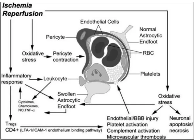
The reperfusion-mediated injury of the blood–brain barrier (BBB) is an important mechanism of the transformation from ischemic to hemorrhagic stroke. In addition to hemorrhage, a subset of patients do not improve after successful recanalization despite restored cerebral circulation (Dalkara and Arsava, 2012). Reperfusion injury is defined as a biochemical cascade causing a deterioration of ischemic brain tissue that parallels and antagonizes the beneficial effect of recanalization (Yang and Betz, 1994).
Figure 4. Schematic model of neurovascular mechanism of post-ischemic reperfusion injury.
During reperfusion after ischemia, overproduction of reactive oxygen species (ROS) causes oxidative stress. The oxidative stress damages the endothelial cells, resulting in an exposure of the sub-endothelial extracellular matrix to blood flow. The exposure triggers adhesion and activation of platelets in microvasculature causing thrombosis. The injured endothelial cells release metalloproteinase that attacks basal lamina causing leakage of the blood–brain barrier (BBB)(Bai and Lyden, 2015).
20
Table 6. Genes involved in the development of reperfusion injury included in the TaqMan®
array analysis presented by the NCBI reference sequence, name and abbreviation.
During post-ischemic reperfusion in a focal transient MCAO rat model, BBB disruption is typically observed to be more severe than when compared with permanent MCAO.
Furthermore, increased cerebral blood flow volume during reperfusion is correlated to worsened BBB disruption (Yang and Betz, 1994). Post-ischemic reperfusion causes enhanced production of free radicals and release of proteases from endothelial cells, astrocytes, microglia, and neurons. The matrix metalloproteinases (MMPs) play a major role mediating attack on the basal lamina in cerebral capillaries (Yang et al., 2007).
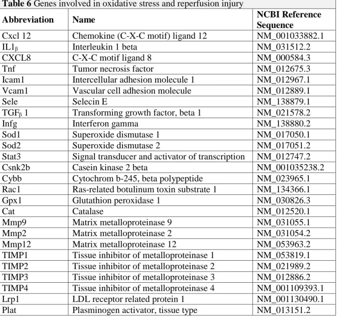
Table 6 Genes involved in oxidative stress and reperfusion injury
Abbreviation Name NCBI Reference
Sequence
Cxcl 12 Chemokine (C-X-C motif) ligand 12 NM_001033882.1
IL1ᵦ Interleukin 1 beta NM_031512.2
CXCL8 C-X-C motif ligand 8 NM_000584.3
Tnf Tumor necrosis factor NM_012675.3
Icam1 Intercellular adhesion molecule 1 NM_012967.1
Vcam1 Vascular cell adhesion molecule NM_012889.1
Sele Selecin E NM_138879.1
TGFᵦ 1 Transforming growth factor, beta 1 NM_021578.2
Infg Interferon gamma NM_138880.2
Sod1 Superoxide dismutase 1 NM_017050.1
Sod2 Superoxide dismutase 2 NM_017051.2
Stat3 Signal transducer and activator of transcription NM_012747.2
Csnk2b Casein kinase 2 beta NM_001035238.2
Cybb Cytochrom b-245, beta polypeptide NM_023965.1
Rac1 Ras-related botulinum toxin substrate 1 NM_134366.1
Gpx1 Glutathion peroxidase 1 NM_030826.3
Cat Catalase NM_012520.1
Mmp9 Matrix metalloproteinase 9 NM_031055.1
Mmp2 Matrix metalloproteinase 2 NM_031054.2
Mmp12 Matrix metalloproteinase 12 NM_053963.2
TIMP1 Tissue inhibitor of metalloproteinase 1 NM_053819.1 TIMP2 Tissue inhibitor of metalloproteinase 2 NM_021989.2 TIMP3 Tissue inhibitor of metalloproteinase 3 NM_012886.2 TIMP4 Tissue inhibitor of metalloproteinase 4 NM_001109393.1
Lrp1 LDL receptor related protein 1 NM_001130490.1
Plat Plasminogen activator, tissue type NM_013151.2
21
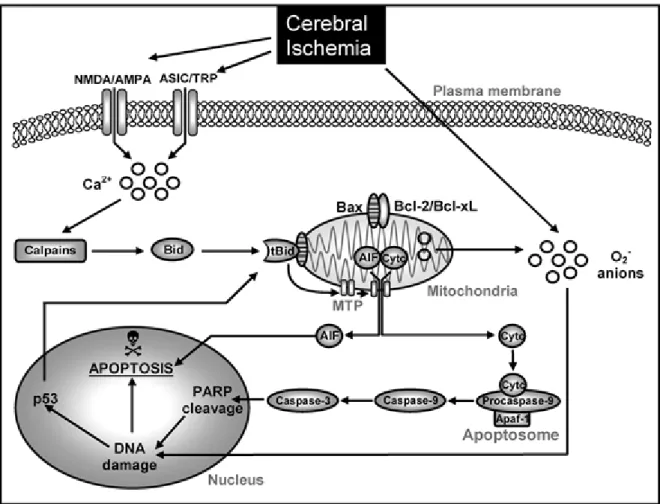
The core of brain tissue exposed to the most dramatic blood flow reduction is fatally injured and subsequently undergoes necrotic cell death within minutes of a focal ischemic stroke. A zone of less severely affected tissue around the necrotic core, also called as the “ischemic penumbra” is rendered functionally silent by reduced blood flow but remains metabolically active (Ginsberg, 1997). Many neurons in the penumbra region may undergo apoptosis after several hours or days, and thus they are potentially recoverable for some time after the onset of stroke. Apoptosis can be initiated by internal events (ie, “Intrinsic Pathway”) (Fig. 5.) involving the disruption of mitochondria and the release of the cytochrome C, or alternatively, cell surface receptors can be activated by specific ligands that bind to “death receptors” (ie, “Extrinsic Pathway”) (Fig. 6.) (Table 7A).
Figure 5. Intrinsic signaling of apoptosis in cerebral ischemia (Broughton et al., 2009) Cerebral ischemia elevates cytosolic calcium levels through the stimulation by glutamate of N-methyl-D-aspartate (NMDA) or D,L-α-amino-3-hydroxy-5-methyl-isoxazolpropionic
22
acid (AMPA) receptors, or by the activation of acid-sensing ion channels (ASICs) . Increased intracellular calcium activates calpains and mediates cleavage of Bid to truncated Bid (tBid) initiating the intrinsic signaling cascade of apoptosis. At the mitochondrial membrane tBid interacts with apoptotic proteins such as Bad and Bax, which is typically neutralized by antiapoptotic B-cell leukemia/lymphoma 2 (Bcl-2) family proteins Bcl-2 or Bcl-xL. After heterodimerization of proapoptotic proteins with tBid, mitochondrial transition pores (MTP) are opened, thus releasing cytochrome c (Cytc) or apoptosis-inducing factor (AIF). Once released into the cytosol, Cytc binds with apoptotic protein-activating factor-1 (Apaf-1) and procaspase-9 to form an “apoptosome,” which activates caspase-9 and subsequently caspase- 3. Activated caspase-3 cleaves nDNA repair enzymes, such as poly (ADP-ribose) polymerase (PARP), which leads to nDNA damage and apoptosis. By contrast, AIF translocates rapidly to the nucleus where it mediates large-scale DNA fragmentation and cell death in a caspase- independent manner. In addition, nuclear pathways of neuronal apoptosis are activated in response to DNA damage, for example, through phosphorylation and activation of p53.
Furthermore, cerebral ischemia and reperfusion generate superoxide anions (O2−), which causes DNA damage (Broughton et al., 2009).
Extrinsic mechanisms of apoptosis involve the engagement of death receptors located on the plasma membrane and is hence also referred as the “death receptor pathway”. Cell surface death receptors belong to the tumor necrosis factor receptor (TNFR) superfamily, and include TNFR-1, Fas, and p75NTR.Forkhead1, a member of the forkhead family of transcription factors, stimulates expression of target genes, such as the Fas ligand (FasL) (Sugawara et al., 2004). The extracellular FasL binds to Fas death receptors (FasR), which triggers the recruitment of the Fas-associated death domain protein (FADD). FADD binds to procaspase- 8 to create a death-inducing signaling complex (DISC), which activates caspase-8. Activated caspase-8 either mediates cleavage of Bid to truncated Bid (tBid), which integrates the different death pathways at the mitochondrial checkpoint of apoptosis, or directly activates caspase-3. At the mitochondrial membrane tBid interacts with Bax, which is usually neutralized by antiapoptotic B-cell leukemia/lymphoma 2 (Bcl-2) family proteins Bcl-2 or Bcl-xL. Dimerization of tBid and Bax leads to the opening of mitochondrial transition pores
23
(MTP), thereby releasing cytochrome c (Cytc), which execute caspase 3-dependent cell death (Broughton et al., 2009).
Figure 6. Extrinsic signaling of apoptosis after cerebral ischemia (Broughton et al., 2009) Interactions between the pro-apoptotic and anti-apoptotic Bcl-2 family proteins on the outer mitochondrial membrane are believed to play an important role in cell survival (Love, 2003).
Typically, anti-apoptotic members (Table 7B) are located on the outer membrane and suppress apoptosis through multiple mechanisms, including conserving mitochondrial membrane potential, inhibiting the release of apoptotic proteins, stabilizing the mitochondrial transition pores, thus preventing its opening, and controlling the activation of caspase proteases (Webster et al., 2006).
24
Table 7. Pro-apoptotic (A) and anti-apoptotic (B) genes included in the TaqMan® array analysis presented by the NCBI reference sequence, name and abbreviation.
Table 7A Pro-apoptotic genes
Abbreviation Name NCBI Reference
Sequence
Bax Bcl2-associated X NM_017059.1
Bid BH3 interacting domain death agonist NM_022684.1
Casp1 Caspase 1 NM_012762.2
Casp2 Caspase 2 NM_022522.2
Casp3 Caspase 3 NM_012922.2
Casp6 Caspase 6 NM_031775.2
Casp7 Caspase 7 NM_022260.2
Casp8ap2 Caspase 8 associated protein 2 NM_001107921.1
Casp9 Caspase 9 NM_031632.1
Card 10 Caspase recruitment domain family NM_001130554.1 Bcl2/11 Bcl2 like 11 transcript variant 1 NM_022612.1
Fadd Fas associated via death domain NM_152937.2
Apaf1 Apoptotic peptidase activating factor 1 NM_023979.1 Diablo Diablo, IAP-binding mitochondrial protein NM_001008292.1
Cycs Cytochrome c NM_012839.2
Bcl3 B-cell CLL/lymphoma 3 NM_001109422.1
Pidd1 P53-induced death domain protein 1 NM_001106318.2 Cradd CASP2 and RIPK1 domain containing adaptor
with death domain NM_001108085.1
Table 7B Anti-apoptotic genes
Abbreviation Name NCBI Reference
Sequence
Proc Protein c NM_012803.1
Hspa1a Heat shock 70kD protein 1A NM_031971.2
Procr Protein c receptor NM_001025733.2
Fr2 Coagulation factor II receptor NM_012950.2
Xiap X-linked inhibitor of apoptosis NM_022231.2
Ikbkg Inhibitor of kappa light peptide gene enhancer
in b-cells, kinase gamma NM_199103.1
Nkap NFKB activating protein NM_001024872.1
Birc3 Baculovira IAP repeat containing 3 NM_023987.2
Naip NLR family apoptosis inhibitor protein XM_226742.4
Bcl2l2 Bcl2-like 2 NM_021850.2
Bcl2l1 Bcl2-like 1 NM_001033671.1
Ripk1 Receptor interactin serin/threonine kinase 1 NM_001107350.1
25 AIMS:
Investigation of the molecular background of impaired recovery associated with gelatinase inhibition in the subacute phase of ELVO
The matrix metalloproteinase (MMP) 2 and 9 enzymes, also called as gelatinases (Fig. 7A), belong to the serine-protease enzyme family, and they are important factors in the hemorrhagic transformation of the ischemic brain lesions: they promote blood-brain barrier injury, and accelerate cerebral cell death (Asahi et al., 2001; Lee and Lo, 2004).Furthermore MMP 9 activity is enhanced by the thrombolytic agent used during reperfusion therapy (Burggraf et al., 2007), therefore the inhibition of MMP enzymes could be an ideal adjunctive treatment to prevent the hemorrhagic complications.
Contrary to their deleterious effect on acute lesions, gelatinases may mediate repair mechanisms during the delayed phases of stroke recovery. Pharmacological gelatinase inhibition 7 days after ischemic brain injury increased lesion size and impaired functional regeneration (Zhao et al., 2006).
Besides their proven interaction with neurovascular remodeling (Zhao et al., 2006), gelatinases may influence recovery through interaction with multiple growth promoting and inhibitory proteins. Gelatinase activity was demonstrated at the edge of the growth cone of dorsal root ganglion neurons (Hayashita-Kinoh et al., 2001), and the expression of phosphorylated neurofilament M, a marker for regenerative elongation, was induced by MMP9 (Demestre et al., 2004). Axonal re-myelination was also shown to require the proteolytic activity of the MMP9 (Oh et al., 1999), while the metalloproteinase mediated cleavage of the Reticulon 4 receptor (Rtn4r) was proposed as a potential disinhibitory mechanism that could enhance axonal regrowth (Walmsley et al., 2004). The same receptor was shown to mediate the astrocytic differentiation of neural progenitor cells, therefore the MMP-Rtn4r interaction may also influence the glial scar formation following central nervous system injury (Wang et al., 2008).
The aim of our study was to explore the molecular background of the impaired recovery associated with sub-acute metalloproteinase inhibition in ischemic stroke.
26
Investigation of the neuro-restorative action of Selegine in ELVO
Selegiline (Fig. 7B) is widely used in the therapy of Parkinson's disease (Tatton et al., 1994).
Its effectiveness is based on the selective inhibition of monoamine oxidase B (MAO-B) and on its anti-apoptotic potential attributed to its ability to eliminate reactive oxygen species and stabilize mitochondrial membrane potential (Simon et al., 2005; Szilagyi et al., 2009).
In in vitro and animal studies our group has demonstrated previously that selegiline attenuates apoptosis and decreases infarct size after permanent occlusion of the middle cerebral artery (Simon et al., 2001). Furthermore, selegiline attenuated spatial learning deficits following focal cerebral ischemia in rats (Puurunen et al., 2001). The clinical use of the administration of low dose selegiline in the sub-acute phase of cerebral infarction has also been tested in a randomized, double-blinded, clinical pilot study. Although significant functional improvement was observed in stroke patients receiving selegiline in combination with physiotherapy, the molecular basis underlying this beneficial effect remained unclear (Sivenius et al., 2001). As the functional improvements were detected with the therapy being initiated days after the onset of stroke, the clinical efficacy was attributed to a potential neurorestorative rather than to the previously known neuroprotective properties of the molecule. This also means, that the selegiline treatment may potentially benefit those stroke patient who could not undergo reperfusion therapy as well.
To understand the mechanisms how selegiline exerts its actions following focal ischemia, we addressed its effects on gene expression.
Figure 7. The crystal structure of the human Gelatinase B (Matrix Metalloproteinase 9) enzyme (A). (Elkins et al., 2002) and the structural formula of the (R) (-) deprenyl (Selegiline) molecule (B) (Knoll et al., 1978).
A B
27 METHODS
The transient Middle Cerebral Artery Occlusion Model
Figure 8. The MCAO filament technique. Schematic diagram displaying the rat carotid artery territories and the positioning of the MCAO filament (Longa et al., 1989).
The experimental model applied in our investigations to model the pathophysiology of thrombectomy in intra-cranial ELVO has been used for simulating stroke in rodents for decades (Longa et al., 1989). Important to note however, that the abrupt restoration of blood flow with the removal of the occluding filament simulate the hemodynamic changes associated with mechanical thrombectomy much better, than the hemodynamics of the gradual reperfusion occurring during thrombolysis.
28
Adult male CD rats (Charles River, Erkrath, Germany) were housed under controlled environmental conditions at an ambient temperature of 22°C with 12h light/dark cycle and free access to food and water. Brain ischemia was induced using slightly modified version of middle cerebral artery (MCA) filament occlusion model (Longa et al., 1989). Briefly, general anesthesia was induced using 5% isofluran in pure O2 at 1 L/min for 4 min in male rats weighing 300-350 g. Animals were kept anaesthetized using 2% isoflurane in pure O2
delivered by snout mask. Animal core (rectal) temperature was maintained constant at 37°C using temperature-controlled heat pad. A midline neck incision was made to expose the right carotid sheath under the operating microscope (Zeiss, Germany) The common carotid artery was isolated with 4-0 silk, and the occipital, pterygopalatine, and external carotid arteries were each isolated, cauterized, and divided. Right MCAO was accomplished by forming a small arteriotomy in the external carotid stump, and advancing a 25 mm 4-0 nylon suture with a 5 mm cast rubber tip (Doccol, Redlands, CA) through the arteriotomy into the internal carotid artery until the tip occluded the MCA (Fig 8.). To ensure adequate levels of cerebral ischemia, transcranial measurements of cerebral blood flow (CBF) were made over the MCA territory using Laser-Doppler Flowmetry (Perimed Inc., Stockholm, Sweden). Placement of the suture was confirmed by reduction of LDF readings to at least 40% of baseline (Fig. 9.).
After 60 min of ischemia, animals were re-anesthetized and the occluding suture was removed.
Figure 9. Trans-cranial measurements of the blood flow during tMCAO show a sudden drop in the cerebral perfusion at the time of the insertion of the occluding filament. (A). TTC staining of the native coronal brain section at bregma level delineates the ischemic lesion (*) by the lack of red staining of the viable mitochondria (B).
29 Stereotactic delivery of gelatinase inhibitor
Figure 10. Stereotaxic injection for the delivery of the gelatinase inhibitor, rat specific coordinates: 0.8 mm caudal to Bregma, lateral: 1.6 mm to midline; depth: 4.1 mm to skull surface (A). Coronal section of a native rat brain demonstrating the successful stereotactic injection of Evans Blue dye into the right lateral ventricle (B).
Due to the excessive amount of inhibitor necessary for achieving sufficient gelatinase inhibitions intra-cranially with the currently available semi-selective compounds during systemic administration, we have decided to apply intra-cranial drug delivery using a dedicated stereotactic device. We performed pilot experiments on similar size rats (n=3) to ensure the adequate calibration of the stereotactic device (Harvard Apparatus, Boston, MA) using Evans Blue dye before applying the inhibitor treatments (Fig 10.) Finally 7 days after the tMCAO, we injected 20 µl of FN-439 metalloproteinase inhibitor (0.59 mg/kg, 720 mM in saline) or saline, into their right lateral ventricle of the animals over 20 min, using the previously calibrated stereotaxic injector under general anesthesia.
Administration of selegiline treatment
Selegiline treatment was started 48 hours following the initiation of the 60 min tMCAO.
Animals in the treatment arm (n=6-6) were infused with 0.2 mg/kg/day of selegiline in a vehicle of 0.9% physiological saline, delivered via implantable osmotic pumps (Alzet, ALZA, Palo Alto, CA) placed intra-peritoneal for seven days (Fig. 11). Control rats (n=6-6) were infused with the vehicle only. The pumps were placed in the abdominal cavity under short general anesthesia. The rats were sacrificed after 7 days of treatment.
A B
30
Figure 11. Experimental protocol of selegiline treatment. Continuous infusions were initiated 48 hours after MCAO. The animals underwent MRI investigation 24h before sacrifice.
Quantification of brain edema
Magnetic resonance imaging (MRI) study was performed on a 1.5 Tesla Philips Achieva clinical scanner using a SENSE-Flex-M coil 6 days after the initiation of treatment. After being anaesthetized with a bolus of ketamine/xilazin cocktail, animals were placed in supine position inside the clinical MRI scanner, and brain edema was visualized with a T2 weighted 3D VISTA sequence (Field of view: (AP/RL/FH) 60/60/45 mm, Voxel size: 0.6/0.6/1.2 mm, Reconstruction matrix = 128, number of slices =75, Echo time was set to the shortest possible, which resulted in TE=301 ms. Flip angle =90°; TR=2250 ms, NSA=5).
The obtained images were contrast enhanced to better differentiate edema from the background. Subsequently, planimetric measurements were performed on the coronal sections 7 mm posterior to the frontal pole using Image J software (NIH, Bethesda, USA).
Animals with extensive cortical infarctions, potentially caused by failed reperfusion (1 animal in each group) were excluded from the statistical analysis.
The Animal Examination Ethical Council of the Animal Protection Advisory Board at the Semmelweis University, Budapest specifically approved these studies. Thus, the procedures involving rats were carried out according to experimental protocols that meet the guidelines of the Animal Hygiene and Food Control Department, Ministry of Agriculture, Hungary, which is in accordance with EU Directive 2010/63/EU for animal experiments EU Directive 2010/63/EU for animal experiments.
The animal experiments were carried out at the Experimental laboratory of the Heart and Vascular Center of Semmelweis University, and the imaging studies were done at the MRI Unit of the Heart and Vascular Center.
31
Tissue sampling and mRNA extraction for the TaqMan® array analysis
Nine days after the tMCAO, the animals were decapitated and their brains were rapidly removed. Coronal sections (12 m thick) were cut from the native brain tissue using a rat brain matrix (Harvard Apparatus, Boston, USA) starting 6 mm caudal to the frontal pole.
Tetraphenyl-tetrazolium chloride (TTC) staining was performed on the first coronal sections (6-8 mm from the frontal pole of the brain) to identify the lesion. After 15 min of incubation at 37°C, 3 samples of brain tissue from the border of the ischemic lesion were dissected using a 2 mm diameter punch biopsy needle. Total RNA was extracted with TRIzol reagent (Invitrogen, Carlsbad, CA) according to the manufacturer’s instructions. The extracted RNA was quantified using UV spectrophotometry; only samples with a 260:280 ratio greater than 1.7 were further processed (Fig 12.). RNA was reverse transcribed in duplicates using Omniscript kit (Qiagen, Valencia, CA). A third RNA sample was incubated without reverse transcriptase as a no-RT control.
Figure 12. UV spectrophotometry analysis of a good quality total RNA sample with a 28s/18s RNA ratio > 1,7.
32 TaqMan® array analysis
Samples were run parallel on a custom designed TaqMan® gene expression array containing probes for 84 representative genes involved in axonal sprouting, apoptosis regulation, reperfusion injury and neurovascular remodeling based on literature data (See Table 4-7).
Gene expressional values were compared between the treatment and control groups after being normalized to a previously established housekeeping control (Psmc4- Rn00821605_g1). Initially 4 housekeeping controls were tested, and the one with the most table expression pattern was chosen for reference. We have only evaluated those probes in the final analysis, that were adequately functioning in both parallel samples of all included subjects (n=5+5 for the gelatinase inhibitor and n=6+6 for selegiline respectively).
RNA isolation and Real-Time Quantitative PCR measurement was performed by UD- GenoMed Medical Genomic Technologies Ltd (Debrecen, Hungary).
Figure 13. Schematic of the TaqMan principle. Polymerization catalyzed by the Taq enzyme proceeds as typical for a PCR reaction using forward and reverse primers. Allele-specific fluorogenic probes, 5′ labeled with a reporter dye and 3′ labeled with a quencher dye, hybridize to complementary templates (1-2). Proximity of the two dyes allows the quencher dye to absorb the emission of the reporter dye. During polymerization, the Taq enzyme displaces and cleaves hybridized probes (3-4), producing an exponential increase in cleaved reporter dye emission. TaqMan probes were named after the videogame Pac- Man (Taq Polymerase + PacMan = TaqMan) as its mechanism is based on the Pac-Man principle (Yuan et al., 2000).
33 In situ gelatin zymography
Successful inhibition of the MMP activity was demonstrated by in situ gelatin zymography.
The frontal part of the brain was cut into 20µm thick slices, and the samples were incubated overnight at 37°C with 40µl/ml DQ gelatin-FITC (Molecular Probes, Eugene, OR) (Amantea et al., 2008). After washing away the excess gelatin, the slides were cover slipped. 5 fields of view were captured in the peri-infarct cortex through a fluorescent microscope (Zeiss, Germany) to be analyzed with the Image J software (NIH, Bethesda, DC).
Antibody selection and tissue preparation for immunolabeling
The information gathered from our array analysis largely resembles of looking for something very small in a large mist at night using a very dime flashlight. Some silhouettes can be made out, but further definition of the objects is definitely needed by another modality.
Immunolabeling, if feasible, is a very reliable method for confirming our initial findings and to localize the protein end-products of the upregulated genes. However the considerable damage to the brain tissue make this technic challenging in the context of experimental stroke, therefore we have limited the use of this rather time consuming modality to study the most relevant gene expressional changes found in our array experiments. We have performed immunolabeling for the intracellular domain of Notch1 (NICD), Jagged 1 and we used (rat endothelial cell antigen) RECA antibodies to analyze micro-vessel density in the Selegiline study. In the MMP inhibitor experiments we applied the Reticulon 4 receptor (Rtn4r) antibody. We also performed double-labeling using standard cellular markers. (See below).
After several attempts to optimize the quality of the labeling, we have decided to process our samples as free floating brain section. 9 days after the MCAO procedure rats were deeply anesthetized and perfused trans-cordially with 150 ml saline followed by 300 ml 4%
paraformaldehyde prepared in phosphate buffer (PB; pH=7.4). Brains were removed and postfixed in 4% paraformaldehyde for 24 h and then transferred to PB containing 20%
sucrose for 2 days. Serial coronal brain sections were cut at 40 m on a cryostat between 4.0 and -6.0 mm bregma levels. Subsequently, the samples were processed for immunohistochemistry.
34 Immunolabeling for Rtn4r
Every fourth free-floating brain section of MCAO-treated animals were immune-stained with antisera raised against Rtn4r (1:300, Abcam, Cambridge, MA; cat. number: ab 26291). The specificity of the labeling is suggested by single bands in western blot experiments provided by the manufacturer.
The sections were pretreated with pH6 citrate buffer at 70°C for 60 minutes, followed by 3%
hydrogen peroxide for 15 min and 1% bovine serum albumin in PB containing 0.5% Triton X-100 for 30 min at room temperature. Then, the antisera (1:300) was applied for 48 h at room temperature followed by incubation of the sections in biotinylated donkey anti-rabbit secondary antibody (1:1000; Vector Laboratories, Burlingame, CA) and then in avidin- biotin-peroxidase complex (1:500; Vector Laboratories) for 1 h. Subsequently, the labeling was visualized by incubation in 0.02% 3,3-diaminobenzidine (DAB; Sigma-Aldrich, St.
Louis, MI), 0.08% nickel (II) sulfate and 0.001% hydrogen peroxide in PB, for 5 min.
Sections were mounted, dehydrated and coverslipped with Cytoseal 60 (Stephens Scientific, Riverdale, CA).
Double immunolabeling of Rtn4r with cell markers
The following antisera were used as cellular markers in double labeling experiments: mouse anti-NeuN as a marker of neuronal nuclei (1∶500; Millipore, Billerica, MA, cat. number:
MAB377), mouse anti-S-100, as a marker of astrocytes (1∶5000 Sigma-Aldrich, St. Louis, MI cat. number: S2532), rabbit anti-ionized calcium-binding adapter molecule 1 (Iba1) as a marker of microglial cells (1:1000; Wako, Japan; cat. number: 019-197419), and rabbit anti- Von Willebrand factor (vWF) as an endothelial marker (1∶1500 Abcam,Cambridge, MI; cat.
number: ab6994).
First, free-floating sections of the 6 rats used for single labeling Rtn4r were immune-labeled for this protein, as described above, except that donkey anti-mouse secondary antibodies, cross-absorbed with IgG of several different species (including rabbit) were used to avoid cross-reactions (1:1000 dilution; Jackson ImmunoResearch, West Grove, CA). Another difference was that the labeling was visualized using fluorescein isothiocyanate-tyramide (1:8000) and H2O2 in Tris hydrochloride buffer (0.1 M, pH=8.0) for 6 min instead of DAB.
Then, the sections intended for double labeling with Iba1 and vWF were incubated in citric
35
acid buffer (pH=6.0) for 30 min to remove rabbit antibodies from the sections in order to avoid cross reactions. Subsequently, all sections were placed in antibodies of the above described cell markers for 48 h at room temperature. The sections were then incubated in Alexa594 donkey anti-mouse (or anti-rabbit for Iba1) secondary antibody (1:500; Molecular Probes, Eugene, OR) for 2 h, washed in PB overnight, mounted and cover slipped in anti- fade medium (Prolong Antifade Kit, Molecular Probes, Eugene, OR).
Immunolabeling of Notch 1 intracellular domain (NICD), and Jagged 1 and RECA Every fourth free-floating brain section of MCAO-treated animals were immune-stained with antisera raised against NICD (ABCAM, Cambridge; cat. number: ab52301), Jagged 1 (LifeSpan BioSciences, Seattle, WA, USA; cat. number: LS-C18929) and RECA (ABCAM, Cambridge, MA, USA; cat. number: ab9774). The specificity of the labeling is suggested by single bands in western blot experiments provided by the manufacturer.
The sections were pretreated with 3% hydrogen peroxide for 15 min followed by 1% bovine serum albumin in PB containing 0.5% Triton X-100 for 30 min at room temperature. Then, the antisera (1:1000 for RECA, 1:500 for NICD, and 1:100 for Jagged 1) were applied for 48 h at room temperature followed by incubation of the sections in biotinylated donkey anti- rabbit (or anti-mouse for RECA) secondary antibody (1:1000; Vector Laboratories, Burlingame, CA) and then in avidin-biotin-peroxidase complex (1:500; Vector Laboratories) for 1 h. Subsequently, the labeling was visualized by incubation in 0.02% 3,3- diaminobenzidine (DAB; Sigma), 0.08% nickel (II) sulfate and 0.001% hydrogen peroxide in PB, for 5 min. Sections were mounted, dehydrated and coverslipped with Cytoseal 60 (Stephens Scientific, Riverdale, NJ).
Double immunolabeling of NICD and Jagged 1 with cell markers
The following antisera were used as cellular markers in double labeling experiments: mouse anti-synaptophysin as a marker of neuronal presynaptic terminals (1:1000; DAKO, Ely, UK;
cat. number SY38), mouse anti-glial fibrillary acidic protein (GFAP) as a marker of astrocytes (1:300; Santa Cruz Biotechnology, Delaware, CA, USA; cat. number: sc-33673), rabbit anti-ionized calcium-binding adapter molecule 1 (Iba1) as a marker of microglial cells (1:1000; Wako, Japan; cat. number: 019-197419) and mouse anti-RECA for endothelium (1:1000, ABCAM, Cambridge, MA, USA; cat. number: ab9774).