Received: 8 August 2017 Revised: 28 December 2017 Accepted: 2 February 2018
T E C H N I C A L A D VA N C E
New flow cytometry-based method for the assessment of the antibacterial effect of immune cells and subcellular particles
Ákos M. L ˝orincz
∗Viktória Szeifert
∗Balázs Bartos Erzsébet Ligeti
Department of Physiology, Semmelweis University, Budapest, Hungary Correspondence
Erzsébet Ligeti, Department of Physiology, Semmelweis University, 1085 Budapest, Üll ˝oi út 26, Hungary.
Email: ligeti.erzsebet@med.semmelweis-univ.hu
∗These authors contributed equally to this work.
Abstract
Techniques currently used for assessment of bacterial count or growth are time-consuming, offer low throughput, or they are complicated or expensive. The aim of the present work was to elabo- rate a new method that is able to detect the antibacterial effect of cells, subcellular particles, and soluble compounds in a fast, cost, and labor effective way. Our proposed technique is based on flow cytometry (FC) optimized for detection of small particles and on fluorescently labeled bac- teria. It allows direct determination of the bacterial count in 3 hours. The effect of various human phagocytes and extracellular vesicles on gram-positive and gram-negative bacteria is investigated in parallel with the new, FC-based method, with colony counting and with our previous, OD- based method. Comparing the killing effect of wild type and NADPH oxidase-deficient murine neutrophils presents an example of detection of a clinically important deficiency. Strong corre- lation was obtained between the results of the different techniques, but the reproducibility of the FC-based test was superior to the OD-based test. The major advantages of the new technique are:
rapidity, low cost, high throughput, and simplicity.
K E Y W O R D S
bacterial killing, extacellular vesicles, fluorescent staining, immunodeficiency, phagocytes
1 I N T RO D U C T I O N
The major function of the immune system is to eliminate microorgan- isms from the host. Estimation of the antibacterial effect of immune cells or factors is important both in research and in the clinical field.
However, fast and reliable measurement of antibacterial effect is still a challenging task. Many different methods have been elaborated and applied hitherto, but as summarized in Table 1, all have serious draw- backs and a simple, fast, and reliable technique is still lacking. Bacterial spreading and counting of CFU is regarded by many scientists as the gold standard, however it requires vast amount of manual labor and provides results only with a delay of 48 hours.
Flow cytometry (FC) is a powerful technique that is widely used for determination of various cellular functions and properties both in research and in clinical diagnostics, but its application in microbiolog- ical testing is underrepresented. The probable reason is that bacteria (approx. 0.5–1𝜇m) are significantly smaller than cells (5–20𝜇m) and this size is on the detection limit of most cytometers. Importantly, fluorescent labeling improves detectability and recent research on
Abbreviations: AO, acridine orange; EV, extracellular vesicles; FC, flow cytometry; LB, lysogeny broth; NOX, neutrophil oxidase; PMN, polymorphonuclear cell
extracellular vesicles has initiated new approaches extending the detection limit of FC down to 200 nm.34
In this study, we present a new and simplified FC-based method to examine the antibacterial effect of any kind of cellular or subcel- lular samples. We compare our new FC-based method to CFU count- ing and to the OD-based assay developed and validated earlier in our laboratory.8By devising the present test, the main goals were relia- bility, high throughput, low cost, low waste production, and results in short time.
2 M AT E R I A L S A N D M E T H O D S 2.1 Materials
HBSS with calcium and magnesium but without glucose was from GE Healthcare Life Sciences (South Logan, UT, USA), Saponin was from Calbiochem-Novabiochem Corporation (San Diego, CA, USA), Zymosan A from Sigma Aldrich (St. Louis, MO, USA), Ficoll-Paque and Percoll from GE Healthcare Bio-Sciences AB (Uppsala, Sweden). The lysogeny broth (LB medium) contained 10 g tryptone, 5 g yeast extract, 10 g NaCl in 1 L distilled water, pH was adjusted to 7.0. Acridine Orange
J Leukoc Biol.2018;103:955–963. www.jleukbio.org c2018 Society for Leukocyte Biology 955
956 L ˝ORINCZET AL.
TABLE1Existingmethodstomeasureantibacterialactivityofimmunecellsorsubcellularunits UsedMeasured parameterTested bacteriaTestedsampleDigital processingLabor intensityThroughputExpert needsContaminated wasteCostsReferences PlateCFUcounting+++BacteriacountAllkindCellular Subcellularminimal+++lowno++++1–7 ODchangesbased methods++BacteriagrowthAllkindCellular Subcellular+++medium+++++8–14 Enzymereactionbased methods++Enzymeactivity (formazan formation) Genetically modif./allCellular Subcellular Lysisdependent
++medium++++15–20 PCRbasedmethod+GeneamountAllkindCellular Subcellular+++medium+++++21,22 Fluorescencequenching basedtechniques+BacteriacountEndogen fluorescentOnlycells++high++++++23–25 FCbasedmethods+Membranepotential orintegrityAllkindCellular Subcellular++high+++++++26–33 Presentmethod-BacteriacountAllkindCellular Subcellular++high++++ Fieldsinboldemphasizetheweakpointsofthemethod.
was from Serva-Feinbiochemica (Heidelberg, Germany). All other used reagents were of research grade. Methicillin-sensitiveS. aureus(ATCC:
29213) andE. coli(ATCC: ML-35) were used. GFP-expressing and chlo- ramphenicol resistantS. aureus(USA300) was a kind gift of Professor William Nauseef (University of Iowa).
2.2 Preparation of human polymorphonuclear cells (PMN), monocytes, and erythrocytes
Venous blood was drawn from healthy human adult volunteers accord- ing to procedures approved by the National Ethical Committee (ETT- TUKEB No. BPR/021/01563-2/2015). Red blood cells were separated from the white blood cells by dextran sedimentation. Thereafter PMN were separated from the monocyte-lymphocyte fraction by 62.5% v/v Ficoll gradient centrifugation (700g, 40 min, 22◦C). Remaining red blood cell contamination was removed by hypotonic treatment. The obtained sample contained 95% or more PMN. Monocytes were sep- arated from the other leukocytes by 46% v/v Percoll gradient centrifu- gation (700g, 40 min, 22◦C). The obtained sediment contained mostly monocytes and some contaminant lymphocytes but no neutrophils. All cell types were used in 107cells/mL concentration.
2.3 Mice and isolation of murine PMN
Neutrophil oxidase 2 (NOX2)-deficient animals on C57Bl/6J background35 were a kind gift from Professor Miklós Geiszt (Semmelweis University, Budapest). All NOX2-deficient mice were males (NOX2−/0) 11–14 week old. Age- and sex-matched C57Bl/6J animals were used as controls. Genotyping was performed by allele- specific PCR.
Murine neutrophils were isolated from the bone marrow of the femurs and tibias of mice by hypotonic lysis followed by Percoll (GE Healthcare) gradient centrifugation as previously described.36 Cells were kept at room temperature in Ca2+- and Mg2+-free medium until use (usually less than 30 min) and prewarmed to 37◦C before acti- vation. Neutrophil assays were performed at 37◦C in HBSS supple- mented with 20 mM Hepes, pH 7.4.
2.4 Opsonization of Zymozan A and bacteria
Zymosan A (5 mg) or 1 mL bacteria (OD600=1.0) were opsonized either with 200𝜇L pooled human serum or with 800𝜇L pooled murine serum for 20 min at 37◦C. Opsonized particles were centrifuged (10,000g, Hermle Z216MK 45◦fixed angle rotor, 10 min, 4◦C), and washed, then resuspended in 1 mL HBSS.
2.5 Preparation of extracellular vesicles (EV) from human PMN
PMN were activated with opsonized Zymosan A particles for 20 min at 37◦C in linear shaking water bath with 80 rpm. After activa- tion, cells were sedimented (500g, Hermle Z216MK 45◦fixed angle rotor, 5 min, 4◦C). Upper 500 𝜇L of the supernatant was filtered through a 5𝜇m pore sterile filter (Sterile Millex Filter Unit, Millipore,
F I G U R E 1 Experimental scheme of CFU counting, the OD and the FC based methods
Billerica, MA, USA). The filtered fraction was sedimented (15,700g, Hermle Z216MK 45◦fixed angle rotor, 5 min, 4◦C), the pellet was carefully resuspended in 500𝜇L HBSS, followed by addition of 20𝜇L LB to each sample. To examine the dose-dependency of EV on bacte- rial survival, a dilution series of the EV fractions was prepared. Corre- sponding to our earlier experiments,14EV derived from 107PMN was taken as reference (1 EV). Compared to this, we prepared diluted (0.1 EV and 0.5 EV) and more concentrated (5 EV) samples. As a negative control we applied heat-inactivated (20 min, 100◦C, orbital shaking 600 rpm) 1 EV.
2.6 Measurement of bacterial survival
Opsonized bacteria (2.5×10⁷) were added to 5×10⁶human cells or to 500𝜇L EV suspended in HBSS containing 4% v/v LB or to 5×10⁶ murine PMN (in 500𝜇L HBSS) (Fig. 1). During a 40 min coincubation step at 37◦C, the bacterial count decreases or increases depending on the samples’ antibacterial effect and the growth of bacteria. At the end of the incubation, 2 mL ice-cold stopping solution (1 mg/mL saponin in HBSS) was added to stop the incubation and lyse cells or EV. After a freezing step at−80◦C for 20 min, samples were thawed to room tem- perature. Complete release of surviving bacteria from the phagocytes was controlled both by flow cytometry and fluorescent microscopy (data not shown). Thus, our test examines true bacterial survival and not phagocytosis. The initial bacterial count was determined by adding the same quantity of bacteria to an extra aliquot of HBSS containing 4% v/v LB at the end of the 40 min incubation, immediately before addition of the ice-cold stopping solution. We termed this sample as
“no growth” sample. Quantification of the samples was performed in parallel using the CFU counting, the OD and the FC-based method.
Finally, the bacterial survival was expressed in percentage as the ratio
of the bacterial count determined in the 40 min and in the “no growth”
samples (Fig. 1).
2.7 CFU counting method
Bacterial suspensions were serially diluted and plated on LB-agar in two parallels to enumerate surviving bacteria. To minimize the impre- cision due to bacterial contamination, agar plates contained 10𝜇g/mL chloramphenicol to ensure selectiveUSA300up-growth.
2.8 Spectrophotometry-based quantification of bacterial count
Bacterial growth was followed as changes in OD using a shaking microplate reader (Labsystems iEMS Reader MF, Thermo Scientific) for 8 hours, at 37◦C, at 650 nm (Fig. 1). After the end of this growth phase the initial bacterial counts were calculated indirectly using an equation similar to PCR calculation, as described previously.8
2.9 Flow cytometry-based quantification of bacterial count
Measurements were executed by a BD FACSCalibur flow cytome- ter, optimized for small particle detection. As both the size and the refractive index of EV and bacteria are very similar, the gating procedure was similar to the EV detection and quantitation used before in.13 Briefly, HBSS was used for setting the thresholds to eliminate instrument noise (Figs. 2A and B). The upper size limit of bacterial detection range was set by 3.8 𝜇m fluorescent beads (SPHERO Rainbow Alignment Particles from Spherotech Inc., USA).
The lower size limit of bacterial detection was set to exclude the
958 L ˝ORINCZET AL.
F I G U R E 2 Gating strategy of flow cytometric measurements. In panel A side scatter is presented against forward scatter, whereas in panels B, C and E–G, green fluorescence is presented against side scatter. (A and B) Representative dot plots on pure HBSS medium to set thresholds. (C) Representative dot plot on fluorescent beads (3.8𝜇m) and GFP-expressingS. aureus(diameter ca. 0.8𝜇m) measured in parallel to set R1 gate; (D) Calibration of the optimal flow rate range of the BD FACScalibur flow cytometer.USA300bacteria were used for calibration. The undiluted sample had an optical density of 1.00 at 600 nm. Bars represent±SEM;n=5 (E). Representative dot plots onUSA300bacteria population at start of killing step, (F) at the end of 40 min incubation in HBSS+4% LB, and (G) at the end of 40 min killing in HBSS+4% LB with human PMN
instrumental noise, thus detected particles were no smaller than 300 nm34 (Fig. 2C). Since the bacteria’s size range (around 500–
1000 nm) is near to the detection limit of a conventional flow cytome- ter, fluorescent labeling was used to improve FC detection of particles.
For fluorescent labeling of nonfluorescent bacteria Acridine Orange (N,N,N′,N′-Tetramethylacridine-3,6-diamine) was used in 5𝜇g/mL final concentration for 5 min at room temperature at pH=3. Samples were very gently sonicated (Bandelin Sonopuls HD 2070, 10% power) for 5 s to disrupt bacterial clumps and doublets. Our control measure- ments agreed with previous findings37and indicated that weak son- ication did not interfere with bacterial viability and acridine orange (AO) staining (data not shown). The fluorescence gate was set above the endogenous fluorescence of nonlabeled bacteria detected by the
“green” fluorescence detector (530/30 nm). Bacteria were enumer- ated in the R1 gate (Fig. 2C). To stay in the reliable detection range of the cytometer and to avoid swarm detection an optimal flow range was defined with a 10-fold dilution scale of fluorescent bacteria. The flow rate was held during measurements under 1000 events/s (3750 events/𝜇L, dashed line in Fig. 2D). Furthermore, all samples were mea- sured again after a 2-fold dilution to control linearity of measurements.
FC data were analyzed with Flowing 2.5 Software (Turku Centre for Biotechnology, Finland).
2.10 Statistics
Statistical analysis was performed using GraphPad Prism 6 for Win- dows (La Jolla, California, USA) andP-values<0.05 were considered as significant.
3 R E S U LT S A N D D I S C U S S I O N
3.1 Bacterial survival in different solutions with or without neutrophils
Antibacterial assays are usually carried out in media not supporting bacterial growth such as PBS or HBSS. Incubation in such media pro- vides the initial bacterial count, but these tests only allow detection of bactericidal but not bacteriostatic effects. In our antibacterial assay initial bacterial count was defined by the “no growth” sample (marked as 100% bacterial survival) and HBSS+4% LB was used as general assay solution. The diluted broth ensures moderate growth that can be inhibited by bacteriostatic effects. Samples containing bacteria under different conditions were divided and bacterial count was determined in parallel by two or three independent methods.
As shown in Fig. 3A, incubation of bacteria for 40 min in pure HBSS had no effect, whereas incubation in HBSS+4% LB resulted in almost doubling of the bacterial count. Next neutrophilic granulocytes (PMN) were coincubated so that the cell to bacteria ratio was altered hun- dredfold. Under all conditions bacterial count was significantly dimin- ished by the end of 20 or 40 min. Analysis of the results obtained by CFU counting compared to the OD and the FC-based methods is pre- sented in Fig. 3B. Very strong correlation was obtained between CFU counting and both other tested methods.
In the following experiments, results of the new FC-based method were only compared to the OD-based method that offers higher throughput and requires less manual labor. First, we tested our assay with different bacterial strains. In case of the GFP-expressingS. aureus
F I G U R E 3 Comparison of the FC-based method with previously applied techniques for measuring bacterial survival in different circum- stances. (A) Survival of GFP expressingS. aureus (USA300)at different PMN to bacteria ratio and killing time. (B) Correlations of OD and FC-based methods to CFU counting. (C–E) Survival in different media with or without PMN present ofUSA300(C), AO stainedS. aureusstrain (D), and AO stainedE. colistrain (E). Black, gray, and white bars represent results obtained by the CFU counting, OD-based and FC-based method, resp.; (n=6;
mean+SEM; Significance was calculated in relation to the assay solution HBSS+4% LB,†, *, and # representP<0.05; two sample Student’st-test)
strain,USA300moderate but clear growth was detected in HBSS+4%
LB with both techniques (Fig. 3C). The weak detergent saponin, used in our assay to release viable bacteria from PMN, did not signif- icantly interfere with the growth of the GFP-expressingS. aureus strain (Fig. 3C). The significant antibacterial effect of neutrophils (PMN/bacteria=1:10) was evident in both assays. As methodological reference points, bacterial survival was also tested in HBSS (no growth) and in LB (maximal growth). In HBSS the expected 100% survival rate was detected and the calculated SD of measurements was very low in both assays. In contrast, significant difference was observed between the compared two techniques when bacteria were grown in LB (Fig. 3C; see later in details).
We carried out the same experiments with two other bacterial strains that did not express endogenous fluorescent protein. These bacteria were stained with AO for the FC measurement. Wild type S. aureusshowed the same survival pattern as the GFP-expressing strain (Fig. 3D). The gram-negativeE. coliwas moderately affected by the detergent saponin and more effectively eliminated by PMN than S. aureusstrains (Fig. 3E). These differences betweenS. aureusandE.
colifit to previous findings on different sensitivity of the two strains to PMN.8Taken together we show that the test works equally well for
gram negative and gram-positive bacteria and with bacteria expressing fluorescent protein or stained with a simple fluorescent dye.
3.2 Antibacterial effect of different blood cells
To test the wider applicability of the new FC-based method, the antibacterial effect of other blood cells was also investigated. Com- pared to the control, both PMN and monocytes decreased the survival ofS. aureusin contrast to erythrocytes that had no antibacterial effect, demonstrating that inert cellular material itself does not modify or fal- sify the measured result (Fig. 4A).
In order to test a clinically relevant alteration of killing ability, we investigated neutrophils from wild type and NADPH oxidase (NOX2) knockout mice deficient in the membrane-localized subunit of the enzyme.35 Both methods showed significant difference in bacterial killing between wild type and NOX2 knock-out murine PMN after a 40 min test (Fig. 4B). Specificity and sensitivity of the two methods were calculated from the data presented in Fig. 4B and proved to be very similar. Killing was regarded effective when bacterial survival was smaller than the lower 95% confidence limit of bacterial survival in HBSS (90.31%). Specificity was 81% for the OD-based technique
960 L ˝ORINCZET AL.
F I G U R E 4 Testing of the antibacterial effect of different blood cells and subcellular particles. Gray and white bars represent results obtained by the OD-based and FC-based method. (A) Effect of human blood cells on AO stainedS. aureus. (n=7 or 6 (in case of erythrocyte), mean+SEM,* and#representP<0.05 in relation to HBSS+4%, two sample Student’st-test). (B) Antibacterial effect of 11–14-week-old male murine wild type (C57Bl/6J) and NOX2 KO (NOX2−/0) PMN on GFP expressingS. aureus (USA300).(n=11 (WT), and 9 (KO) mean+SEM,*representsP<0.05, two sample Student’st-test). The dotted line represents the lower 95% confidence limit of bacterial survival in HBSS (90.31%). (C–E) Dose-dependent antibacterial effect of PMN-derived EV: (C) Survival ofUSA300(n=8) (D) AO stainedS. aureusstrain (n=6), and (E) AO stainedE. colistrain (n=6);
mean+SEM,*and#representP<0.05 in relation to heat inactivated EV (H.I. EV)
and 91% for the FC-based technique. Sensitivity was identical for both techniques (89%). The present findings are in agreement with previous observations on superoxide-dependent elimination of S. aureus by Rada et al.8] and lack of superoxide production in NOX2-deficient neutrophils.35
3.3 Dose-dependent effect of PMN-derived extracellular vesicle (EV) on bacteria
Next, we asked the question whether our new FC-based method was suitable for detection of the antibacterial effect of subcellular parti- cles. As we and others showed before, activated PMN produce EV that impair the growth of both gram positive and negative bacteria.14,38,39 However, dose dependency of EV-mediated antibacterial effect was not showed before. In our previous experiments, we tested the antibacterial effect of EV produced by 1×10⁷ activated PMN (referred to as “1 EV”). In the current experiments, we tested a broader range from tenfold diluted to fivefold concentrated EV preparations on all three bacterial strains (Fig. 4C–E). The antibacterial effect was evident with all three tested bacteria, but their sensitivity proved to be different. When testing theUSA300strain statistically significant
effect was attained at “1 EV” concentration. In case of the two other bacterial strains, statistically significant antibacterial action was only brought about by fivefold concentrated EV preparation.
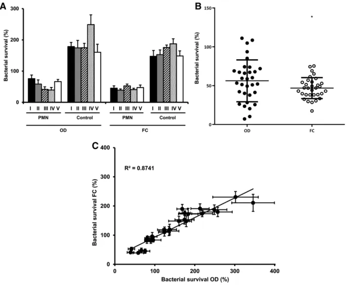
3.4 Reproducibility of the results
To examine the reproducibility of our new FC-based assay we com- pared survival ofS. aureusin HBSS+4% LB in the presence and absence of PMN as determined in five separate experimental series (in each series 6 or 8 identical experiments were carried out within one month) (Fig. 5A). PMN effectively reduced bacterial survival in all five exper- imental series detected by either technique, although the FC-based method seemed to produce more homogenous results. In the absence of PMN very similar bacterial growth was detected by both techniques, although the deviation was smaller in case of the FC-based method (Fig. 5A).
In Fig. 5B the result of every single experiment of the five series is presented by one dot. The mean of the total of 32 experiments carried out with the two different methods fits very well but the deviation of individual data is significantly lower in case of the FC-based method.
F I G U R E 5 Comparison of the reproducibility of the FC-based and OD-based methods. (A) Bacterial survival ofS. aureusin five separate exper- imental series with or without PMN present (test medium was HBSS+4% LB in all cases). In each series 6–8 experiments were carried out (mean+SEM). (B) Scattering of every single measurement ofS. aureussurvival in the presence of PMN (n=32, mean±SD).F-test showed signifi- cant difference in variances of the two methods (signed as*); however,t-test with Welch’s correction showed nonsignificant difference in means.
(C) Summarized linear regression of bacterial survival measured by OD- and FC-based methods (n=37,±SEM)
Figure 5C summarizes all the data ofS. aureussurvival presented in Figs. 3–5. The high value of the linear regression coefficient indicates good comparability of the two methods up to approx. 300% growth rate. At very high growth rate, incubation of bacteria for 40 min in nutrient rich medium resulted in an active reproductive state com- pared to the quiescent “no growth” reference sample. This difference significantly influenced the growing up time in the OD-based method and led to overestimation of the initial bacterial count.
4 C O N C L U S I O N
In our opinion, our new method offers advantages over all techniques summarized in Table 1. However, the most important advantage is in the time factor. Applying the new FC-based method, bacterial count of the sample can be determined in 3 hours as compared to 16 hours with the OD-based test and 48 hours of CFU counting. In cases of symptomatic immunodeficiencies of unknown origin (congenital or
acquired), investigation of phagocytic functions, including bacterial killing is recommended.40In these cases, the time difference in obtain- ing diagnostic results is critical. The proposed assay determines the number of bacteria, thus no conversion or further processing of the measured data is required. Depending on the property of the used medium, i.e. whether it allows bacterial growth or not, both bacterio- static and bactericidal effects can be measured.
Taken together, we propose a new, fast, easy, reliable, and repro- ducible FC-based test that is suitable for high throughput quantitation of bacteriostatic or bactericidal effect of any immune cell or noncel- lular material and provides large potential both for research and for clinical purposes.
AU T H O R S H I P
L.M.A. and E.L. devised the method, V.S. and L.M.A. carried out the experiments on human PMN, B.B. carried out the experiments on murine PMN, L.M.A. and E.L. summarized the data and prepared the manuscript.
962 L ˝ORINCZET AL.
AC K N O W L E D G M E N T S
The authors are indebted to Profs Miklós Geiszt and William Nauseef for access to the NOX2-deficient mice and the GFP-expressing bacte- rial strain, resp., to Professor Dóra Szabó, Drs. Eszter Ostorházi, Gábor Sirokmány, and Roland Csépányi-Kömi for stimulating discussions and critical reading of the manuscript and Regina Tóth-Kun for expert and devoted technical assistance. Experimental work was supported by research grants from the Hungarian Research Fund (OTKA/NKFIH) No. K108382 and K119236 to E.L.
D I S C LO S U R E
The authors have no conflict of interest to declare.
R E F E R E N C E S
1. Brinkmann V, Reichard U, Goosmann C, et al. Neutrophil extracellular traps kill bacteria.Science. 2004;303:1532–1535.
2. Hartl D, Latzin P, Hordijk P, et al. Cleavage of CXCR1 on neu- trophils disables bacterial killing in cystic fibrosis lung disease.Nat Med. 2007;13:1423–1430.
3. Ellson CD, Davidson K, Ferguson GJ, O’Connor R, Stephens LR, Hawkins PT. Neutrophils from p40phox-/- mice exhibit severe defects in NADPH oxidase regulation and oxidant-dependent bacterial killing.
J Exp Med. 2006;203:1927–1937.
4. Drewniak A, Gazendam RP, Tool AT, et al. Invasive fungal infection and impaired neutrophil killing in human CARD9 deficiency.Blood.
2013;121:2385–2392.
5. Hartl D, Krauss-Etschmann S, Koller B, et al. Infiltrated neutrophils acquire novel chemokine receptor expression and chemokine responsiveness in chronic inflammatory lung diseases. J Immunol.
2008;181:8053–8067.
6. Decleva E, Menegazzi R, Busetto S, Patriarca P, Dri P. Common methodology is inadequate for studies on the microbicidal activity of neutrophils.J Leukoc Biol. 2006;79:87–94.
7. Cho H, Jeong DW, Liu Q, et al. Calprotectin increases the activity of the SaeRS two component system and murine mortality duringStaphy- lococcus aureusinfections.PLoS Pathog. 2015;11:e1005026.
8. Rada BK, Geiszt M, Kaldi K, Timar C, Ligeti E. Dual role of phagocytic NADPH oxidase in bacterial killing.Blood. 2004;104:2947–2953.
9. Moskwa P, Lorentzen D, Excoffon KJ, et al. A novel host defense sys- tem of airways is defective in cystic fibrosis.Am J Respir Crit Care Med.
2007;175:174–183.
10. Futosi K, Nemeth T, Pick R, Vantus T, Walzog B, Mocsai A. Dasa- tinib inhibits proinflammatory functions of mature human neutrophils.
Blood. 2012;119:4981–4991.
11. Nemeth T, Futosi K, Hably C, et al. Neutrophil functions and autoim- mune arthritis in the absence of p190RhoGAP: generation and analysis of a novel null mutation in mice. J Immunol. 2010;185:
3064–3075.
12. Lorincz AM, Schutte M, Timar CI, et al. Functionally and morphologi- cally distinct populations of extracellular vesicles produced by human neutrophilic granulocytes.J Leukoc Biol. 2015;98:583–589.
13. Lorincz AM, Timar CI, Marosvari KA, et al. Effect of storage on physical and functional properties of extracellular vesicles derived from neu- trophilic granulocytes.J Extracell Vesicles. 2014;3:25465.
14. Timar CI, Lorincz AM, Csepanyi-Komi R, et al. Antibacterial effect of microvesicles released from human neutrophilic granulocytes.Blood.
2013;121:510–518.
15. Drevets DA, Canono BP, Campbell PA. Measurement of bacte- rial ingestion and killing by macrophages. Curr Protoc Immunol.
2015;109:14.6.1–17.
16. Moussa SH, Tayel AA, Al-Hassan AA, Farouk A. Tetrazolium/formazan test as an efficient method to determine fungal chitosan antimicrobial activity.J Mycol.2013;2013:7.
17. Casalino-Matsuda SM, Nair A, Beitel GJ, Gates KL, Sporn PH.
Hypercapnia inhibits autophagy and bacterial killing in human macrophages by increasing expression of Bcl-2 and Bcl-xL.J Immunol.
2015;194:5388–5396.
18. Atosuo J, Lehtinen J, Vojtek L, Lilius EM.Escherichia coliK-12 (pEGF- PluxABCDEamp): a tool for analysis of bacterial killing by antibacterial agents and human complement activities on a real-time basis.Lumines- cence. 2013;28:771–779.
19. Atosuo JT, Lilius EM. The real-time-based assessment of the microbial killing by the antimicrobial compounds of neutrophils.ScientificWorld- Journal. 2011;11:2382–2390.
20. Hamers MN, Bot AA, Weening RS, Sips HJ, Roos D. Kinetics and mechanism of the bactericidal action of human neutrophils against Escherichia coli.Blood. 1984;64:635–641.
21. Aellen S, Que YA, Guignard B, Haenni M, Moreillon P. Detection of live and antibiotic-killed bacteria by quantitative real-time PCR of specific fragments of rRNA.Antimicrob Agents Chemother. 2006;50:
1913–1920.
22. Nocker A, Cheung CY, Camper AK. Comparison of propidium monoazide with ethidium monoazide for differentiation of live vs. dead bacteria by selective removal of DNA from dead cells.J Microbiol Meth- ods. 2006;67:310–320.
23. Schwartz J, Leidal KG, Femling JK, Weiss JP, Nauseef WM. Neutrophil bleaching of GFP-expressing staphylococci: probing the intraphagoso- mal fate of individual bacteria.J Immunol. 2009;183:2632–2641.
24. Pang YY, Schwartz J, Thoendel M, Ackermann LW, Horswill AR, Nauseef WM. agr-Dependent interactions ofStaphylococcus aureus USA300 with human polymorphonuclear neutrophils.J Innate Immun.
2010;2:546–559.
25. Greenlee-Wacker MC, Rigby KM, Kobayashi SD, Porter AR, DeLeo FR, Nauseef WM. Phagocytosis ofStaphylococcus aureusby human neu- trophils prevents macrophage efferocytosis and induces programmed necrosis.J Immunol. 2014;192:4709–4717.
26. Mason DJ, Allman R, Stark JM, Lloyd D. Rapid estimation of bacte- rial antibiotic susceptibility with flow cytometry.J Microsc. 1994;176:
8–16.
27. Deere D, Porter J, Edwards C, Pickup R. Evaluation of the suitability of bis-(1,3-dibutylbarbituric acid) trimethine oxonol, (diBA-C4(3)-), for the flow cytometric assessment of bacterial viability.FEMS Microbiol Lett. 1995;130:165–169.
28. Mason DJ, Lopez-Amoros R, Allman R, Stark JM, Lloyd D. The ability of membrane potential dyes and calcafluor white to distinguish between viable and non-viable bacteria.J Appl Bacteriol. 1995;78:309–315.
29. Hoefel D, Grooby WL, Monis PT, Andrews S, Saint CP. Enumera- tion of water-borne bacteria using viability assays and flow cytom- etry: a comparison to culture-based techniques.J Microbiol Methods.
2003;55:585–597.
30. Berney M, Hammes F, Bosshard F, Weilenmann HU, Egli T. Assess- ment and interpretation of bacterial viability by using the LIVE/DEAD BacLight kit in combination with flow cytometry.Appl Environ Microbiol.
2007;73:3283–3290.
31. Leuko S, Legat A, Fendrihan S, Stan-Lotter H. Evaluation of the LIVE/DEAD BacLight kit for detection of extremophilic archaea and visualization of microorganisms in environmental hypersaline samples.
Appl Environ Microbiol. 2004;70:6884–6886.
32. Martin E, Bhakdi S. Quantitative analysis of opsonophagocytosis and of killing ofCandida albicansby human peripheral blood leukocytes by using flow cytometry.J Clin Microbiol. 1991;29:2013–2023.
33. Jepras RI, Paul FE, Pearson SC, Wilkinson MJ. Rapid assessment of antibiotic effects onEscherichia coliby bis-(1,3-dibutylbarbituric acid) trimethine oxonol and flow cytometry.Antimicrob Agents Chemother.
1997;41:2001–2005.
34. van der Pol E, van Gemert MJ, Sturk A, Nieuwland R, van Leeuwen TG. Single vs. swarm detection of microparticles and exosomes by flow cytometry.J Thromb Haemost. 2012;10:919–930.
35. Pollock JD, Williams DA, Gifford MA, et al. Mouse model of X- linked chronic granulomatous disease, an inherited defect in phago- cyte superoxide production.Nat Genet. 1995;9:202–209.
36. Mocsai A, Zhang H, Jakus Z, Kitaura J, Kawakami T, Lowell CA. G- protein-coupled receptor signaling in Syk-deficient neutrophils and mast cells.Blood. 2003;101:4155–4163.
37. Kerstens M, Boulet G, Van Kerckhoven M, et al. A flow cytomet- ric approach to quantify biofilms.Folia Microbiol (Praha). 2015;60:
335–342.
38. Oehmcke S, Westman J, Malmstrom J, et al. A novel role for pro- coagulant microvesicles in the early host defense against streptococ- cus pyogenes.PLoS Pathog. 2013;9:e1003529.
39. Herrmann IK, Bertazzo S, O’Callaghan DJ, et al. Differentiating sep- sis from non-infectious systemic inflammation based on microvesicle- bacteria aggregation.Nanoscale. 2015;7:13511–13520.
40. de Vries E, European Society for Immunodeficiencies, m. Patient- centred screening for primary immunodeficiency, a multi-stage diag- nostic protocol designed for non-immunologists: 2011 update.Clin Exp Immunol. 2012;167:108–119.
How to cite this article:L ˝orincz ÁM, Szeifert V, Bartos B, Ligeti E. New flow cytometry based method for the assessment of the antibacterial effect of immune cells and subcellular parti- cles.J Leukoc Biol. 2018;103:955–963.https://doi.org/10.1002/
JLB.4TA0817-317R