Neuroanatomy and function of brain structures involved in the regulation of prolactin secretion and milk yield
Ph.D. Theses
FLÓRA SZABÓ M.D.
Department of Human Morphology and Developmental Biology Faculty of Medicine, Semmelweis University
János Szentágothai Doctoral School of Neuroscience Program of Neuroendocrinology (6)
Tutor: Prof. Katalin Köves M.D., D.Sc.
Referees Associate Prof. Szabolcs Várbíró M.D., Ph.D.
Prof. Klára Matesz, M.D., D. Sc.
Chairman of examining committee Prof. János Sólyom, M.D., D.Sc.
Members of examining committee Prof. Jenő Egyed, M.D., D.Sc.
Associate Prof. Andrea Székely, M.D., Ph.D.
List of content
Page
ABBREVIATIONS 5
1. INTRODUCTION 7
2. BACKGROUND 8
2.1. Biochemistry of PRL 8
2.2. Structures producing PRL 9
2.3. PRL receptors (PRL-R) 13
2.4. Function of PRL 14
2.5. Regulation of PRL secretion 15
2.6. Autonomic innervation of the mammary gland 24
3. AIM OF EXPERIMENTS 26
3.1. Morphological studies on SIPR pathway 26
3.2. Physiological studies 26
3.3. Studies on autonomic innervation of the mammary gland 27
4. MATERIALS AND METHODS 28
4.1. Animals 28
4.2. Non-transynaptic tract tracing ex periments 28
4.2.1. Stereotaxic interventions 28
4.2.2. Perfusion and tissue sectioning 29
4.2.3. Immunohistochemistry 29
4.2.3.1. Single stains 29
4.2.3.2. Double stains 30
4.3. TH and ENK expression in TIDA neurons 32
4.3.1. In situ hybridization 32
4.3.1.1. Probe preparation 32
4.3.1.2. Hybridization and visualization 33
4.3.2. Immunohistochemistry 34
4.3.3. Image analysis of mRNA 35
4.3.4. Image analysis of ENK peptide immunoreactivity in the ME 35
4.4. Transynaptic tract tracing experiments 36
4.4.1. Preparation of virus 36
4.4.2. Tissue preparation 36
4.4.3. Immunohistochemistry 37
5. RESULTS 38
5.1. Morphological studies of SIPR pathway 38
5.1.1. Experiment 1. Anterograde tracer (BDA) injection in the rostral mesencephalon 38
5.1.2. Experiment 2. Retrograde tracer (FG) injection in the lateral ARC 42
5.2. Physiological studies 46
5.2.1. Quantitative analysis of TH mRNA in ARC 46
5.2.2. Quantitative analysis of ENK mRNA in ARC 48
5.2.3. Histological appearance of TH and ENK mRNA expressing cells in ARC 50
5.2.4. Quantitative analysis and histological appearance of ENK peptide in ME 54
5.3. Autonomic innervation of the mammary gland 58
5.3.1. Specificity test for retrograde transportation of virus 58
5.3.2. Virus labeling at various levels of the nervous system 59
5.3.3.Chemical characterization of the virus labeled neuronal perikarya in PvG 59
5.3.4. Chemical characterization of neuronal cell bodies in the lateral horn and PV 60 5.3.5. Chemical characterization of the nerve fibers in the mammary
6.2. Physiological findings: Effect of suckling stimulus on TH and ENK 67
6.3. Morphological findings for the autonomic innervation of the mammary gland 71
7. BRIEF CONCLUSION OF THE THREE EXPERIMENTS 75
8. NEW RESULTS 76
9. SUMMARY 77
10. ÖSSZEFOGLALÁS 79
11. REFERENCES 81
12. LIST OF PUBLICATIONS 95
RELATED TO THE THESIS 95
NOT RELATED TO THE THESIS 95
11. ACKNOWLEDGEMENT 96
ABBREVIATIONS
ACTH adrenocorticotrop hormone ARC arcuate nucleus
ATP adenosine triphosphate BDA biotinylated dextran amine
Cas catecholamines
CART cocaine and amphetamine regulated transcript CGRP calcitotin gene related peptide
CNS central nervous system CTP cytosine triphosphate
Cy2 cyanine dye excited at 492 nm, more photostable than FITC
Cy5 indodicarbocyanine dye excited at 650 nm, more photostable than rhodamine D2 dopamine receptor 2
DA dopamine
DOPA dihydroxy phenilalanine
DAB diamino benzidine tetrahydrochloride DBH dopamine--hydroxylase
DEPC diethylpyrocarbonate DYN dynorphine
EDTA ethylenediaminetetraacetic acid ENK enkephaline
FG fluorogold
GAL galanine
GH growth hormone
GH3 lactomammotrope cell line GTP guanine triphosphate
KPBS potassium phosphate buffer-saline LH luteinizing hormone
LHRH luteinizing hormone-releasing hormone
ME median eminence
NEDA neuroendocrine dopaminergic NMDA N-metil-D-aspartate
OD optical density OXY oxytocin
PCR polymerase chain reaction PFA paraformaldehyde
PHDA periventriculo-hypophysial dopaminergic PPN peripeduncular nucleus
PRL prolactin
SIPR suckling induced prolactin release SPFpc subparafascicular parvocellular nucleus TH thyrosin hydroxylase
TIDA tuberoinfundibular dopaminergic
Th thoracic
THDA tuberohypophyseal dopaminergic TIP-39 tuberoinfundibular peptide-39 TRNA torula RNA
UTP uracil triphosphate
VAChT vesicular acethylcholine transporter VLM ventrolateral medulla
VMN ventromedial nucleus VR ventral rootlet
1. INTRODUCTION
The offsprings of mammalian species are fed by milk produced in the mammary gland of their mother. Secretion of milk is a process called lactation. It has two major phases: 1. initiation of the milk secretion is the lactogenesis; 2. maintaining of the secretion is the galactopoiesis, in rats it is also called midlactation (see Tucker 1981).
The lactogenesis is further devided into stages I and II. Stage I. The mammary alveolar cells differentiate histologically and enzymatically. During this stage colostrum is formed. It is rich in fat, proteins and immunoglobulins. It does not contain lactose. In rats Stage I. begins 30 hours before delivery. Stage II. Copious amount of milk is produced.
Begining of lactogenesis coincides with decline in the level of progesterone and transient elevation of estradiol, prolactin (PRL), growth hormone (GH), and glucocorticoids. In rats this stage occurs immediately prior to and at the time of parturition. Fig. 1. shows changes in the concentration of hormones in blood during periparturient period. In the Stage II. the milk contains proteins, fat, carbohydrates (lactose), minerals, and vitamins.
Fig. 1. Hormonal changes in the serum around parturition (Tucker HA. Neuroendocrinology in Physiology and Medicine, 2000, p. 169).
2. BACKGROUND
Because my theses include studies of brain structures regulating PRL secretion I will describe the biochemistry and receptors of PRL, distribution of PRL and its receptors and the function and regulation of this hormone.
2.1.Biochemistry of PRL
Rat PRL is a protein hormone composed of a polypeptide chain of 197 amino acids (Nicoll et al 1986) (Fig. 2). Its molecular weight is 23 kD. A 22 kD variants is also described (Antony et al 1993). PRL was first discovered in the pituitary gland (Stricker and Greuter 1928; Riddle et al 1932). The rat PRL was characterized by Parlow and Shome in 1976. It is encoded by chromosoma 17 (Alam et al 2006).
.
Fig. 2. Amino acid sequence of rat anterior pituitary PRL. A: alanin; C: cystein; D: aspartic acid; E: glatamic acid, F: phenylalanine; G: glycine; H: histidine; I: isoleucine; K: lysine; L: leucine; M: methionine; N:
asparagine; P: proline; Q: glutamine; R: arginine; S: serine; T: threonine; V: valine; W: tryptophan; Y:
tyrosine. Three disulfide bridges are found between cysteines. From: Neill, J.D. and Nagy, Gy.M. Prolactin secretion and its control (In The physiology of Reproduction, Eds: Knobil, E.K. and Neill, J.D., 1994).
Fig. 3. Three dimensional image of PRL.
The three dimensional image of PRL molecule was constructed by Goffin and his coworkers (1995). The model contains the four-helix bundle and binding loops joining the helices.
Extensive posttranslational modifications of PRL have been reported. PRL may be cleaved, deamidated, phosphorylated, glycosylated, sulfated and disulfide dimerized (Sinha 1995).
In human serum two high molecular mass forms of circulating PRL were identified, macroprolactin (big-big prolactin) with 100kD MW or over and big prolactin with 40-60 kD MW (Fahie-Wilson et al 2005). Their clinical significance is not clarified but they may be responsible for hyperprolactinemia.
2.2. Structures producing PRL
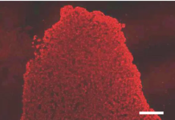
PRL is secreted in various tissues. In the anterior pituitary acidophilic cells, known
Fig. 4. Immunofluorescence staining shows PRL immunoreactive cells in the anterior pituitary of a diestrous female rat. The cells are evenly distributed in the anterior lobe and they show a cup-shaped appearance. Scale:
200 m. (Courtesy of Andrea Heinzlmann, Assistant Professor, Department of Human Morphology and Developmental Biology.)
During lactation the mammotropic cells proliferate. They loose their cup-shaped appearance. Fig. 5 shows PRL staining of an anterior pituitary deriving from a midlactating rat (sacrificed 10 days after parturition).
Fig. 5. Immunofluorescene staining showing PRL immunoreactivity in the anterior pituitary of a midlactating rat. PRL cells are extremly proliferated. Scale: 200m.
In the last thirty years PRL and PRL-like immunoreactivities or PRL mRNA was demonstrated over ten tissues other than the pituitary gland (Sinha 1995). Table 1 shows the tissues where PRL or PRL-like molecules were demonstrated and the related references.
The richest source of peripheral PRL is the placenta. This molecule is called placental-lactogene produced by decidual cells (Golander et al 1978; Rosenberg et al 1980;
Handwerger et al 1990; Alam et al 2008). PRL-like molecules were also demonstrated in
the endometrium, myometrium and smooth muscle fibroid of uterus (Masler and Riddick 1979; Walters et al 1983; Nowak et al 1993).
PRL mRNA was shown in immunocells including tymocytes, T-lymphocytes and T-lymphoblasts (DiMattia et al 1988; Montgomery et al 1992; Pellegrini et al 1992; De Bellis et al 2005).
Table 1. Tissues other than pituitary in which PRL-like molecules have been reported.
Occurrence of PRL References
________________________________________________________________________
_______________________________________________________________________
Placenta Golander et al, 1978; Rosenberg et al, 1980 Handwerger et al, 1990; Alam et al, 2008 Uterus Masler and Riddick, 1979; Walters et al, 1983;
Eyal et al, 2007
Immune system Di Mattia et al, 1988; Montgomery et al, 1992;
Pellegrini et al, 1992; De Bellis et al, 2005 Mammary gland Kurtz et al, 1993; Koizumi et al, 2003 Adrenal gland* Nolin, 1978
Corpus luteum Nolin, 1978; Erdmann et al, 2007
Prostate Harper et al, 1981; Nevalainen et al, 1997 Untergasser et al, 2001
Testes Roux et al, 1985; Untergasser et al, 1996 Imaoka et al, 1998
Urethral gland* Tsubura, 1986 Lacrimal gland Wood et al, 1999 Sweat gland* Roberstson et al, 1989
The synthesis of PRL in mammary gland was first demonstrated by Kurtz and coworkers in 1993. PRL mRNA was localized in the epithelium of alveoli and ducts of lactating mammary gland by in situ hybridization. It suggests that the mammary gland might contribute to PRL in milk by de novo synthesis. Another research group was able to show PRL mRNA in the sebaceous glands of nipple (Koizumi et al 2003).
The presence of PRL in other peripheral organs including adrenal gland and the corpus luteum, uterus (Nolin et al 1978; Erdmann et al 2007; Eyal et al 2007), the prostate gland (Harper et al 1981; Nevalainen et al, 1997; Untergasser et al, 2001), testes (Roux et al, 1985; Untergasser et al, 1996; Imaoka et al 1998) and urethral gland (Tsubura et al 1986) lacrimal gland (Wood et al 1999), sweat gland (Robertson et al 1989), pancreatic islets (Meuris et al 1983) was also demonstrated but the presence of it in adrenal gland, urethral gland, sweat gland, pancreatic islets, and gut was not confirmed by the mRNA study.
PRL was discovered in the brain as well. The concentration of PRL in brain tissues much lower than in the anterior pituitary. Fuxe and his coworkers (1977) demonstrated at the first time that PRL-like immunoreactivity is present in some hypothalamic nerve terminals. The origin of these fibers were not explored by this research group. Twelve years later Harlan and his coworkers (1989) demonstrated PRL immunoreactive cell bodies in the hypothalamic arcuate nucleus (ARC) and in adjacent areas ventral to the ventromedial nucleus (VMN). Fiber projections extended rostrally to the anterior hypothalamus, preoptic area, nucleus accumbens, septum, diagonal band of Broca, caudate putamen, frontal cortex, and accessory olfactory bulb and to the central amygdala. Another team (Paut-Pagano et al 1993) published contraversary results. They have only found PRL immunoreactive cell bodies in the lateral hypothalamic area surrounding the fornix, not in the ARC; however, fibers dispersed all over the brain. After hypophysectomy amount of PRL was not decreased indicating that PRL demonstrated in the brain areas is produced locally. With the use of reverse transcript-polymerase chain reaction (RT-PCR) it was shown that PRL mRNA in the brain is identical to anterior pituitary PRL mRNA (Wilson et al 1992).
2.3. PRL receptor (PRL-R)
PRL-Rs belong to cytokine receptor family (Cosman et al1990). They have three domains: a ligand-binding extracellular, a single hydrophobic transmembrane, and a cytoplasmic domain. In rats two isoforms are described, short and long forms with 291 and 592 amino acids, respectively (Kelly et al 1993). The two isoforms differ in the length of intracellular domain. Intracellular domain is necessary for signal transduction. The two isoforms are the products of a single gene and they are generated by alternative splicing.
PRL-R is encoded by chromosome 2q16. Signal tranduction pathway is the tyrosine kinase Jak-2 system.
PRL-Rs are widely distributed in rat tissues. With the use of quantitative PCR technique Nagano and Kelly (1994) mapped the distribution of PRL-R isoforms in 17 tissues (cerebral cortex, choroid plexus, hypothalamus, pituitary, heart, lung, thymus, spleen, liver, pancreas, kidney, adrenal, ovary, uterus, skeletal muscle, skin and mammary gland) of adult female rats during estrous cycle, pregnancy and lactation. In the hypothalamus and pituitary both receptors are expressed but the predominant form is the long isoform. It was also seen that the amount of receptors is higher in diestrous than in proestrous stage of the cycle. On the contrary, in the cerebral cortex and the peripheral reproductive organs the amount of the long form was higher in proestrus than in diestrus.
The liver is the only organ where the short form is clearly dominant. In the pancreas a high level of PRL-R was found in the islets. The first immunohistochemical detection of the PRL-R immunoreactivity (Roky et al 1996) revealed that the receptor is associated with nerve cell bodies where a dense PRL immunoreactive fiber staining was found earlier (Fuxe et al 1977). PRL-R-like immunoreactive neurons were found in pyramidal cell layer of the cerebral cortex, septal nuclei, amygdaloid complex, hypothalamic nuclei (suprachiasmatic, supraoptic, paraventricular and dorsomedial), substantia nigra, habenula and in the subcommissural organ.
Mammary gland is the primary target tissue for PRL action. Both at the end of
Immunohistochemistry revealed that the PRL-Rs are associated with all endocrine cell populations in the anterior pituitary (Morel et al 1994). The highest level of receptors was found on somatotropes, and decreasing number on lactotropes, then thyrotropes, corticotropes and gonadotropes. These findings suggest that PRL influences the pituitary hormone secretion via auto- and paracrine manners.
With RT-PCR, Northen and Western blot analysis the presence of PRL-Rs was revealed in gonads of both sexes (Zhang et al 1995; Guillaumot and Benahmed 1999), and in situ hybridization identified receptors on the interstitial Leydig cells, Sertoli cells and on the spermatogenetic cell line as well (Zhang et al 1995; Hondo et al 1995). In situ hybridization also revealed PRL-Rs in the acinar epithelium and in the interstitium of the lacrimal gland (Wood et al 1999). In the liver PRL-Rs and their regulation by sexual steroids were also demonstrated (Tanaka et al 2005).
2.4. Functions of PRL
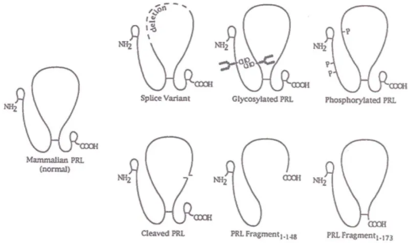
Posttranslational modification gives rise to different variants of PRL molecules (Fig.
6) which have different functions when bind to the receptors.
Fig. 6. Schematic diagram of PRL molecule and some structural variants. CHO refers to the N-linked carbohydrate moieties. P represents the site of phosphorilation. Broken line indicates the delition of amino acid residues. The nick in the large disulfide loop shows a proteolytic cleavage site. From Sinha, Y.N.
Structural variants of prolactin: Occurrence and physiological significance. Endocrine Reviews 16: 358, 1995.
Most throughly investigated variant is the short PRL fragment (1-148). It lacks the fourth helix. This is a potent inhibitor of capillary endothelial cell proliferation (Ferrara et
al 1991). Cleaved PRL increased thymidine incorporation into DNA in gonadotropes and thyrotropes, but not in other pituitary cell types. This results indicate that cleaved PRL is a potent paracrine growth regulator in the pituitary tissue (Andries et al 1992).
Phosphorylated PRL inhibited the secretion of nonphosphorylated PRL from a lactomammotrope cell line (GH3). In this way it serves as autocrine regulator of PRL secretion (Ho et al 1989). PRL complexed with immunoglobulins produces growth response in peripheral blood lymphocytes. Similar growth promoting effect of PRL- immunoglobulin complex was demonstrated in malignant B-lymphocytes (Walker et al 1995). It means that the modified PRL may play a role as growth factor for these cells (Walker et al 1993).
PRL knock-out in mice induces infertility, but does not prevent the maternal behavior. In the mammary gland a normal ductal tree develops, but the ducts fail to develop lobular decorations, which is characteristic of the normal virgin adult mammary gland (Horseman et al 1997).
2.5. Regulation of PRL secretion
It was demonstrated by Everett (1954, 1956) more than fifty years ago that a pituitary autograft without hypothalamic connections can maintain the pseudopregnancy and corpora lutea. He postulated the existence of a hypothalamic factor which was released into the portal blood and inhibited the PRL secretion. A few years later Talwaker in Meites’
laboratory (Talwalker at al 1963) confirmed the existence of a PRL inhibiting factor in hypothalamic extracts. Soon it was realized that this inhibiting factor was dopamine (DA) (Macleod 1974).
DA is one of catecholamine neurotransmitters. There are neurons which use it as neurohormone. These neurons take up tyrosine and tyrosine hydroxylase (TH) converts it into dihydroxy-phenilalanine (DOPA). This is the immediate precursor of DA. This is further converted into DA by aromatic L-amino acid decarboxylase. In other neurons DA
Figure 7. Schematic illustration of the dopaminergic neurons and their projections to various parts of the central nervous system. 1. Meso- (nigro-) striatal pathway. 2. Meso- (nigro-) hypothalamic-median eminence pathway. 3.
Ventral mesolimbic pathway. 4. Mesopontine pathway. 5. Tubero-infundibular (TIDA) and tubero-hypophyseal pathway (THDA). 6. Incertohypothalamic pathway. 7. Olfactory bulb (probable projection to AON). 8.
Dopaminergic fibers innervating the dorsal vagal complex. Abbreviations: A = amygdala; AON = anterior olfactory nucleus; CCN = central cerebellar nuclei; DBB = diagonal band of Broca; DVC = dorsal vagal complex; HIPP F = hippocampal formation; LC = locus coeruleus; MIDB = midbrain; MO = medulla oblongata; OB = olfactory bulb;
PHIPP G = parahippocampal gyrus; PPC = prepiriform cortex; PV = hypothalamic paraventricular nucleus; S = septum; SN = substantia nigra; TH = thalamus. From: Köves and Heinzlmann, Neurotransmitters and Neuropeptides in Autism (in: New Autism Research Developments, Ed.: B. S. Mesmere, 2008, p. 35)
DA is produced in several brain regions such as substantia nigra, zona incerta, olfactory bulb and the hypothalamic ARC. Fig. 7 schematically illustrates the most important dopaminergic pathways in the rat central nervous system.
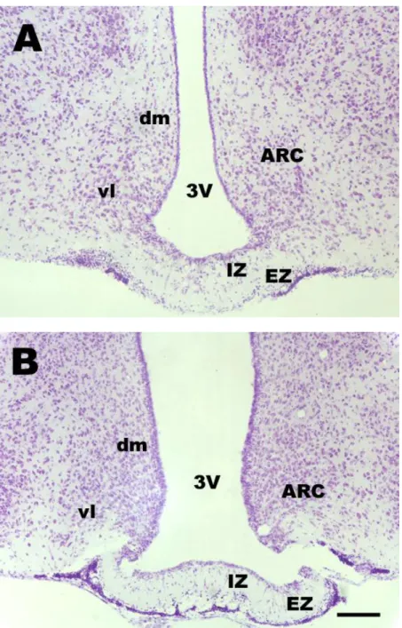
The ARC begins just rostral to the hypothalamic infundibular recess and lies along the walls of this recess troughout its length. Two distinct subdivisions are generally recognized: dorsomedial part which has a small cell population and the ventrolateral part which has medium-sized neurons (Meister and Hökfelt 1988; Simerly and Young 1991).
Fig. 8 shows the ARC at rostral (A) and caudal (B) levels by cresyviolette staining.
Fig. 8. Microphotographs demonstrating ARC in the frontal sections of a midlactating rat hypothalamus at two rostrocaudal levels. A is at A6,8 and B is at A5,8 to interaural line. Cresyviolette staining.
Abbreviations: 3V = third ventricle, ARC = arcuate nucleus; dm = dorsomedial part of ARC; EZ = external
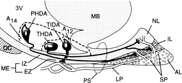
The hypothalamic neuroendocrine dopaminergic (NEDA) neurons are involved in the regulation of PRL secretion. Three populations are identified to the rostro-caudal direction: 1. the periventriculo-hypophysial dopaminergic (PHDA), 2. the tubero- hypophyseal dopaminergic (THDA) and 3. the tubero-infundibular dopaminergic (TIDA) systems. The cells of origin of these pathways are located in the periventricular-ARC region. PHDA neurons are located in the most rostral subdivision and terminate in the intermediate lobe. THDA neurons occupy the middle region and terminate in both intermediate and neural lobes. TIDA neurons are located in the middle and posterior subdivisions and terminate around the capillaries in the external zone of the median eminence (ME) (see Freeman et al 2000; Tóth et al 2002).
Fig. 9 schematically illustrates PHDA, THDA and TIDA neurons and their termination in the pituitary gland and in external zone (EZ) of ME. PHDA neurons terminate in the intermediate lobe, THDA neurons in both neural and intermediate lobes, and TIDA neurons in EZ of ME.
Fig. 9. Schematic illustration of PHDA, THDA and TIDA pathways in the sagittal section of the hypothalamus and the pituitary gland (From Freeman, Kanyicska, Léránth and Nagy: Physiological Review 80, 1523-1631, 2000). Abbreviations: 3V = third ventricle; A12 and A14 = dopaminergic cell groups; AL = anterior lobe; EZ = external zone; IL = intermediate lobe; IZ = internal zone; LP = long portal vessels; MB
= medial basal hypothalamus; ME = median eminence; NL = neural lobe; OC = optic chiasm; PS = pituitary stalk; SP = short portal vessels.
Fig. 10 shows TH immunostaining in ARC at mid antero-posterior level (A6,4 to interaural line).
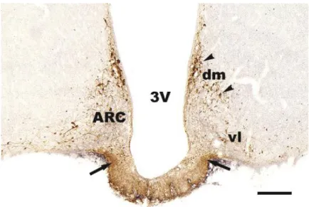
Fig. 10. TH immunostaining in ARC nucleus. TH immunopositive cells are mainly located in the dorsomedial part of ARC. In the ventrolateral part of this nucleus the TH cells are scattered. TH cells located in the dorsomedial part of ARC project to the ME forming the tubero-infundibular dopaminergic pathway. They are called TIDA neurons. In ME the dopaminergic fibers are denser in the lateral part of the infundibular recess then in the middle portion and located in the external zone of the ME. Arrowheads show TIDA neuronal cell bodies, arrows show TIDA fibers in the external zone. Abbreviations: 3V = third ventricle; ARC = arcuate nucleus; dm = dorsomedial part of ARC; vl = ventrolateral part of ARC. Scale:
250m.
Under non-lactating conditions, these neurons produce DA and continuously and tonically release it into the hypophysial portal circulation (Ben-Johnatan 1977). DA acts on D2 receptors of lactotropes to inhibit PRL release. When DA release is inhibited, PRL is rapidly released into the general circulation (Ben-Jonathan and Hnasko 2001). PRL can be controlled in this manner by a host of stimuli such as stress, sexual activity and stimuli to the breast (Pena and Rosenfeld 2001).
There are ample evidence that suggest that afferent activity to the TIDA neurons is a powerful regulator of TIDA neuronal activity and thus, of PRL secretion. Mammary
produces a 63% decline in pituitary stalk and ME DA levels preceding the rise in plasma PRL (De Greef et al 1981; Plotsky and Neill 1982; Plotsky et al 1982). Further evidence to support the importance of afferent activity to the TIDA neurons is the observation that prevention of suckling on teats of only one side up-regulates TH expression in TIDA neurons on the contra-lateral side to blocked nipples (Berghorn et al 2001). This indicates that the sensory stimulus prompted by suckling is responsible for the TH suppression in TIDA neurons.
In the past years a lot of attention has been focused on the importance of suckling for successful lactation. There have been numerous debates about the ideal duration and frequency of breastfeeding episodes to ensure adequate milk supply. Over a million infant deaths have been attributed to the lack of breastfeeding in the world (McVea et al 2000), so in recent years there has been an increase in movements that advocate breastfeeding. Understanding the circuits that are responsible for this process is critical in understanding the physiological changes that take place in the body and their impact on maternal and infant health. This could also provide an explanation to how certain central nervous system (CNS) disorders such as tumors, head injury, infection (tuberculosis, histoplasmosis), or infiltrative diseases (sarcoidosis, hemochromatosis, lymphocytic hypophysitis) disrupt the process of lactation (Pena and Rosenfeld 2001).
The most widely studied neuroendocrine reflex responsible for milk production is the suckling induced PRL release (SIPR). It is clear that PRL secretion and release by mammotropes in lactating rats are mainly controlled by dopaminergic neurons of the medial basal hypothalamus (Leong et al 1983). DA acts as the main inhibitory transmitter, responsible for tonically inhibiting PRL production and release in non- lactating rats. At the beginning of lactation, suckling stimuli by the pups eventually reach the hypothalamus, inhibiting the activity of TIDA neurons which form one of catecholaminergic cell groups (Fuxe and Hökfelt 1972), thus allowing the release of PRL from the pituitary into the general circulation and in turn, PRL stimulates milk secretion.
The exact pathway from the nipples to the neurons of the medial basal hypothalamus that conveys the suckling stimulus to the TIDA neurons is not well characterized. Suckling also stimulates oxytocin (OXY) release from the magnocellular
supraoptico-paraventriculo-hypophyseal system. Previous reports have suggested that the release of PRL and OXY during suckling are coordinated (Samson et al 1986). By monitoring milk let-down reflexes due to OXY release or electrical activity of identified OXY neurons after brain stimulation or following suckling after lesions, a profile of brain sites involved in the suckling induced neuroendocrine axis has emerged. Studies indicate that the suckling stimulus from the mechanoreceptors of the nipples is delivered to the spinal cord with a relay in the cervical spinal nucleus (Dubois-Dauphin et al 1985). After ascending from this nucleus, a projection to the mesencephalic tegmentum (Dubois- Dauphin et al 1985; Hansen and Kohler 1984; Tindal and Knaggs 1971; Tindal and Knaggs 1969) rather than the more classical thalamic sites (Dubois-Dauphin et al 1985) conveys suckling signals to the hypothalamus for milk ejection control. There appears to be at least one additional relay before hypothalamic neuroendocrine neurons are reached.
The peripeduncular nucleus (PPN), nestled among the medial geniculate nucleus, the posterior intralaminar thalamic nucleus and the cerebral peduncle, has been suggested to be an important mediator of the suckling stimulus for successful lactation. Such observations were made based on studies in which these areas were lesioned (Factor et al 1993, Hansen and Kohler 1984) and lactation was impaired. Experiments using stimulation paradigms (Tindal and Knaggs 1969; 1975) noted that the lateral-most region of the midbrain tegmentum, likely within the PPN as defined by commonly used atlases (Paxinos and Watson 1986) were effective in releasing PRL. Previous studies (Tindal et al 1969) had determined that electrical stimulation of the more medial parts of the midbrain tegmentum also released PRL, but it is unclear whether that is due to stimulation of fibers of passage or of neurons. To resolve this, it is important to distinguish the PPN from the subparafascicular parvocellular nucleus (SPFpc) or more medial regions of the tegmentum.
An interneuronal relay from the mesencephalon to the hypothalamus is proposed, but has yet to be identified for either projection to OXY or TIDA system. Since the
midbrain site. It is likely that the signals travel together until they reach the brain stem where the neuronal pathways for milk ejection and PRL regulation diverge.
In a previous study suckling stimulus induced c-Fos expression in the ventrolateral medulla (VLM), locus ceruleus, lateral parabrachial nucleus, lateral and ventrolateral portions of the caudal part of the periaqueductal gray matter, and caudal portion of the paralemniscal nucleus (Li et al 1999b). This experiment also suggest the role of brain stem structures in relaying the suckling stimulus to the hypothalamus. In another study by the same research group it was found that fluorogold (FG) tracer injected in the ARC was retrogradely transported to the midbrain. The tracer appeared in some cell groups in which c-Fos was activated by suckling stimulus. These cell groups were mainly found in the PPN and VLM (Li et al 1999a). In this latter study the tracer spred over the border of the ARC. It is not sure that the neurons in the PPN and VLM project directly to the ARC.
It was observed that soon after the initiation of suckling, DA turnover and release are markedly reduced (Demarest et al 1983; Mena et al 1976; Merchenthaler 1993;
Selmanoff and Wise 1981). Overall, inhibition of the TIDA system assumes the dominant feature during suckling via marked down-regulation of the rate limiting enzyme for DA synthesis, TH (Wang et al 1993). However, the expression of TH mRNA in TIDA neurons seems to be very dynamic, reflecting the changes in suckling activity. Previous studies determined that within 1.5 hrs of termination of suckling, the TIDA neurons showed early signs of up-regulation of TH mRNA reflected by the appearance of 1 or 2 sites of heteronuclear RNA in the nucleus of TIDA neurons (Berghorn et al 2001). An increase in cytoplasmic TH mRNA was seen about 6 hours after the termination of suckling (Berghorn et al 2001) and mRNA levels peaked by 12-24 hr. Evidence of increased protein synthesis was also noted in ME terminals at 6 hr (Berghorn et al 1995).
From these data, it is uncertain if the early signs of up-regulation of TH represent a trigger for full up-regulation of TH mRNA or whether continuous stimulation of these neurons is necessary to achieve high TH levels.
Another peptide whose expression varies in TIDA neurons under non-lactating and lactating conditions is enkephaline (ENK). ENK is barely detectable in cycling rats,
while its levels dramatically increase in the ARC and ME of lactating animals (Ciofi et al 1993; Merchenthaler 1993). The data in the literature indicate that this up-regulation of ENK is due to the hyperprolactinemia of lactation (Merchenthaler 1994; 1995).
It is not clear what role ENK in TIDA neurons plays during lactation. Although existing data show that the TH producing activity of TIDA neurons is definitely suppressed, this does not mean that these neurons are not active in synthesizing other transmitters. Inactivity of the TIDA neurons during lactation would not allow existing DA to be released at all. However, although TH expression is low during suckling, some TH is still present in the cells and in the ME (Wang et al 1993) and thus some DA release is possible (Ben-Johnatan et al 1980). A study (Arbogast and Woogt 1998) has proposed that ENK could be co-released with DA and serve to attenuate the effect of DA on lactotropes, raising the possibility that ENK also contributes to PRL secretion. Other neuroendocrine systems could also be targets of ENK produced in the ARC. During suckling a number of neuroendocrine systems operates differently than in a cycling animal: among others, luteinizing hormone (LH) control changes dramatically and stress responses are greatly attenuated. ENK has been suggested to play a direct role in the regulation of luteinizing hormone-releasing hormone (LHRH) secretion by innervation of LHRH neurons. Recently Pimpinelli and his coworkers [2006] were able to demonstrate
-opioid receptors in a subpopulation of LHRH nerve terminals of the ME. -opioid receptors can bind ENK suggesting that ENK fibers directly innervate LHRH nerve terminals. The data for the role of ENK in the regulation of LHRH and LH secretion and release are controversial. In in vitro studies, ENK inhibited the release of LHRH from mediobasal hypothalamus fragments (Drouva et al 1981; Higuchi and Kawakami 1981;
Motta and Martini 1982; Moult et al 1981). Other studies show that opiate antagonists stimulate LH secretion in rats (Blank et al 1980; Cicero et al 1979; Higuchi and Kawakami 1981), sheep (Schillo et al 1985), primates (Gosselin et al 1983; Van Vugt et
LH secretion in anterior pituitary cell cultures (Slama et al 1990). Therefore, the role of ENK in the regulation of LHRH or LH still needs to be further studied.
Several neuropeptides and neurotransmitters were identified in the cell groups which are the potential relay stations of the SIPR and not yet proved. In the ventrolateral part of ARC pro-opiomelanocortin (POMC), dynorphin (DYN), alpha-melanocyta stimulating hormone (-MSH), cocaine and amphetamine regulated transcript (CART) and acetylcholin was also demonstrated in neuronal cell bodies. Recently it was shown that DYN is present in about 30% of POMC neurons (Maolood and Meister 2008) and 20% of POMC neurons also contain pituitary adenylate cyclase activating polypeptide (PACAP) mRNA (Dürr et al 2007). SPFpc was demostrated as the main source of the tuberoinfundibular peptide (TIP39) (Dobolyi et al 2003) which was isolated from the tuberoinfundibular region (Usdin et al 1999). In this region cell bodies were not identified, but the most dense TIP39 fiber network was shown here (Dobolyi et al 2003).
Calcitonin gene related peptide (CGRP) cell bodies were also demonstrated in the SPFpc and PPN (Yasui et al 1989). In this region there is dense galanin (GAL) and ENK fiber networks which are termination of ascending pathways from the lumbar spinal cord (unpublished data).
The role of a serotoninergic mechanism located in the parvocellular portion of PV in the regulation of SIPR was also suggested by Bodnár and her coworkers (2002). They found that frontal cuts in front and behind the hypothalamus blocked SIPR.
Administration of a serotonin blocker (5, 7-dihydroxitriptamin) into PV or the lesion of its parvocellular part prevented the SIPR. However, 6-hydroxidopamine (-adrenerg blocker) administration into PV had no effect.
2.6. Autonomic innervation of the mammary gland
The nipple and the mammary gland receive not only sensory, but autonomic innervation as well. As it was discussed in details milk secretion is iniciated by PRL release and milk ejection is induced by OXY release (Bisset et al 1967; Mena et al 1979;
Song et al 1988); however, the milk yield at the beginning of the suckling is basically influenced by noradrenergic input. It was demonstrated by Findlay and Grosvenor (1969)
that catecholamines depressed the milk yield at the beginning of suckling antagonizing the effect of OXY. It was also shown that -adrenergic blocker propranolol given icv enhanced the milk yield (Morales et al 2001). It was supposed that the -adrenergic blocker relieved the effect of OXY on the ductal constriction in the mammary gland.
Trans-section of the spinal cord between thoracic (Th)3 and Th4 segments or pharmacological sympathectomy, but not adrenalectomy and hypophysectomy, results in a faster rate of milk flow (Mena et al 1979; 1995; Morales et al 2001). The above- mentioned results suggest that the inhibition of milk yield at the beginning of suckling is mediated by a reflex pathway closed in the central nervous system. The afferent limb may be the same as the SIPR pathway, the efferent limb may be provided by the sympathetic pre- and postganglionic neurons present in the intermedio-lateral cell- column in the spinal cord and in the sympathetic trunk, respectively. The central synaptic station or stations may be in the brain stem and in the hypothalamus, probably in PV.
It is well known that the postganglionic parasympathetic nervous system consists of cholinergic neurons. Cholinergic neurons also comprise a small population of sympathetic postganglionic neurons that innervate the sweat glands (Landis and Fredieu 1986; Schäfer et al 1997). It was demonstrated a few years ago that the vast majority of sympathetic postganglionic neurons innervating the porcine mammary gland were located in Th10 and Th11 sympathetic ganglia (Franke-Radowiecka 2007). Description of the autonomic innervation of the rat mammary gland was published by Gerendai at al (2001). It is not clarified at this moment what kind of neurotransmitters mediates the sympathetic stimulus to the alveoli and ductal wall of the mammary gland, which is cholinergic and which is adrenergic.
3. AIM OF EXPERIMENTS
3.1. Morphological studies on SIPR pathway
The aim of these morphological studies was to further clarify which of the mesencephalic nuclei provides a relay to the ARC or to a cell group in the vicinity of ARC. We conducted a series of tract tracing studies in non-lactating rats to determine if any direct PPN to ARC connections existed and if not, where the relay from the mesencephalon to the ARC neurons was located. The neurochemical nature of those arcuate neurons which receive ascending fibers from the mesencephalon and those which send fibers from the mesencephalon to ARC was also investigated.
3.2. Physiological studies
The aim of the physiological studies was to compare the dynamics of changes in the expression of TH and ENK mRNA in TIDA neurons following a brief interruption of suckling (3-4 hours). We wished to elucidate 1) whether such brief interruption triggers full TH up-regulation that continues for a time after pup return then it declines (Hypothesis I) or 2) whether reinitiation of suckling immediately stops this process of up- regulation as a switch (Hypothesis II) (Fig. 11).
Fig. 11. Diagram showing our Hypothesis I and II. Pup rem = pup removal. Arrow indicates the time of the pup-return 4 hrs after pup removal.
We also planned to investigate 3) whether the time course of changes of ENK expression shows an opposite pattern as that of TH mRNA expression in the same animals and 4) whether the changes in the ENK expression well correlate with the ENK protein synthesis.
The progression of TH expression was followed up to 24 hrs after pups were returned to their dams. The dynamic changes in TH mRNA of TIDA neurons were compared with those of dams whose pups were permanently removed. In the same experimental animals we also investigated the ENK mRNA levels and followed the changes in the ENK peptide in the ME. Two more additional groups were included in this latter experiment. The animals of these groups were sacrificed 48 and 72 hrs after the removal of pups.
3.3. Studies on autonomic innervation of the mammary gland
The aim of this part of the work was to further explore the multisynaptic autonomic neuronal chain that innervates the nipples and the mammary glands of lactating rats using retrograde virus labeling and to chemically characterize the neurons of the neuronal chain which may participate in the regulation of the milk yield at the beginning of suckling.
4. MATERIALS AND METHODS
4.1. Animals
Sprague-Dawley female rats purchased from Zivic-Miller Laboratories, Inc.
(Zelienople, PA) or Sprague-Dawley and Wistar female rats purchased from Gödöllő were used for the experiments. The University of Maryland’s committee on Animal Care and Use approved the experimental paradigms according to NIH guidelines for the non- transsynaptic tracing and physiological experiments. The treatment of the animals using for virus labeling was in accordance with the rules of the „European convention for the protection of vertebrate animals used for experimental and other scientific purposes”, Strasbourg, 1986. Our protocol was approved by the Department of Animal Health Care, permission number: 22.1/1158/3/2010. The rats (3-4 months old) were housed on a 12 hour light/12 hour dark schedule and given free access to food and water. Temperature was maintained at 22±2°C.
4.2. Non-transynaptic tract tracing experiments 4.2.1. Stereotaxic interventions
Experiment 1 (7 animals): To determine the projections of neurons of the PPN, the primarily anterograde tracer biotinylated dextrane-amine (BDA) (MW 10,000, Sigma- Aldrich, St. Louis, MO) was administered into this area using iontophoresis and labeled fibers were looked for in the medial basal hypothalamus.
Experiment 2 (5 animals): The primarily retrograde tracer FG (Fluorochrome Inc., Englewood, CO) was administered iontophoretically into the region of the ARC and labeled cell bodies were looked for in the peripeduncular region of the midbrain.
On the day of the surgery, animals were anesthetized using 4% chlorohydrate (1ml/100g) and placed into a stereotaxic instrument (Benchmark Digital Stereotaxic – myNeurolab, St. Louis, MO). A single midline scalp incision was made to visualize the surface of the skull. The points of bregma and lambda were leveled and a small window was cut in the skull to expose the brain at Paxinos-Watson (1986) atlas coordinates: PPN – A 4,2mm, V 3.10mm and L 3.50mm or ARC – A 6.20mm, V 0.25mm and L 0.15mm, to
the interaural line and the midline, respectively. The sagittal sinus was removed to expose the interhemispheric fissure, which was used as the reference midline. A glass micropipette (30µm tip diameter) filled with 10% BDA or 2% FG dissolved in 0.9% saline was used to apply the tracers using positive pulses of 2uA, alternating 5 seconds on and 5 seconds off for a total of 5 minutes. It was then removed and the scalp was closed with wound clips.
Following the surgery, the animals were returned to the animal colony.
4.2.2. Perfusion and tissue sectioning
Ten to 14 days following surgery the animals were anesthetized with an overdose of sodium pentobarbital (100mg/kg), the blood was flushed out by physiological saline containing 2% sodium nitrite solution and perfused transcardially with 0.1M potassium- phosphate buffered saline (KPBS) containing 4% paraformaldehyde (PFA) solution and 2.5% acrolein (pH 6.8). An additional rinse of physiological saline was used to remove residual PFA and acrolein. Brains were immersed into a 30% sucrose solution and later brains were cut in coronal plain on a freezing sliding microtome at 25μm and collected into an ethylene-glycol containing cryoprotectant/anti-freeze solution (Watson et al 1986).
Sections were stored at -20˚C until use.
4.2.3. Immunohistochemistry 4.2.3.1.Single Stains
Experiment 1 (visualization of BDA): Sections were removed from the cryoprotectant/anti-freeze solution, rinsed with KPBS several times, and then incubated in 1% sodium borohydride/KPBS for 20 minutes to remove residual aldehydes and acrolein.
The sodium borohydride solution was rinsed out of the tissue with KPBS. The sections were incubated in goat anti-biotin (Vector Laboratories, Burlingame, CA) at a dilution of 1:70,000 made up in KPBS with 0.4% Triton X-100 for 48 hours at 4°C. Following primary incubation, sections were rinsed with KPBS and then immersed into donkey anti- goat biotinylated secondary antibody solution (Vector Laboratories, Burlingame, CA) at a dilution of 1:600 for an hour at room temperature. The tissue was then rinsed and placed
diaminobenzidine tetrahydrochloride (DAB) chromogen with H2O2 in 0.175M sodium acetate. The reaction was stopped by rinses with sodium acetate. The specificity of BDA labeling was demonstrated by omiting primary and secondary antibodies using only an ABC Kit. This method also resulted in labeling; however, the application of anti-biotin antibody and biotinylated secondary antibody extremly enhanced the intensity of labeling.
With the use of this technique, a high dilution of anti-biotin antibody (1:70,000) was effective. The above-mentioned technique was usually used in our laboratory.
Experiment 2 (visualization of FG): The staining procedure is similar to Experiment 1, but sections were incubated in rabbit FG antibody (Chemicon, Temecula, CA) at a dilution of 1:100,000 for 48 hours at 4°C, then in goat anti-rabbit biotinylated secondary antibody solution, then in ABC complex and nickel-DAB chromogen. The specificity of FG labeling was demonstrated omiting primary and secondary antibodies and ABC Kit. FG alone resulted in weak fluorescence signal. With the use of primary and secondary antibodies, ABC Kit and nickel intensified DAB chromogen the labeling was extremly enhanced.
Only those animals were included in the experiments, where the stereotaxic interventions were located in the targeted areas.
4.2.3.2. Double Stains
Experiment 1: To demonstrate the relation of BDA labeled fibers ascending from the injection site to TIDA and DYN neurons residing in the ARC, we conducted BDA and TH, and BDA and DYN double labeling. The visualization procedure of BDA has been described above. After completing the BDA stain, sections were rinsed and incubated with monoclonal TH antibody (Chemicon, Temecula, CA) at a dilution of 1:500,000 for 24 hours. The sections were rinsed with KPBS and then immersed into horse anti-mouse biotinylated secondary antibody solution (Vector Laboratories, Burlingame, CA) at 1:600 for an hour at room temperature. Following rinses, the tissue was placed into avidin-biotin complex solution (ABC Elite Kit, Vector Laboratories, Burlingame, CA) for 1 hour at room temperature. TH was visualized using DAB chromogen with H2O2 in Tris buffer (pH 7.5) without nickel intensification. Another set of BDA stained sections was incubated with rabbit DYN antiserum (Peninsula
Laboratories, Belmont, CA) at a dilution of 1:20,000 for 24 hours, then with goat anti- rabbit biotinylated antiserum. After tyramide amplification (Tyramide Amplification Kit was purchased from New England Nuclear, PerkinElmers, Waltham, MA), the final reaction product was visualized with streptavidin Cy3 (Jackson ImmunoResearch Laboratories Inc., West Grove, PA). To demonstrate the relation of DYN and TIDA neurons, a series of sections containing ARC was stained for DYN and TH immunoreactivities using ABC technique and nickel intensified DAB chromogen for demonstrating DYN and only DAB chromogen to demonstrate TIDA neurons. DYN antibody was purchased from Peninsula Laboratories (Belmont, CA) and used at a dilution of 1:20,000 for 24 hours.
Experiment 2: To chemically characterize the FG containing neurons retrogradely labeled from the ARC, we stained the midbrain sections for CGRP, TIP39 or GAL immunoreactivity.
A. FG/CGRP double labeling. The vizualization of FG has been described above.
Midbrain sections containing FG retrograde labeling were incubated in rabbit CGRP antiserum (Chemicon, Temecula, CA) at a dilution of 1:30,000 for 24 hours, then in goat biotinylated antibody at a dilution of 1:600 for an hour. The final reaction product was visualized by ABC complex and DAB chromogen only.
B. FG/TIP39 double labeling. FG and TIP39 were stained by double immunofluorescence staining. The sections were first incubated in rabbit FG antiserum, then in biotinylated secondary antibody, and finally in streptavidin Cy2 conjugate (dilution 1:500) for an hour. After rinsing, the sections were incubated in rabbit TIP39 antiserum raised and characterized by Usdin et al 1999 at a dilution of 1:15,000 for 48 hours. The sections were then incubated in goat anti-rabbit serum conjugated with Cy5 (Jackson ImmunoResearch Laboratories Inc., West Grove, PA) at a dilution of 1:500 for 24 hours.
Inc., San Carlos, CA) at a dilution of 1:5,000. We used ABC technique, and DAB chromogen was applied without nickel intensification.
4.3. TH and ENK expression in TIDA neurons
Adult timed-pregnant rats were housed on a 12 hour light/12 hour dark light schedule (lights on at 3am and off at 3pm) with unlimited access to food and water. The females gave birth on gestation day 21 or 22 and pups were culled to 8 on post partum day (pp) 2. The dams were divided into 3 main groups with 5-6 rats investigated for each time point. Group I: Dams continued to suckle throughout the experiment.
Group II. Pups were removed on pp10 and the dams were perfused at 3-4, 6, 7-8, 10-12, 16-20, or 24-28 hours after removal.
Group III. Pups were removed on pp10 for 4 hours (the time at which previous studies indicated clear heteronuclear TH mRNA up-regulation) and then pups were returned for 3-4, 6-8, 12-16, or 20-24 hours. In the case of ENK study, based on initial patterns of change (which indicated a very slow decline in ENK expression) additional groups in which pups were removed for 48 and 72 hours were added.
Group IV. Females with diestrous II stage of estrous cycle were also included in the experiment in which ENK mRNA were determined.
To prepare the dams for mRNA analysis, dams were anesthetized with sodium pentobarbital (100mg/kg) and perfused transcardially with a solution of 0.9% sodium chloride with 2% sodium nitrite followed by 4% PFA solution containing 2.5% acrolein (pH 6.8). Brains were removed and transferred to a 30% sucrose solution. The brains were sectioned on a freezing sliding microtome at 25µm and collected into a cryoprotectant/anti-freeze solution and stored at –20˚C. This procedure enables collection of tissue over prolonged periods of time and then storage of the sections with full maintenance of mRNA levels for over 12 years with no decay (Hoffman and Le 2004).
4.3.1. In situ hybridization 4.3.1.1. Probe preparation
The pGEM3-TH3’ construct contains a 475 base pair EcoRI/HindIII fragment corresponding to amino acids 219-377 of the rat TH enzyme. This TH fragment was
derived from the RR1.2 plasmid obtained from Dr. D.M. Chikaraishi (Duke University).
For antisense TH riboprobes (cRNA), the plasmid was linearized with HindIII and transcribed with T7 RNA polymerase, yielding a 509 nucleotide complementary RNA (cRNA).
A 693-bp rat ENK cDNA construct gift of Stanly Watson (University of Michigan and permission from Dr. Audrey Seasholtz) subcloned in SphI-SmaI site of pGEM3-3Z plasmid was linearized with HindIII and transcribed with T7 RNA polymerase to synthesize antisense proenkephalin cRNA probe in vitro. For sense probe, this plasmid was linearized with Ava1 and transcribed with SP6 RNA polymerase.
The in vitro transcription reaction mixture contained 1.0mM Biotin-16-uracil triphosphate (UTP) (Roche, Indianapolis, IN), 1g HindIII-linearized-pGEM3z-TH3, 4mM DTT, 40 units T7 RNA polymerase (Roche, Indianapolis, IN), 0.35mM UTP, and 1.0mM each of adenosine triphosphate (ATP), guanine triphosphate (GTP), and cytosine triphosphate (CTP). The transcription reaction was stopped by the addition of 1l of ethylenediaminetetraacetic acid (EDTA). For an RNA sense probe, the pGEM3-TH3
plasmid was linearized with EcoRI and transcribed with Sp6 RNA polymerase. The targeted sequences of nucleotides for the antisense probe are all contained within a single exon thus enabling the probe to bind to either mature or heteronuclear RNA in the tissue.
4.3.1.2. Hybridization and visualization
Day 1 (RNAse free). Sections were removed from the cryoprotectant/anti-freeze solution and rinsed KPBS made with 0.1% Diethyl Pyrocarbonate water (DEPC H20), then incubated in 1% sodium borohydride/KPBS+DEPC H2O to remove residual aldehydes and acrolein. Sections were then rinsed repeatedly. The tissue was rinsed with 0.1M triethanolamine buffer (TEA, pH 8.0) followed by an incubation in 0.25% acetic anhydride in TEA at room temperature. Sections were then washed with 2 x SSC (0.3M NaCl, 0.33M NaCitrate, pH 7.0) solution and prehybridized at 50°C for 2 hours by using
concentration of 600ng/kbp/ml), the probe and TRNA were heat denatured, mixed with hybridization buffer, placed on the tissue, and incubated overnight at 50°C.
Day 2. Tissue was rinsed with 4 x SSC for 30 minutes, once with RNAse buffer (10mM Tris pH 8.0, 500mM NaCl, 0.75mM EDTA, pH 8.0), heated to 37°C and incubated in RNAse (20ug/ml) in RNAse buffer at 37°C. Following rinses with RNAse buffer, the tissue was incubated in RNAse buffer at 37°C. After an hour of 2 x SSC, 1x SSCs, and 0.1 x SSC rinses, the tissue was incubated in 0.1 x SSC for 60 minutes at 55°C. After incubation the tissue was rinsed with KPBS, then incubated in goat anti- biotin (Vector Laboratories, Burlingame, CA) at a concentration of 1:100,000 in KPBS+0.4% Triton X-100 at 4°C for 48 hours.
Day 4: Following incubation of the sections with primary antiserum, sections were rinsed with KPBS and then immersed into donkey anti-goat secondary antiserum solution (Vector Laboratories, Burlingame, CA) at 1:600 in KPBS with 0.4% Triton X- 100 at room temperature for 1 hour. The tissue was then placed into avidin-biotin complex solution (ABC Elite Kit, Vector Laboratories, Burlingame, CA). Following rinses with KPBS and 0.175M sodium acetate solution, the TH or ENK mRNA was visualized using a nickel sulfate 3, 3 DAB chromogen with H2O2 in 0.175M sodium acetate. The reaction was stopped by rinsing with the sodium acetate solution followed by rinses with KPBS. The sections were then placed into saline and mounted onto gelatin- subbed slides and later coverslipped with Histomount (National Diagnostics, Atlanta, GA).
4.3.2. Immunocytochemistry
Day 1: Sections were removed from the cryoprotectant/anti-freeze solution, rinsed with KPBS several times, and then incubated in 1% sodium borohydride/KPBS for 20 minutes to remove residual aldehydes and acrolein. Sections were then rinsed to remove the sodium borohydride solution and were incubated for 48 hours in rabbit anti-ENK (Incstar, Stillwater, MN) at a concentration of 1:300, made up in KPBS with 0.4% Triton X-100.
Day 3: Following incubation in the primary antiserum, sections were rinsed with KPBS and then immersed into donkey anti-rabbit secondary antibody solution (Vector
Laboratories, Burlingame, CA) at 1:800. The tissue was then rinsed and placed into avidin-biotin complex solution (ABC Elite Kit, Vector Laboratories, Burlingame, CA), then rinsed with KPBS, followed by rinses with 0.175M sodium acetate solution. The reaction product was visualized using a nickel sulfate 3, 3-DAB chromogen with H2O2 in 0.175M sodium acetate. The reaction was stopped by sodium acetate.
4.3.3. Image Analysis of the mRNA
Three sections containing representative areas of the ARC were included in the analysis. The slides were coded so the observer was blind to the animal’s treatment. The sections were placed under a Nikon Eclipse 800 microscope linked to a Cooke camera and two levels of the ARC were examined, with two images captured at 60x on the left side of the third ventricle. IP Spectrum Software (Vienna, VA) installed on a Macintosh G4 computer was used for capturing and analyzing the images. The entire thickness of the sections was photographed in the Z plane in 0.2m intervals. Only the 10 middle frames were then collapsed into a flattened image to visualize all mRNA clusters present within a 2m thickness of a section. To determine the optical density (OD) of the mRNA grains, the OD of the background was subtracted from each image. Each cell was outlined as the regions of interest, segmented, and the OD for each cell containing TH or ENK mRNA determined separately. Values were expressed as means ± SEM for each experimental group. Differences in the mean grey levels between pup returned, removed but not returned and control groups were determined by One Way Analysis of Variance.
the Tukey-Kramer post hoc comparison analysis for all pairs was performed, p<0.05 was considered statistically significant.
4.3.4. Image Analysis of the ENK peptide immunoreactivity in the ME
Three sections of the ME were analyzed per animal. The slides were coded so the observer was blind to the animal’s treatment. The sections were placed under a Nikon Eclipse 800 microscope linked to a Cooke camera. IP Spectrum Software (Vienna, VA)
immunohistochemistry was analyzed using One Way Analysis of Variance, complemented with the Tukey-Kramer post hoc comparison analysis for all pairs.
4.4. Transynaptic tract tracing experiments
Four Wistar (W) and nine Sprague Dawley (S-D) female primiparous (2-3 month old) rats between 7 and 15 postpartum days were used for the experiments. After delivery the number of litters was reduced to eight. They were kept in a light (14 h light and 10 h dark schedule, light on from 7 am till 7 pm) and temperature controlled (maintained at 22±2C) vivarium. The interventions were carried out under general anesthesia by ketamine-hydrochloride (75mg/100g bw) (Sigma, St. Louis, MO).
The dams were inoculated with a virus labeled with green fluorescence protein (GFP) in 2l physiological saline (8x10⁸ plaque forming unit). The virus was injected in the first and second nipple areas at right side. The animals were sacrificed at different times (2-4 days) after the inoculation.
4.4.1. Preparation of virus
A genetically modified pseudorabies (PRV) strain termed memGreen-PRV was used for the tracing experiments. The consruction of memGreen-PRV was described by Boldogkői and his coworkers (2000). The wild type strain Kaplan of PRV was modified by elimination the gE and gI genes of the virus, which resulted in a viral tracer spreading in an esclusively retrograde direction. The gE and gI genes were replaced by a gene expression cassette encoding a membrane-bound green fluorescence protein which makes easy the identification of the virally-infected cells.
4.4.2. Tissue preparation
All animals were again anesthetized at the end of the experimental period and the blood was flushed out through the ascending aorta, then the animals were perfused by 4%
paraformaldehyde (PFA) (Merck, Darmstadt, Germany) in potassium phosphate buffer (KPB) (0.1M, pH 7.4). The components were purchased from Sigma-Aldrich (St. Louis, MO). After 24 hour postfixation the right 2nd nipple area with the underlying mammary tissue, the right sympathetic trunk, the upper thoracic segments of the spinal cord and the brain were blocked and placed in ascending sucrose solution (10-20-30%), then
embedded in Cryomatrix (Thermo Shandon, Pittsburg, PA). Twenty m thick sections were cut on cryostate (Cryotome, Thermo Shandon, Pittsburg, PA). In all parts of the nervous system, what we took out, GFP labeling was looked for to demonstrate the presence of retrogradely transported virus. Sympathetic trunk, the spinal cord, medulla oblongata and the hypothalamus were immunostained for both dopamine--hydroxylase (DBH) and vesicular acetylcholin transporter (VAChT). The hypothalamus was also stained for OXY. The mammary tissues were stained for S-100 protein, CGRP, DBH and VAChT immunoreactivities.
4.4.3. Immunohistochemistry
After washing, the slides were treated with 1% Triton X-100 for better penetration of antibodies. The following primary antisera were used for immunostaining against: 1) S-100 (a protein present in the peripheral nerve sheath), raised in rabbit and purchased from DAKO Co. (Carpinteria, CA) in a 1:1000 dilution. 2) VAChT (present in cholinergic nerve fibers) raised in guinea pig and purchased from Milipore (Boston, MA) in a 1:500 dilution. 3) DBH (an enzyme present in adrenergic and noradrenergic nerve fibers) raised in mouse and purchased from Milipore (Boston, MA) in a 1:1000 dilution.
4) CGRP raised in rabbit by Görcs and characterized by Jakab and his coworkers (1990) in 1:2000 dilution. Antigen-antibody complex was visualized using indirect immunofluorescence technique. Fluorescence labeled secondary antibodies against rabbit, guinea pig and mouse immunoglobulins were purchased from Sigma and applied in a dilution 1:400.
The specificity test: Omitting the primary antibodies prevented the immunostaining. We also used positive controls such as the brain stem in the case of DBH and S-100 and anterior horn of the spinal cord in the case of VAChT, where the positive staining was well established.
5. RESULTS
5.1. Morphological studies of the SIPR pathway
5.1.1. Experiment 1: Anterograde tracer (BDA) injection in the rostral mesencephalon
Connection between the mesencephalon and ARC
BDA administered exclusively into the PPN failed to label any fibers inside the ARC; however, labeled fibers were found in the vicinity of the ipsilateral ARC and the ventromedial hypothalamic (VMN) nuclei (not shown). Administration of the tracer ventral and medial to the PPN actually did label fibers in the ipsilateral ARC. According to the stereotaxic rat brain atlas (Paxinos and Watson, 1986) this mesencephalic area was identified as the parvocellular part of the subparafascicular nucleus (SPFpc). Fig. 12A and B shows the PPN and SPFpc in the cross-section of the mesencephalon at A4,2 to the interaural line and the localization of the injection site of BDA in the mesencephalon.
From the injection site BDA was anterogradely transported into the ARC where we have observed BDA labeled fibers (Fig. 12C). BDA was demonstrated by immunohistochemistry.
The neurons in the proximity of BDA labeled fibers in the ARC did not seem to be TIDA neurons, the fibers were mostly located lateral to these cells (Fig. 13). The BDA/TH double label immunostaining proved our observation correct. BDA labeled axons did not contact TH containing TIDA neurons in either case. An example is shown by the insert of Fig. 13.
Interaction between BDA fibers of mesencephalic origin and DYN neurons
When the hypothalamic slides containing BDA fibers were stained for DYN immunoreactivity, it was realized that BDA fibers were close apposition on DYN immunoreactive cell bodies which are located in the ventrolateral part of ARC (Fig. 14).
Fig. 12. Site of BDA injection in the mesencephalon is shown by A and B. C shows BDA fibers in the ipsilateral ARC. A. Schematic illustration of trans-section of the mesencephalon according to Paxinos and Watson (1986) at A4,2 to interaural line. B. Microphotograph shows the trans-section of mesencephalon
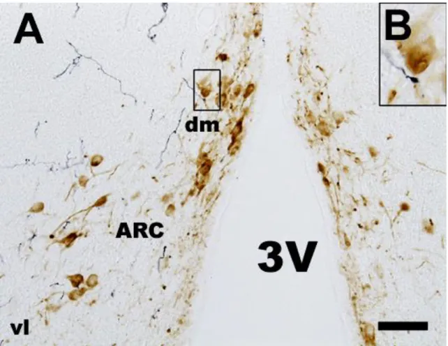
Fig. 13. BDA and TH double labeling in the frontal section of the medial basal hypothalamus (A). Insert (B) shows a high power detail of ARC (in rectangle) ipsilateral to the site of BDA injection in the mesencephalic SPFpc. TH immunopositive cells (indicated by brown colour) in the dorsomedial part of ARC do not show any contact with BDA labeled fibers (indicated by blue colour). Abbreviations: 3V = third ventricle; ARC = arcuate nucleus; dm = dorsomedial part of ARC; vl = ventrolateral part of ARC.
Scale: 100m in A and 25m in B.
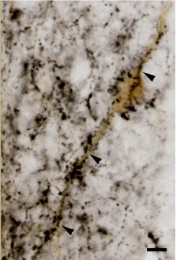
Fig. 14. Microphotograph shows DYN immunoreactive cell bodies (orange colour) and BDA fibers (black colour). BDA fibers exhibit close apposition on DYN immunoreactive cell bodies and dendrites (indicated by white arrowheads). Scale: 25m.
In turn, DYN fibers innervate TIDA neurons which are located in the dorsomedial part of ARC (Fig. 15).
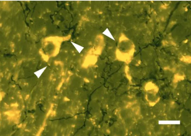
Fig. 15. Microphotograph shows a TIDA neuron (brown colour) which is heavely innervated by DYN fibers (black colour). Arrowheads show close apposition of DYN fibers and dendrites and cell bodies of TIDA neurons. Scale: 20m.
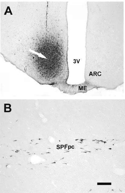
5.1.2. Experiment 2. Retrograde tracer (FG) injection in the lateral ARC
In the midbrain of those animals where FG administration included the antero- ventro-lateral portion of the ARC (Fig. 16A) there was FG labeling in an area located in the rostral mesencephalon that consisted of horizontally oriented cells (Fig. 16B) extending from rostromedial to caudolateral direction fusing with the posterior intralaminal nucleus and PPN. This region is SPFpc.