Prediction of intestinal absorption and brain penetration of drugs
Ph.D. Thesis
Éva Hellinger Pharm.D.
Semmelweis University
Doctoral School of Pharmaceutical Sciences
Supervisor: Dr. Károly Tihanyi Ph.D.
Consultant: Dr. Monika Vastag Ph.D.
Official reviewers: Prof. Dr. Kornélia Tekes Ph.D.
Dr. Katalin Jemnitz Ph.D.
Head of the Final Exam Committee: Prof. Dr. Tamás Török D.Sc.
Members of the Final Exam Committee: Prof. Dr. Imre Klebovich D.Sc.
Dr. István Krizbai Ph.D.
Budapest
2012
Table of content
Abbreviations ... 5
1. Introduction ... 7
1.1. Role of in vitro ADME predictions ... 11
1.2. Intestinal absorption and its modelling ... 12
1.2.1. Small intestine structure and function ... 13
1.2.2. Factors influencing drug absorption ... 14
1.2.2.1. Biological variables of the small intestine that influence drug absorption ... 15
1.2.2.2. Drug properties influencing absorption ... 16
1.2.3. Models of intestinal absorption ... 17
1.2.3.1. In silico models ... 18
1.2.3.2. Non-cell based in vitro systems ... 18
1.2.3.3. Cell culture based in vitro models ... 19
1.2.3.4. Excised tissues (ex vivo models) ... 22
1.2.3.5. In situ models ... 25
1.2.3.6. In vivo animal models ... 25
1.2.3.7. Conclusions to models of absorption ... 26
1.2.4. Improving the properties of Caco-2 cultures ... 28
1.3. Brain penetration and models of blood-brain barrier permeability ... 29
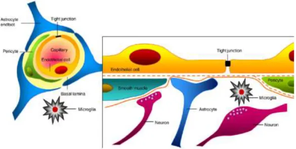
1.3.1. Barriers of the CNS ... 29
1.3.2. The neurovascular unit ... 30
1.3.3. The barrier function ... 31
1.3.4. Elements of the barrier ... 32
1.3.4.1. Transport pathways ... 32
1.3.4.2. Intercellular junctions ... 35
1.3.5. In vitro BBB models ... 35
1.3.5.1. Critical features of in vitro cell-based BBB penetration models... 36
1.3.5.2. Primary brain endothelial cell-based BBB models ... 38
1.3.5.3. Brain endothelial cell line-based BBB models ... 40
1.3.5.4. Epithelial cell based surrogate BBB models ... 41
1.3.6. Modelling CNS permeation ... 42
1.3.6.1. Factors influencing brain penetration in vivo ... 42
1.3.6.2. In vivo models of brain penetration ... 43
1.3.6.3. In vitro - in vivo correlations using BBB models ... 44
2. Aims ... 46
3. Materials and methods ... 47
3.1. Chemicals ... 47
3.2. Cell cultures ... 47
3.2.1. Caco-2 cells ... 47
3.2.2. VB- Caco-2 cells ... 47
3.2.3. MDCK cells ... 48
3.2.4. Rat BBB model ... 49
3.3. Cell morphology ... 49
3.4. Electron microscopy ... 49
3.5. Immunostaining ... 50
3.6. Real-time PCR ... 51
3.7. Western blot ... 51
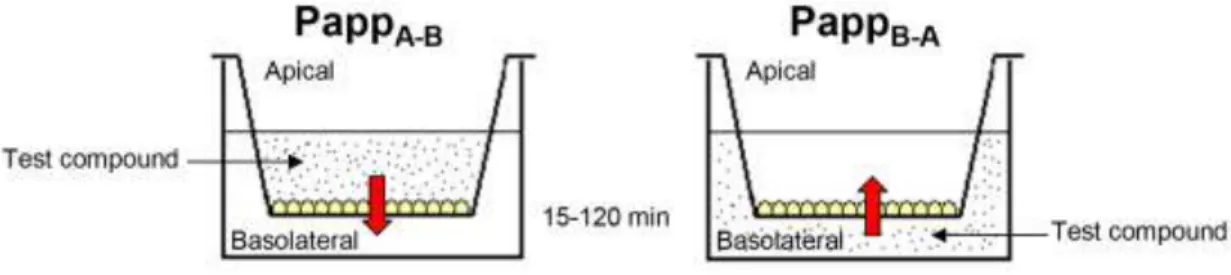
3.8. Bidirectional transport assay ... 52
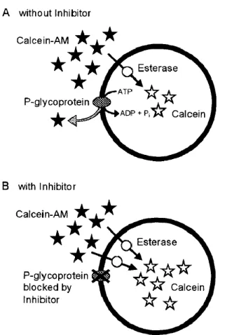
3.9. Calcein-AM extrusion assay ... 54
3.10. CYP activity ... 56
3.11. Equilibrium dialysis measurements ... 56
3.12. In vivo studies of drug permeability in mice ... 57
4. Results ... 58
4.1. Drug penetration model of vinblastine-treated Caco-2 (VB-Caco-2) cultures.... 58
4.1.1. VB-Caco-2 culture ... 58
4.1.2. P-glycoprotein mRNA and protein levels in VB-Caco-2 and in Caco-2 cultures ... 59
4.1.3. Functionality of VB-Caco-2 in comparison to Caco-2: passive penetration 60 4.1.4. Prediction of human absorption by VB-Caco-2 ... 60
4.1.5. P-gp functionality of VB-Caco-2 compared to Caco-2 in bidirectional transport assay ... 63
4.1.6. The effect of vinblastine withdrawal on P-gp level and functionality in VB- Caco-2 cultures ... 64
4.1.7. Screening of NCEs using VB-Caco-2 and Caco-2 bidirectional transport assay ... 66
4.1.8. P-glycoprotein functionality of VB-Caco-2 in Calcein AM assay ... 67
4.1.9. CYP enzyme activity ... 68
4.2. Challenging brain penetration modelling with VB-Caco-2: ... 69
Comparison of brain capillary endothelial cell-based and epithelial cell-based surrogate BBB penetration models ... 69
4.2.1. Morphology: electron microscopy and immunohistochemistry ... 69
4.2.1.1. Rat brain capillary endothelial cells co-cultured with pericytes and astrocytes (rat BBB) ... 69
4.2.1.2. Native human Caco-2 and VB-Caco-2; dog kidney epithelial cell lines: MDCK and MDCK-MDR1 ... 73
4.2.2. P-glycoprotein expression in rat BBB EPA and in epithelial cell lines (native Caco-2, VB-Caco-2, MDCK and MDCK-MDR1) ... 74
4.2.3. Comparison of paracellular tightness of the models ... 74
4.2.4. Comparison of efflux of P-gp substrate drugs and permeability of mixed mechanism drugs ... 75
4.2.5. Correlation of in vitro and in vivo drug permeability in rat BBB and in native Caco-2, VB-Caco-2, MDCK and MDCK-MDR1 models ... 77
4.2.5.1. Brain tissue and plasma protein binding of reference drugs ... 77
4.2.5.2. Effect of tissue binding and P-gp functionality on in vitro – in vivo permeability correlation ... 78
4.2.6. Comparison of high P-gp activity models: VB-Caco-2 versus MDCK-MDR1 ... 80
5. Discussion ... 82
5.1. Drug penetration model of vinblastine-treated Caco-2 cultures ... 82
5.2. Challenging brain penetration modelling with VB-Caco-2: ... 85
Comparison of brain capillary endothelial cell-based and epithelial cell-based surrogate BBB penetration models ... 85
6. Conclusions and novel findings ... 90
7. Summary ... 92
8. Összefoglalás ... 93
9. References ... 94
10. Publications ... 127
Acknowledgements ... 129
Appendix (Paper I-IV) ... 130
Abbreviations
ABC ATP binding cassette
ADME absorption, distribution, metabolism and excretion AJ adherens junctions
ATP adenosine triphosphate BBB blood brain barrier
BBCEC bovine brain capillary endothelial cells BBMEC bovine brain microvessel endothelial cells BCRP breast cancer resistance protein
BCSFB blood-cerebrospinal fluid barrier
BCECF 2‟,7‟-bis(2-carboxyethyl)-5(6)-carboxyfluorescein
BCECF-AM 2‟,7‟-bis(2-carboxyethyl)-5(6)-carboxyfluorescein acetoxymethyl ester Caco-2 colon adenocarcinoma cell line
calcein-AM calcein–acetoxymethylester CEC cerebral endothelial cell CNS central nervous system CSF cerebrospinal fluid CYP cytochrome P450
EPA triple co-culture of rat brain capillary endothelial cells with pericytes and astrocytes
fu free (unbound) fraction GI gastrointestinal tract GLUT glucose transporter
HA human absorption
HPT human peptide transporter 1
IAM immobilized artificial membrane assay IRF initial rate of fluorescence
ISF interstitial fluid
KHB Krebs-Henseleit buffer KM Michaelis constant
Kp total brain to total blood concentration ratio
Kp,uu unbound brain to unbound blood concentration ratio at a steady state
LAT large neutral amino acid transporter LDL low-density lipoprotein
MBUA mouse brain uptake assay MCT monocarboxylic acid transporter MDCK Madin-Darby canine kidney
MDCK-MDR1 MDCK cells transfected with the human MDR1 gene MDR1 multidrug resistance protein
MOAT multispecific organic anion transporter MRP multidrug resistance protein
mRNA messenger RNA MW molecular weight NaF fluorescein sodium OAT organic anion transporter
OATP organic anion transporting polypeptide OCTN organic cation transporter
PAMPA parallel artificial membrane permeation assay PBCEC porcine brain capillary endothelial cells PBMEC porcine brain microvessel endothelial cell Papp apparent permeability
PBS phosphate buffered saline PCR polymerase chain reaction
PD pharmacodynamics
Pe permeability coefficient PEPT peptide transporter 1
P-gp P-glycoprotein, encoded by ABCB1, also termed MDR1 PK pharmacokinetics
PS permeability surface area product SLC solute carrier
TEER trans epithelial/endothelial electric resistance TJ tight junction
VB-Caco-2 vinblastine-treated Caco-2
ZO zonula occludens
1. Introduction
A prerequisite of drug action is their presence in the molecular surroundings of the target in sufficient concentration and for sufficient length of time. Living organisms prevent drugs and other xenobiotics from entering a body by means of efflux transporters and eliminate them by means of highly adaptive metabolizing enzymes.
The ADME properties of drugs (absorption, distribution, metabolism and excretion) will define their pharmacokinetic properties. Nowadays, all factors and traits that determine the pharmacokinetic profile of drugs are subject to modelling and serve as a ground of prediction. Pharmacokinetic properties of drug candidates are decisive data, either promote or block the development of NCEs.
The absorption, distribution, metabolism, excretion, and action of a drug all involve the crossing of cell membranes. In most cases, a drug must transverse the plasma membranes of many cells to reach its site of action. Bypassing cells by paracellular passage through intercellular gaps is a physiological mechanism e.g. in filtration across the glomerulus in the kidney. Generally, large lipophilic drugs do not penetrate paracellularly; they need to pass barriers through the cell membrane (transcellular diffusion). The capillaries of the central nervous system (CNS) and a variety of epithelial tissues have tight intercellular junctions, so paracellular passage through them is limited at best. Most drugs cross membranes by passive processes driven by the concentration gradient, but mechanisms involving the active participation of transporters or carriers also play an important role. Active transport is characterized by a direct requirement of energy, movement against a concentration gradient, saturability, selectivity and competitive inhibition by co-transported compounds (1).
Transporters are membrane proteins that control the influx of essential nutrients and ions and the efflux of cellular waste, environmental toxins, drugs, and other xenobiotics. Regarding drug transport, two groups of transporters are known; ABC (ATP binding cassette) and SLC (solute carrier) transporters. Most ABC proteins are primary active transporters, which need ATP hydrolysis to actively pump their substrates across membranes. These efflux transporters likely evolved as a defense mechanism against harmful substances, therefore, they have broad substrate specificity,
consequently many drugs qualify as substrates for them. The SLC superfamily includes genes that encode facilitated transporters and ion-coupled secondary active transporters that generally mediate the cellular uptake of nutrients such as glucose and amino acid, and drugs that structurally resemble the endogenous ligands of the transporter.
Therefore, efflux and uptake transporters play substantial role in the pharmacokinetic and pharmacodynamic pathways of their substrate drugs, including pathways involved in both, therapeutic and adverse effects.
Transporters that are important in pharmacokinetics are generally located in the intestinal, renal, and hepatic epithelia, as well as in the capillary endothelia of blood- brain barrier (BBB). Transporters expressed in the liver and kidney work in concert with drug-metabolizing enzymes to eliminate drugs and their metabolites, thereby affecting exposure, and hence toxicity, in all organs (2). At the same time, efflux transporters control the tissue distribution and have a role in tissue penetration, therefore, may serve as protective barriers to particular organs and cell types. Access of solutes to several tissues such as the brain and testes is restricted and the efflux transporters in these barrier endothelia may limit penetration of drugs.
Among the best recognized transporters in the ABC superfamily are P- glycoprotein (P-gp, encoded by ABCB1, also termed MDR1) (Fig. 1) (3), which extrudes a large variety of xenobiotics (Table 1). The overexpression of P-gp (with other efflux transporters) is responsible for the multidrug resistance of tumours to some cancer chemotherapeutic agents. But P-gp is physiologically present on the apical surface of intestinal and kidney epithelial cells, on the luminal surface of brain capillary endothelial cells forming the BBB and on the canalicular membrane of hepatocytes. P- gp mediated efflux has been recognized as a serious limiting factor in brain entry of several substrate drugs (4,5,6), such as the HIV protease inhibitors and loperamide.
Loperamide is a potent opioid that lacks the central effects characteristics of other opioids and therefore used for diarrhoea (1). P-gp also influences the intestinal absorption of low permeability or dissolution limited P-gp substrates. Consequently, these drugs have reduced or variable plasma levels and non-linear pharmacokinetics (7,8,9,10). The degree of absorption or the intestinal efflux of cyclosporine, talinolol and tacrolimus reportedly correlates with the intestinal MDR1 mRNA level in humans,
and a decreased absorbed fraction has been reported in response to P-gp induction by carbamazepine or rifampicin or St. John‟s wort (11,12,13,14,15).
Fig. 1. Model of substrate transport by P-gp. A: Substrate (magenta) partitions into the bilayer from outside of the cell to the inner leaflet and enters the internal drug-binding pocket through an open portal.
B: ATP (yellow) binds to the nucleotide-binding domains causing a large conformational change presenting the substrate and drug-binding site(s) to the outer leaflet/extracellular space (3).
Table 1. Reported substrates and inhibitors of P-gp (16).
In the intestine the saturation of P-gp mediated efflux transport can occur due to high local concentration of dissoluted drugs and the simple diffusion may overcome P- gp activity (especially by high solubility - high diffusion drugs), still variable and non- linear pharmacokinetics along doses can occur and make drug development difficult.
Saturation of P-gp function is less likely at BBB as usually low drug levels are present in the plasma; therefore, P-gp can limit severely brain penetration of P-gp substrate drugs (9).
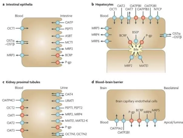
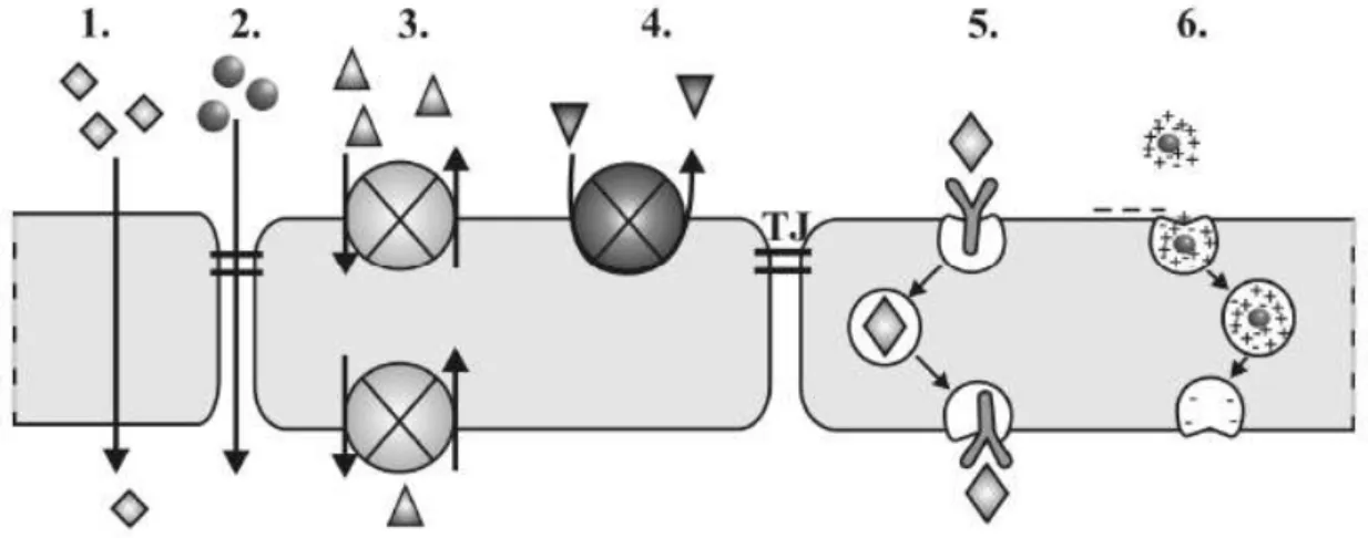
Additionally, MRP2 (multidrug resistance protein, ABCC2), and BCRP (breast cancer resistance protein, ABCG2) are also expressed in the apical side of the intestinal epithelia, where they serve to pump out xenobiotics, including many orally administered drugs. At the BBB, beside P-gp, there is accumulating evidence for the roles of BCRP and MRP4 in limiting the brain penetration of drugs (17,2). Fig. 2 shows the transporters in plasma membrane domains of intestinal epithelia, hepatocytes, kidney proximal tubules and brain capillary endothelial cells.
Fig. 2. Selected human transport proteins for drugs and endogenous substances. a: Intestinal epithelia contain in their apical (luminal) membrane several uptake transporters including one or more members of the organic anion transporting polypeptide (OATP) family; peptide transporter 1 (PEPT1;SLC15A1); ileal
apical sodium/bile acid co-transporter (ASBT; SLC10A2); and monocarboxylic acid transporter 1 (MCT1; SLC16A1). The apical ATP-dependent efflux pumps include multidrug resistance protein 2 (MRP2; ABCC2); breast cancer resistance protein (BCRP; ABCG2); and P-glycoprotein (P-gp; MDR1, ABCB1). The basolateral membrane of intestinal epithelia contains organic cation transporter 1 (OCT1;
SLC22A1); heteromeric organic solute transporter (OSTα-OSTβ); and MRP3 (ABCC3). b: Human hepatocyte uptake transporters in the basolateral (sinusoidal) membrane include the sodium/taurocholate co-transporting peptide (NTCP;SLC10A1); three members of the OATP family (OATP1B1(SLCO1B1), OATP1B3 (SLCO1B3) and OATP2B1 (SLCO2B1)); organic anion transporter 2 (OAT2; SLC22A7)and OAT7 (SLC22A9); and OCT1. Efflux pumps in the hepatocyte basolateral membrane include MRP3, MRP4 (ABCC4) and MRP6 (ABCC6). Apical (canalicular) efflux pumps of the hepatocyte comprise P-gp; bile-salt export pump (BSEP or SPGP; ABCB11); BCRP (ABCG2); and MRP2. In addition, multidrug and toxin extrusion protein 1 (MATE1; SLC47A1) is located in the apical hepatocyte membrane. c: Kidney proximal tubules contain in the apical (luminal) membrane OAT4 (SLC22A11);
urate transporter 1 (URAT1; SCL22A12); PEPT1 and PEPT2 (SLC15A2); MRP2 and MRP4; MATE1 and MATE2-K (SLC47A2); P-gp; organic cation/ergothioneine transporter (OCTN1; SLC22A4); and organic cation/carnitine transporter (OCTN2; SLC22A5). Basolateral uptake transporters in proximal tubule epithelia include OATP4C1 (SLCO4C1); OCT2; and OAT1, OAT2 and OAT3 (SLC22A8). d:
Apical (luminal) transport proteins of brain capillary endothelial cells contributing to the function of the blood–brain barrier include the uptake transporters OATP1A2 and OATP2B1; and the efflux pumps P-gp, BCRP, MRP4 and MRP5 (ABCC5) (17).
1.1. Role of in vitro ADME predictions
In the early phase of drug discovery, PD- and PK-related aspects of molecules must be considered. Beyond the affinity and selectivity for the target pharmaceutical properties (stability, solubility, question of formulation), permeability across cell membranes and other ADME traits may require optimization (18). The cost and time taken to develop new medicines has continued to rise over recent times while the number of new drug approvals has declined. Therefore, the pharmaceutical industry is generally looking to improve success rates and reduce candidate attrition during the drug development process. One of the options for that is the early termination of drug development programmes that would ultimately fail, consequently resources can be focused on those compounds most likely to succeed (19).
Inappropriate pharmacokinetic behaviour, one of the causes of failure in drug development includes such factors as low bioavailability due to high extraction or poor
absorption characteristics, short elimination half-life leading to short duration of action and excessive variability due to genetic or environmental factors (19). In 1991, adverse pharmacokinetic and bioavailability results were the most significant cause of attrition in the clinic, and accounted for ~40% of all attritions (20). This observation has led to an increased emphasis on pharmacokinetic input to the drug discovery process throughout the pharmaceutical industry. Much progress has been made in developing tools for the prediction of drug absorption, drug clearance and drug–drug interactions, in addition to the scaling of pharmacokinetic parameters from animals to man. The use of pharmacokinetic predictions in the drug discovery phase has resulted in fewer compounds failing as a result of inappropriate clinical pharmacokinetics (19). By 2000, adverse pharmacokinetic and bioavailability had dramatically reduced as a cause of attrition in drug development, and contributed to it less than 10% (20). Moreover, consideration of the pharmacokinetic profile of a new chemical entity can also be beneficial in the assessment of safety and efficacy. Generally it is possible to make fairly robust predictions of the pharmacokinetic profile in man using in vitro systems and preclinical pharmacokinetic studies (19).
1.2. Intestinal absorption and its modelling
The oral route is a convenient way to administer drug products and it still remains the route of choice for a high patient compliance in most indications.
Absorption across the intestinal barrier is a prerequisite for an orally administered drug to take effect. Absorption is the result of a complex process that begins when the dosage formulation is swallowed and proceeds through the liberation of the drug molecules from the formulation, crossing the biological membranes of enterocytes by passive diffusion and/or active transport, and metabolization by enzymes. Absolute bioavailability is the fraction of a dose that reaches systemic circulation following oral administration. The key determinants of the systemically available drug level are the fraction of the dose that enters the enterocytes and the fraction that escapes first pass metabolism. Current physiological properties of the gastrointestinal system, the physicochemical properties of the drug and its formulation will have impact on the systemic availability.
1.2.1. Small intestine structure and function
The primary function of the gastrointestinal tract (GI) is the absorption of nutrients and water, but it also serves for drug absorption following oral administration.
Furthermore, the protection of an organism from systemic exposure of various toxins, antigens, and microorganisms is also an important function. Therefore, the GI is also an interface between the outer world and an organism.
Drug absorption occurs in each anatomical segments of the GI, starting from the buccal mucosa, moving through the stomach, the small intestine (duodenum, jejunum and ileum) and ending in the colon. The pancreas and the biliary system of the liver secrete enzymes and detergents into the duodenum. Pancreatic secretions contain hydrolytic enzymes (proteases, lipases, amylases) that digest food, but also decompose drugs with sensitive groups. Biliary acids improve drug solubility and dissolution through micellization and wetting (21). Some metabolites and xenobiotics are subjects to entero-hepatic circulation, a process of intestinal reabsorption and repeated biliary excretion.
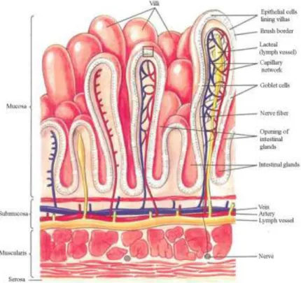
The functional layers of the small intestine are the mesothelium (serosa), muscularis propria, submucosa and the mucosa-containing absorptive epithelium. The intestine is richly supplied with blood vessels, nerve endings and lymphatics (Fig. 3).
The layer of columnar epithelial cells represents the most important barrier that nutrients and drugs must traverse to enter a body. The epithelium displays fingerlike projections, called villi, which provide increased surface area in order to achieve maximal absorption. The villi house the self-renewing population of epithelial cells with different functions, including secretory cells, endocrine cells and absorptive enterocytes.
There are membrane invaginations at the feet of the villi, known as crypts, which consist of continuously dividing stem cells. Stem cells produce daughter cells, which are the source of both epithelial and crypt cells (22,23,24).
Fig. 3. Structure of the small intestine wall (25).
The microvilli-covered apical surface of the epithelium constitutes the brush border with enzymes such as sucrase, lactase and alkaline phosphatase. Brush border enzymes are involved in the digestion of carbohydrates. These enzymes hydrolyse sensitive drug substrates such as esters, amides etc. The brush border morphology is a special characteristic of the intestine that greatly increases the surface area of the absorptive cell and so provides increased contact with substances in order to achieve maximally efficient absorption (26,27). Special morphological features such as the brush border, villi and circular folds, together with the long transit time and variable pH, make the small intestine the primary site for drug absorption in the GI.
1.2.2. Factors influencing drug absorption
Drug absorption is influenced by the biological variables of gastrointestinal tract, like transit time, pH, presence of food, secreted detergents, gut motility and the expression level of transporters and enzymes. Moreover, the net pharmacokinetic
performance of the drug is also the consequence of the drug properties determining its solubility, penetrability and affinity to transporters and enzymes.
1.2.2.1. Biological variables of the small intestine that influence drug absorption
Residence time
Gastric residence and intestinal transit time influence drug absorption. This is especially true for dissolution-limited drugs. The mean gastric residence time is shorter than that of the intestine (28). Relatively high inter-individual variability also exists.
Fed/fasted state, dosage form (tablet, capsule, solution, suspension etc.), drug particle size (an important physical parameter of the dissolution rate) and the volume of co- administered water all impact residence time.
pH
There is a pH gradient along the GI tract, which ranges from more acidic in the stomach to acidic/neutral in the small intestine. The solubility, dissolution and penetrability of ionizable drugs are sensitive to these pH variations. The pH range in the stomach and the jejunum varies greatly depending on the fasted/fed state (29).
Active transports
Drug absorption is particularly strongly influenced by both active influx and efflux transport processes for compounds with low passive penetrability. Based on gene expression levels, the most abundant active transporters in the human intestine are HPT1 (human peptide transporter 1), PEPT1 (peptide transporter 1), BCRP, MRP2, MDR1 and MCT1 (monocarboxylic acid transporter 1) (30). A compound with P-gp liability may overcome the intestinal barrier due to saturation at high intestinal dose and display a non-linear PK. The P-gp level increases distally along the segments of the small intestine, with relatively low interindividual variation (≤ 2-fold) compared to CYP3A4 (> 10-fold) (31,32).
Metabolism
Drug metabolism in the gut wall can severely reduce the drug level available for absorption. CYP3A4 is a major metabolic enzyme in the intestine (33). In the upper intestinal segments, it is assumed that there is interplay between P-gp and metabolic enzymes. P-gp and 3A4 have overlapping substrate specificity and so they act in concert: P-gp reduces intracellular drug levels, therefore CYP3A4 can act more effectively at lower substrate level.
Some drugs show varying solubility and stability in different regions of the intestine as a result of changes in environmental pH, degradation by enzymes present in the lumen of the intestine or interaction with endogenous solubilising agents such as bile. In other instances active efflux or uptake transport mechanisms will modify drug absorption to various extent in certain regions of the GI tract. Such drugs display region-specific absorption i.e. they can only be absorbed efficiently in specific segments of the GI tract that are named "absorption windows" (34).
1.2.2.2. Drug properties influencing absorption
Solubility and permeability are two major features of a drug that determine its oral absorption. The Biopharmaceutical Classification System has categorized drugs in terms of their solubility and intestinal permeability. Class I compounds are defined as those with high solubility and high permeability, and predicted to be well absorbed when given orally. All other compounds (classes II-IV) suffer either from low solubility, low permeability or both, and will present challenges to the development of products with acceptable oral bioavailability (34).
Penetration of xenobiotics is a virtually continuous process of traversing hydrophilic and lipophilic phases, so the lipophilic and hydrophilic features of drugs are important in this process. The Lipinski Rule defines the likelihood of poor absorption in humans, using the structural properties of over 2000 compounds that survived Phase I clinical trials (35). The „Rule-of-Five‟ predicts that poor absorption or permeation is more likely when the number of hydrogen bond donors is more than 5, there are at least 10 hydrogen bond acceptors, the molecular weight is greater than 500 and the calculated
Log P is greater than 5. The „Rule-of-Five‟ should be considered as a qualitative predictor of absorption and permeability. With large molecular size, solubility and passive penetrability decreases. If the number of hydrogen bonds and thus the drug‟s polar feature increases, aqueous solubility will increase, but passive penetration (partitioning in the lipid bilayer of the cell membrane) will decrease. Lipophilicity above a certain level limits molecular penetration, as drugs are simply trapped in the cell membranes. For optimal absorption, a good balance of permeability and aqueous solubility is deemed in the range of 0 < logP < 3 or 1 logD7.4 3.
Veber et al. has defined additional critical properties that are required in order to achieve good oral bioavailability in rats (36). They reported the findings from a study of rat bioavailability data for 1100 drug candidates. They found that molecules possessing fewer than 10 rotatable bonds and having a polar surface area less than 140 Å2 (or H- bond count less than 12) generally showed oral bioavailability in rats exceeding 20%.
Ionizability is also a critical drug property that influences drug absorption.
Charged molecules display higher solubility than their neutral forms, yet they show lower rates of penetration. As there is a pH gradient along the gastrointestinal tract, the solubility and penetration of chargeable drugs may differ accordingly. Most drugs (75%) are weak bases, therefore less ionized and penetrate faster in the more basic segments of the GI, preferably in the small intestine.
High lipophilicity and therefore low aqueous solubility of new chemical entities is a common problem in recent drug research.
1.2.3. Models of intestinal absorption
In order to predict the absorption of new chemical entities, preclinical research generates data using in silico, in vitro, ex vivo and in vivo models previously correlated with human data. The availability of suitable human data is one of the most critical points of human prediction. High enough bioavailability (> 80%) may reflect good absorption and is a useful source of human data. The ratio of the cumulative urinary excretion of the drug and metabolites following oral and intravenous administration is another important form of data used for the human prediction endpoint. Low solubility, formulation-, salt- and dose-dependent absorption, and metabolism complicate the
evaluation of absorption (37). Some human single-pass perfusion data are also available, and present a good correlation with the extent of absorption determined by other pharmacokinetic studies (38).
1.2.3.1. In silico models
In silico methods use calculated physicochemical drug properties (e.g.
lipophilicity, hydrogen bonding, molecular surface properties, solubility, solvatation energy, charge distribution) as descriptors for drug permeation, and frequently directly predict drug pharmacokinetic (PK) properties such as human absorption (39,40,41,42,37). The accuracy and the predictive value of in silico models are critically dependent on the reliability of the biological data on which the model is based (43).
PSA, NPSA (non-polar surface area) and dynamic PSAd appear to be important descriptors for the prediction of passive penetration (44,45). Prediction for active transports still needs to be proven (46,47). Otherwise, in silico models are high throughput, cost-effective techniques (requiring minimum usage of resources and manpower), as they are normally used for screening of virtual libraries, and a molecule will only be synthesized if it shows a satisfactory level of absorption.
1.2.3.2. Non-cell based in vitro systems
There are several methods that use artificial membranes or liposomes to predict drug absorption or drug-membrane interaction (48,49,50). The most widely used methods are IAM (immobilized artificial membrane assay) chromatography and PAMPA (parallel artificial membrane permeation assay). In IAM an artificial membrane is employed as the stationary phase of chromatography. A monolayer made up of phospholipid analogues is covalently bonded to the surface of silica particles. The technique can be performed coupled with a conventional HPLC instrument. Based on chromatographic retention time, it is possible to rank and classify a large number of drugs in a cost effective way (51). The method runs with small amounts of test articles and does not require either high purity samples or quantification. However, IAM models
good lipid partition in this model do not necessarily cross the epithelial cell layer. The results simply represent passive transcellular uptake.
The PAMPA is a high throughput assay for permeability screening in early drug research. It is performed in two-part multi-well plates with an artificial lipid membrane impregnated with mixture of lecithin/phospholipids dissolved in an inert organic solvent (52,53). The drug concentration in the acceptor plate is followed by HPLC, or simply by a more efficient system coupled UV-plate reader. Due to its high throughput capacity, PAMPA can be used to pre-screen large sets of compounds for passive penetrability prior to cellular assays, and can provide information about ionization status, pH dependence of the absorption, and the influence of the unstirred water layer on drug permeability. PAMPA tolerates lower pH levels and a higher content of solubilizing agents than cell-based assays (54). The composition of the artificial membranes, type of filter material and the applied pH make PAMPA useful both for intestinal and BBB penetration measurement. Similarly to IAM, only passive transcellular transport can be predicted using PAMPA, as there is no consideration of paracellular pathway, carrier- mediated transport, drug metabolism, and active transporters, therefore, false positives and negatives may occur for active transporter substrates and for drugs capable of metabolism at the site of permeation.
1.2.3.3. Cell culture based in vitro models
For the majority of oral drugs, the intestinal epithelium forms the main barrier that must be permeated in order to enter the body. Epithelial cell-based systems are therefore expected to be the best in vitro models of oral drug absorption. In vitro models of drug penetration that display adequate passive permeability and efflux transporter functionality, especially those that offer both P-gp and good human predictability simultaneously, are much-needed tools for high throughput application in early drug research. Primary cultures of enterocytes have very poor viability (55,56,57), but immortalized cell lines such as Caco-2, HT-29, T-84, MDCK and 2/4/A/1 grow rapidly into confluent monolayers, and model absorption at some level. Reference compounds with known human absorption serve for validation of the models. Compared to the simpler IAM and PAMPA systems, the cell-based systems more closely resemble in
vivo conditions. Depending on the type and differentiation of the monolayers, these models display not only passive transcellular but also paracellular and active transport features. They have an advantage over in vivo animal models in that they require neither high amounts of test compounds nor animal test subjects.
Caco-2
Caco-2 is the preferred cell-based model for human drug absorption (58) (59).
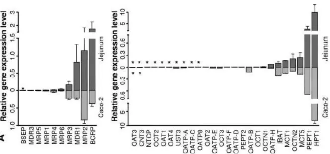
Caco-2 cells are derived from human colonic adenocarcinoma, but they have morphological and functional similarities to small intestinal enterocytes (60). The cells undergo spontaneous differentiation on permeable filters, depending on culture conditions. The advantage of the human origin of the cells is underlined by the results that many of the active transporter genes that are present in the human intestine, such as MDR1, MRP-2, -3, -5; BCRP, OCTN-1, -2, MOAT-C, PepT1 and OATP-B, are expressed at some level in Caco-2 cells (Fig. 4) (61,30,62,63). The functionality of several transporters were shown in native Caco-2 by measuring adequate substrates in bidirectional transport assay, for example: talinolol (64), cimetidine, vinblastine, colchicine, cyclosporine (65), digoxin, (66) for P-gp, dactinomycin, daunorubicin, dipirydamole, domperidone (65) for MRP2, adefovir, (67) for MRP4, daunorubicin, ciprofloxacin, furosemide, sulfasalazine (65,68) for BCRP, estrone-3-sulfate, (69), levofloxacin, (70), imatinib, (71) for OATP1A2 and estrone-3-sulfate (72,73,74) for OATP2B1.
A number of research labs have established a correlation between the permeability across Caco-2 monolayers and the human dose fraction absorbed for different sets of compounds (for reviews, see (75,76,77)).
The Caco-2 transport assay enables the measurement of permeability of both the absorptive (apical-to-basolateral) and secretive (basolateral-to-apical) directions. The apical compartment models the intestinal lumen, and the basal compartment models the blood stream. As an output, apparent permeability (Papp) is calculated and efflux ratio can be characterized by the ratio of Papp basolateral to apical vs. Papp apical to basolateral. A ratio greater than 2.0 is generally accepted as an indicator of efflux (78). Preincubation of the cells with verapamil or quinidine is frequently used to confirm P-gp activity.
Fig. 4. Relative expression levels of ABC transporters (left panel) and SLC (right panel) transporters in human tissues isolated from jejunum mucosa. Each corresponding gene expression level in the Caco-2 cell line is shown in the opposing direction on the graph (light gray bars) for visualization of homologies and discrepancies in transporter expression profiles. The bars represent the mean relative expression levels; error bars indicate the standard deviation. *, absence of gene expression (30).
The Caco-2 cell model is widely accepted for permeability assessment, although it suffers from shortcomings such as long term cultivation of cells and the under- expression of active influx and efflux transporters and metabolic enzymes such as CYP3A4 (79,80,81,82,83). The permeability of hPEPT1 substrates such as cephalexin and amoxicillin are reported to be underestimated in Caco-2 (84). There is also a variable and low expression of P-gp in Caco-2 cultures, which seems to limit its use for screening P-gp substrates or studying P-gp related interactions (85,86). A number of laboratories have attempted to overcome some of the shortcomings of Caco-2.
Improvement some functions of Caco-2 was our aim too, this is discussed later.
Other cell culture models of drug penetration
The HT-29 is a human intestinal colon cancer cell line that only differentiates after modifications of culture media, e.g. replacing of various components leads to clone selection and differentiation. The presence of galactose instead of glucose causes the formation of polarized cells. Clones differentiate into mucus-secreting goblet cells that can be used for studying the effect of mucin on intestinal absorption (87,88).
The T84 cell line, like Caco-2, differentiates spontaneously in culture after reaching confluency. However, the cells express fewer biochemical and morphological markers of differentiation (89). This cell line also expresses P-glycoprotein. It has been demonstrated that it is a good model for studying the induction of efflux transporters (90).
Madin-Darby canine kidney (MDCK) cells differentiate spontaneously into columnar epithelial cells and form tight junctions when cultured on semi-permeable membranes. A major advantage of MDCK cells over Caco-2 is the shorter cultivation period (3 days vs. 3 weeks) (91). Permeability data obtained for a large set of compounds (n=55) in MDCK model correlated well with human absorption and also with Caco-2 penetrability (92). MDCK cells derived from dog kidney cells may express transporters that are grossly different from those in the human intestine, which limits its usefulness for the prediction of human intestinal absorption. MDCK cells transfected with human MDR1 is a well accepted surrogate model of blood-brain barrier permeability (93).
2/4/A1 is a foetal rat intestinal epithelial cell line conditionally immortalized with a temperature-sensitive mutant of SV40 (94). 2/4/A1 is only useful for passively transported compounds (42). The transport rate of typical hydrophilic, poorly permeable paracellular compounds (e.g. mannitol and creatinine) in 2/4/A1 monolayers is comparable to that in the human jejunum, and much faster than in the Caco-2 cell model.
1.2.3.4. Excised tissues (ex vivo models)
The isolated tissues have the advantage of working with intact organs with physiological cell-cell contacts and normal intracellular matrices. In these models, the effect of variable tissue properties such as the distribution of transporters in the different segments of the GI may be examined (95).
In vivo, compounds passing the enterocyte are distributed to the systemic blood circulation by the blood and lymph vessels of the adjacent lamina propria. The underlying muscle layer does not represent a barrier to the absorptive process. The musculature can therefore be stripped off the tissues, consequently the tissue can be
oxygenated more efficiently. If the tissues are sufficiently supported with oxygen, they can be used for several hours. However, as the tissue is separated from its in vivo blood circulation, its viability is hampered and may lead to incorrect and non-reproducible permeability data (96), which is the main disadvantage of these ex vivo models.
Isolated-perfused intestinal segments
The entire small intestine, or alternatively, just a segment of it, is cannulated at both ends. After removal, the intestine or intestinal segment is placed in a perfusion apparatus and circulated luminally with an oxygenated buffer containing the test compound (97). In a more elaborate version of this method, the intestine is perfused both luminally and vascularly, so the superior mesenteric artery and the portal vein are cannulated and perfused (98,99). Wei et al. recently adapted this technique for human intestinal segments (100). The drug disappearance is calculated and an equal level of drug absorption is assumed, which is only valid when apical uptake is a rate-limiting step in drug absorption. If drug metabolism or accumulation occurs in the mucosa, then this approach leads to an overestimation of drug absorption (101). Using luminally and vascularly perfused method, both drug appearance in the vascular perfusate and its disappearance from the lumen can be followed.
The technique is valuable in the elucidation of transport mechanisms and may also be useful in evaluating the absorption of poor solubility drugs that require formulation (101).
Everted gut sacs
The small intestine of rodents (mostly rats) is removed, flushed and placed into an oxygenated special tissue culture medium. The intestine is everted, divided into sacs, and then laced up at both ends. In one end of the sac, a blunt needle syringe is inserted to inject the required volume into the sac lumen. The sac is placed in a tank of oxygenated buffer with the test compound (102). The drug content is analyzed in the solution of the serosal side and in the digested tissue/sacs. The transport of drugs, nutrients, liposomes, proteins and macromolecules can be studied with this method (103), and kinetic parameters can be determined with high reliability and
reproducibility. Examination of the paracellular transport of hydrophilic molecules makes possible to study the effect and toxicity of potent enhancers (104).
Drug absorption can be measured at different parts of the intestine, and the region, where drug absorption is maximal, may be identified. (105). It has been demonstrated that this method is suitable to study the effect of P-gp on xenobiotic transport by co-administered P-gp inhibitors (106,107), the role of influx transporters like PepT1, LAT2 (105,108), intestinal drug metabolism (109), hydrolysis of prodrugs (110,111), food effect (112) and the effects of excipients and different formulations on absorption (113,78).
Even using well-oxygenated buffers, the destruction of the intestinal mucosa is rapid. Apart from this issue, it is an inexpensive, relatively simple technique that is useful in examining site specific intestinal absorption. Paracellularly-transported compounds had similar permeability values to those obtained with human perfusion studies.
Ussing chambers
Ussing and Zerahn (114) first introduced Ussing chambers to study the active transport of sodium in isolated frog skin. Since then, the method has been used to study ion transport, drug absorption and permeability across the intestinal sheets of rats, rabbits, hamsters and mice (115,116). Using human biopsies of gastric, jejunal and colon mucosa barriers, the functions of the human GI tract can also be studied (117,118,119). Ussing chambers have also been used to study the intestinal metabolism of xenobiotics (120,121).
The technique is applied to opened, isolated intestinal sections mounted between two halves of a diffusion chamber. The intestinal section, as a flat sheet, separates the buffers from the drug containing buffers so that the transport of molecules across the tissue and between the chambers can be measured.
Both mucosal-to-serosal (m-s) and serosal-to-mucosal (s-m) directional fluxes can be studied, enabling the differentiation of passively and actively absorbed drugs (122). Different segments of the GI can be used, enabling the evaluation of site-specific absorption. Even the electro-physiological properties of the intestinal membrane during drug absorption can be monitored (123). Determination of species differences with
respect to intestinal characteristics may support the selection of suitable animal models for drug bioavailability studies (124,125). The short viability of the tissues in simple media due to the lack of blood flow and the sensitivity of the mounted sheets in the diffusion cells to mechanical damage may limit the generation of reliable data.
1.2.3.5. In situ models
In situ experiments involve perfusion of the drug solution through isolated cannulated intestinal segments. The perfused organ stays connected with the systemic blood circulation of the animal (126). Drug permeability, absorption kinetics, intestinal transport and metabolism, regional absorption and drug secretion into the intestinal lumen after i.v. administration are frequently studied in in situ models (127,128,129).
Using in situ models, drug absorption is generally estimated on the basis of drug disappearance from the intestinal lumen, but the rate of decrease in the drug concentration in the perfusate is not always equal to the rate of absorption into the portal vein. Sampling from mesenteric vessels or the portal vein can give a better estimation of drug absorption (130). Perfused flow rate and anaesthesia can influence the rate of drug absorption (89,131,132). Non- or low-absorbable volume markers such as PEG 4000, mannitol or Lucifer Yellow should be used to take into consideration the change in the luminal drug solution due to absorption or secretion of water. This method requires a large number of animals to obtain statistically significant absorption data and relatively high amounts of test compounds ( > 10 mg).
A particular advantage of this method is that by bypassing the stomach acidic compounds are not likely to be precipitated, and the process is not complicated with biliary excretion and entero-hepatic circulation as is the case in in vivo methods. The presence of an intact blood supply and intact innervation mimics in vivo conditions well, and the model provides useful kinetic data.
1.2.3.6. In vivo animal models
Animal models are commonly used to predict the extent of the absorption of drug candidates. In practical terms, oral bioavailability is determined through this
method. However, drug bioavailability is influenced by far more factors than intestinal absorption alone. Sampling from the portal vein may rule out the metabolism in the liver, and intra duodenal administration of dissolved compounds makes possible to eliminate the role of dissolution, gastric acidity and emptying (124).
In vivo models integrate all of the biological factors that may affect drug absorption, such as the mucus layer, the dynamic components of the mesenteric blood circulation and all of the other factors that can influence drug dissolution. However, there are numerous species differences that influence absorbed drug levels. The gastric volume available for drug dissolution and gastric pH differ between species. Rodents have a less acidic pH than humans (133). There are also significant species differences in the activity, type and distribution of metabolic enzymes and also in the expression of transporters in the different species. GI volume, motility and transit time also show species specificity. Dogs are known to overpredict the human bioavailability of hydrophilic drugs. Rats seem to be better predictors of the bioavailability of compounds with paracellular and carrier-mediated processes (133,134,135,125). Generally, for low solubility, dissolution-limited, metabolism-subjected drugs, the bioavailability may not reflect absorption alone, and may contain numerous species-related factors.
1.2.3.7. Conclusions to models of absorption
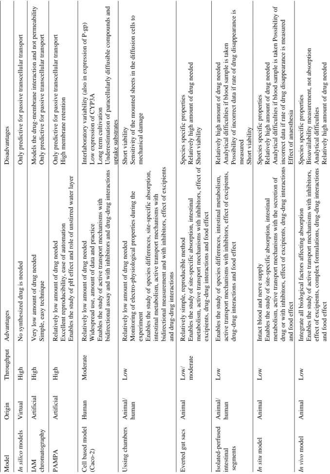
Predictive, cost-effective, high-throughput absorption models are necessary tools for drug research (Table 2). The choice of method depends largely on the questions to be answered and the stage of intended application. At more advanced levels of drug research, the questions are more complex. In the design phase, in silico approaches using relatively simple rules can orient chemistry thinking; later, when drug substances are available, fast screens such as PAMPA followed by the relevant cell-based assay can reveal drug candidates with highly limiting passive and/or active penetrability and metabolic liabilities.