Article
Antimicrobial, Antioxidant and Antiproliferative Secondary Metabolites from Inonotus nidus-pici
Zsófia Garádi1,2 , Miklós Dékány3 ,Ágnes M. Móricz4 , AnikóGaál5, Viktor Papp6 , Szabolcs Béni1 and Attila Ványolós1,*
Citation: Garádi, Z.; Dékány, M.;
Móricz, Á.M.; Gaál, A.; Papp, V.; Béni, S.; Ványolós, A. Antimicrobial, Antioxidant and Antiproliferative Secondary Metabolites fromInonotus nidus-pici.Molecules2021,26, 5453.
https://doi.org/10.3390/molecules 26185453
Academic Editors: Magdalena Ligor and Aleksandra Szydłowska- Czerniak
Received: 3 August 2021 Accepted: 29 August 2021 Published: 7 September 2021
Publisher’s Note:MDPI stays neutral with regard to jurisdictional claims in published maps and institutional affil- iations.
Copyright: © 2021 by the authors.
Licensee MDPI, Basel, Switzerland.
This article is an open access article distributed under the terms and conditions of the Creative Commons Attribution (CC BY) license (https://
creativecommons.org/licenses/by/
4.0/).
1 Department of Pharmacognosy, Semmelweis University, Üll˝oiút. 26, H-1085 Budapest, Hungary;
garadi.zsofia@pharma.semmelweis-univ.hu (Z.G.); beni.szabolcs@pharma.semmelweis-univ.hu (S.B.)
2 Directorate of Drug Substance Development, Egis Pharmaceuticals Plc, P.O. Box 100, H-1475 Budapest, Hungary
3 Spectroscopic Research, Gedeon Richter Plc., Gyömr˝oiút 19-21, H-1103 Budapest, Hungary;
m.dekany@richter.hu
4 Plant Protection Institute, Centre for Agricultural Research, ELKH, Herman Ottó út 15, H-1022 Budapest, Hungary; moricz.agnes@atk.hu
5 Biological Nanochemistry Research Group, Institute of Materials and Environmental Chemistry, Research Centre for Natural Sciences, Magyar Tudósok Körútja 2, H-1117 Budapest, Hungary; gaal.aniko@ttk.hu
6 Department of Botany, Hungarian University of Agriculture and Life Sciences, Villányiút 29-43, H-1118 Budapest, Hungary; papp.viktor@uni-mate.hu
* Correspondence: vanyolosa@pharmacognosy.hu
Abstract: Inonotus nidus-piciis a sterile conk which produces macrofungus, a neglected Central- Eastern European relative of the prizedInonotus obliquus, also known as chaga. Investigation of the methanol extract of the poroid fungusI. nidus-piciresulted in the isolation of citropremide (1), 3,4-dihydroxybenzalacetone (2) , lanosterol (3), ergost-6,8,22-trien-3β-ol (4), and ergosterol peroxide (5). The structures of fungal compounds were determined on the basis of one- and two-dimensional NMR and MS spectroscopic analysis. Compounds1–2and4–5were evaluated for their antioxidant and antimicrobial properties against several bacterial and fungal strains. 3,4-dihydroxybenzalacetone (2) and ergost-6,8,22-trien-3β-ol (4) demonstrated moderate antimicrobial activity, while the former possessed notable antioxidant activity in DPPH assay. The antiproliferative examinations performed on three human cancer (MES-SA, MES-SA/Dx5, A431) cell lines demonstrated that compounds4and 5have notable cytotoxic activity with IC values in micromolar range. The current study represents the first report on the chemical profile ofI. nidus-pici, providing a comprehensive study on the isolation and structure determination of bioactive secondary metabolites of this macrofungus.
Keywords:Hymenochaetaceae; chaga; sclerotium; steroids; ceramide; antimicrobial; antiproliferative;
doxorubicin resistant
1. Introduction
Higher macrofungi, also known as mushrooms, have been used as a source of food and for their medicinal (anticancer, anti-inflammatory, and immunomodulatory, etc.) prop- erties since ancient times, particularly in the traditional medicines of the Far East. Recent mycochemical studies unequivocally demonstrate that mushrooms represent an almost unlimited reservoir of biologically active natural products with a high structural diver- sity: aromatic cadinane sesquiterpenoids fromPhellinus piniwith potential SARS-CoV-2 inhibitory activity [1], meroterpenoids fromAlbatrellus yasudaewith amyloid-βaggregation inhibitory properties [2], and antibacterial diterpenoids fromPsathyrella candolleana[3], among others. Fungal sclerotia are also notable sources of biologically active secondary metabolites, and many of these fungal structures are considered as prospective medicines or as a food source [4–6]. For instance, the sclerotia of some edible mushroom species
Molecules2021,26, 5453. https://doi.org/10.3390/molecules26185453 https://www.mdpi.com/journal/molecules
(e.g.,Laccocephalum mylittae,Pleurotus tuber-regium) are known as important food compo- nents, while others (e.g.,Lignosusspp.,Pachymaspp.,Polyporus umbellatus) are utilized by traditional medicine and consumed principally for their positive healing effects [7–10]).
The so-called chaga mushroom (Inonotus obliquus) is a wood-decay fungus that pro- duces a massive, black, crusty conk (i.e., sclerotium) mainly on living birch (Betulaspp.) trees. The sterile conk ofInonotus obliquus(Ach. ex Pers.) Pilát is a well-known source of traditional medicine, especially in Russia, Poland, and the Baltic countries, and has a long history of use by indigenous people to treat cancer, cardiovascular diseases, viral and bacterial infections, and gastro-intestinal disorders [11–13]. Currently,I. obliquusis one of the most intensely studied medicinal mushrooms, and has been shown to contain a variety of biologically active compounds, e.g., polysaccharides, triterpenoids, and styrylpyrone derivatives [14–17]. In contrast, no data are available in the literature concerning the chemical constituents of a closely related species,I. nidus-piciPilát ex Pilát. Despite this, in previous research we highlighted the antioxidant and xanthine oxidase activities of this fungus [18]. Based on the similar sterile conks produced by this species,I. nidus-piciwas previously considered as only a form ofI.obliquusand discussed in the former literature under the namePoria obliquaf. “sur chêne” [19], orXanthochrous obliquusf.cavernatus[20].
However, the two species are well-separated by morphological features and phylogenetic evidence [21,22]. In contrast withI. obliquus, both the poroid form and the sterile conks ofI.
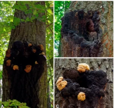
nidus-picidevelop on the same host and colonize mainly livingQuercusspp. (i.e.,Q. cerris), or occasionally other angiosperms, e.g., Fagus,Juglans,Aesculus,Platans, andFraxinus species [23,24]. The resupinate basidiomes ofI. nidus-picidevelop inside the cavities of the host tree, while the yellowish-brown sterile conks (becoming black, hard, and rimose) grow on cortical layers around the cavity (Figure1).
Figure 1.Basidocarp and the imperfect stage (sterile conk) ofInonotus nidus-picion livingQuercus cerrisin Hungary. (A) Young and old sterile conks of the studied specimen (PV1043); (B) old sterile conks and sporulating basidiocarp (Börzsöny Mts, 19. Jun 2021; (C) young and old sterile conks (Visegrád Mts, 1 October 2011). Photos: V. Papp.
According to the literature,I. nidus-pici(Hymenochaetaceae,Basidiomycota) is considered to be a mainly Central-Eastern European species, but has also been identified in Spain and Iran [23–25]. Given the lack of any information on the chemical profile ofI. nidus-pici, we
decided to conduct a research project to explore the mycochemical and pharmacological potential of this species.
2. Results and Discussion
Thorough investigation of the methanol extract obtained from the sterile conks ofI.
nidus-piciresulted in the identification of five compounds (Figure2).
Figure 2.Compounds isolated fromInonotus nidus-pici.
The fungal extract was first subjected to solvent–solvent partition between aqueous MeOH andn-hexane, followed by extraction with chloroform and ethyl acetate. The resultingn-hexane and chloroform extracts were purified using normal and reversed phase flash column chromatography. Compounds1–5were structurally characterized on the basis of NMR and MS spectroscopic data and confirmed by comparing them to those reported earlier in the literature.
All fungal metabolites isolated have been identified for the first time in sterile conks ofI. nidus-pici.
The1H NMR spectrum of compound1in THF-d8showed two overlapping resonances with triplet multiplicity atδ0.89 (t,J= 7.0 Hz, 6H, CH3-180, CH3-24) ppm which indicated the presence of two methyl groups (Table1). The methylene resonances atδ1.22–1.60 (m,
~68H, H-30-H170, H-5-H-23) indicated the presence of two saturated aliphatic chains. The
1H resonance atδ7.45 (d,J= 8.5 Hz,1H, NH-10) ppm lacking13C HSQC correlation and the13C resonance atδ175.1 ppm revealed the presence of an amide moiety. According to its correlation in the COSY spectrum, the amide group is adjacent to a CH group atδ 4.06 (m, 1H, H-2) ppm. The13C NMR spectrum showed four well-separated resonances atδ77.1 (C-3), 73.1 (C-4), 72.7 (C-20), and 62.3 (C-1) ppm. These resonances and their HSQC correlations suggested the presence of a CH2and three CH groups attached to hydroxyl groups. Moreover, the resonances of the four hydroxyl groups also appeared in the1H NMR spectrum atδ4.92 (d,J= 5.2 Hz, 1H, 20-OH), 4.38 (m, 1H, 3-OH), 4.37 (m, 1H, 1-OH), and 3.98 (d,J= 6.4 Hz, 1H, 4-OH) ppm. Based on the aforementioned NMR characteristics combined with their homo- and heteronuclear correlations, the compound was suggested to be a ceramide derivative in which the fatty acid chain contains a hydroxyl group at the alpha position, and the long chain base contains three hydroxyl groups. Due to the overlapping methylene resonances, MS experiments were necessary to unequivocally confirm the exact structure. MS experiments showed that the accurate mass found for the [M + H]+ ion was 684.65127, while the calculated mass for [C42H85O5N + H]+was
684.65005 in positive ESI mode resulted in a relative mass difference of 2.5 ppm. Mass accuracy was between 2.5 and 3.5 ppm for the fragment ions. The resulting protonated species [M + H]+underwent a neutral loss (H2O), resulting in the fragment ionm/z666 in the MS/MS spectrum. Further to this, a subsequent loss of water produced the fragment ion 648 (M + H−H2O−H2O = 648). A minor fragment ion peak (m/z300; C18H38O2N) can prove the number of methylene groups next to the amide group (i.e., the number of carbon atoms in the fatty acid residue), whilem/z282 ion (C18H36ON) can be explained as water loss fromm/z300. The measured NMR and MS spectra for compound1can be found in the Supplementary Material (Table S1, Figures S1–S10).
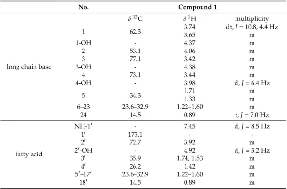
Table 1.Complete1H and13C NMR resonance assignments for compound1(THF-d8).
No. Compound 1
long chain base
δ13C δ1H multiplicity
1 62.3 3.74 dt,J= 10.8, 4.4 Hz
3.65 m
1-OH - 4.37 m
2 53.1 4.06 m
3 77.1 3.42 m
3-OH - 4.38 m
4 73.1 3.44 m
4-OH - 3.98 d,J= 6.4 Hz
5 34.3 1.71 m
1.33 m
6–23 23.6–32.9 1.22–1.60 m
24 14.5 0.89 t,J= 7.0 Hz
fatty acid
NH-10 - 7.45 d,J= 8.5 Hz
10 175.1 - -
20 72.7 3.92 m
20-OH - 4.92 d,J= 5.2 Hz
30 35.9 1.74, 1.53 m
40 26.2 1.42 m
50–170 23.6–32.9 1.22–1.60 m
180 14.5 0.89 m
Thus, the structure of compound1was identified as citropremide, which has a poly- hydroxylated ceramide structure. It has been isolated for the first time from a fungal species, previously being identified only in the stem bark of the evergreen shrubCitropsis gabonensisfrom theRutaceaefamily [26]. NMR spectra were also measured in pyridine-d5 for comparison with data in the previous literature (Figures S7 and S8). The identity of the NMR chemical shifts and the optical rotation of compound1(α26D + 8, pyridine,c0.25) also suggested the same absolute configuration as the known compound: (2S,3S,4R)-2-[(2R)- 2hydroxyoctadecanoylamino]tetracosane-1,3,4-triol. This configuration is the most typical for related ceramides from natural sources in which the fatty acid and the long chain base lengths vary, but the molecular formula is the same [27,28].
Compound2is 3,4-dihydroxybenzalacetone (osmundacetone by its vernacular name), and was previously identified in several medicinal mushrooms, e.g.,InonotusandPhellinus species [29,30], and also in the Far Eastern fern,Osmunda japonica, from where its name originates [31]. The NMR characteristics of compound2are provided in the Supplementary Material (Table S2, Figures S13 and S14), which are similar to the characteristics exhibited in previous publications [24].
Osmundacetone exhibits significant antioxidant properties. Moreover, it is neuropro- tective based on its activity against oxidative glutamate toxicity in an HT22-immortalized hippocampal cell line, according to a recent study published by Trinh et al. [32].
Compound3was found to be a lanostane, while4and5were ergostane type triter- penes. The 1D and 2D NMR spectra of the isolated compounds suggested that they were lanosterol (3), ergost-6,8,22-trien-3β-ol (4), and ergosterol peroxide (5). The1H NMR data
of compound3was similar to previously published data, and the NMR spectra of4and 5were also identical to that of a previous report [28,29]. The complete1H and13C reso- nance assignments of the compounds are given in the Supplementary Material (Table S3, Figures S15–S20).
In the used concentrations, compounds1and5were inactive in the antimicrobial and antioxidant assays. An antioxidant effect was observed only in the case of2, with EC50 29.7±1.3µM that was similar to the (+)-catechin positive controls EC50 14.6±0.4µM.
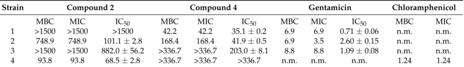
Neither compound2nor4could inhibit mycelial growth of the tested fungi at the used concentration, but they showed moderate antibacterial activity (Table2). Osmundacetone (2) was effective against all bacterial strains exceptB. subtilis, while compound4inhibited all bacterial strains exceptA. fischeri. Comparing their antibacterial potencies to those of antibiotics, the strongest activity was attributed to compound4againstB. subtiliswith a five times higher MIC value than that of gentamicin (Table2).
Table 2.The MBC, MIC and IC50values of compounds2and4and two antibiotics inµM against 4 bacterial strains.
Strain Compound 2 Compound 4 Gentamicin Chloramphenicol
MBC MIC IC50 MBC MIC IC50 MBC MIC IC50 MBC MIC
1 >1500 >1500 >1500 42.2 42.2 35.1±0.2 6.9 6.9 0.71±0.06 n.m. n.m.
2 748.9 748.9 101.1±2.8 168.4 168.4 41.9±0.5 6.9 3.5 2.60±0.15 n.m. n.m.
3 >1500 >1500 882.0±56.2 >336.7 >336.7 203.0±8.1 8.8 8.8 1.09±0.08 n.m. n.m.
4 93.8 93.8 68.5±2.8 >336.7 >336.7 >336.7 n.m. n.m. n.m. 1.24 1.24
1Strains: (1) Bacillus subtilis subsp. spizizenii (Gram +), (2) Rhodococcus fascians (Gram +), (3) Pseudomonas syringae pv. maculicola (Gram−), (4) Aliivibrio fischeri (Gram−). n.m.—not measured.
The cytotoxicity experiments revealed that among the isolated compounds, ergosterol peroxide (5) was the most effective metabolite, providing the lowest IC50 (36.18µM) value against the doxorubicin resistant cell line, being comparable with the IC50 of 18.4µM of the reference compound, doxorubicin (Table3). Compared to the triterpenes (4–5) and the phenolic compound (2), citropremide (1) exhibited a noticeably decreased activity against all the investigated human cancer cell lines. Considering the results of compound4, a substantially decreased cytotoxic property was detected against the epidermoid cancer cell line. In general, among the three examined cell lines, MES-SA proved to be the most susceptible to the applied fungal metabolites.
Table 3.IC50cytotoxicity values (µM) of isolated compounds and the reference compound against uterine sarcoma (MES-SA, MES-SA/Dx5) and epidermoid carcinoma (A431) cell lines.
Compound MES-SA MES-SA/Dx5 A431
1 92.1±1.1 195.0±9.8 142.1±3.2
2 57.5±3.1 109.6±5.2 101.3±4.1
4 41.9±2.9 42.2±2.1 83.2±5.4
5 37.6±2.6 36.2±3.2 45.7±3.9
doxorubicin 0.65±0.04 18.4±1.98 1.12±0.11
3. Materials and Methods
Optical rotation was measured on a Jasco P-2000 digital polarimeter at the NaD
line. The chemicals used in the experiments were supplied by Sigma-Aldrich, Budapest, Hungary and Molar Chemicals, Halásztelek, Hungary. Flash chromatography was car- ried out on a CombiFlash®Nextgen 300+ instrument with 200–800 nm UV-VIS variable wavelength detection using RediSep Rf Gold normal-phase silica and reversed-phase C18 Flash columns (4, 12, 40 and 60 g) (Teledyne Isco, Lincoln, NE, USA). NMR spectra were recorded in deuterated chloroform (chloroform-d, 99.8 atom% D, contains 0.03% (v/v) TMS, Sigma-Aldrich), methanol (methanol-d4, 99.8 atom% D, contains 0.03% (v/v) TMS, Sigma-Aldrich), tetrahydrofuran (tetrahydrofuran-d8, 99.5 atom% D, Sigma-Aldrich) or pyridine (pyridine-d5, 99.8 atom% D, Sigma-Aldrich) on a BRUKER AVANCE III HD 600
(600/150 MHz) instrument equipped with a Prodigy cryo-probehead at 295 K. The pulse programs were taken from the Bruker software library (TopSpin 3.5). 13C and1H chemical shifts (δ) are given in ppm relative to the NMR solvent or relative to the internal standard (tetramethylsilane), while the coupling constants (J) are given in Hz. The complete1H and
13C assignments were achieved by widely accepted strategies based on1H NMR,13C NMR,
1H-1H COSY,1H-1H NOESY,1H-13C HSQC, and1H-13C HMBC measurements. HRMS analyses were performed on a Thermo Velos Pro Orbitrap Elite (Thermo Fisher Scientific, Bremen, Germany) system. The ionization method was EI (with 70 eV) and ESI operated in positive ion mode. The protonated molecular ion peaks were fragmented by CID at a normalized collision energy of 35–45%. For the CID experiment, helium was used as the collision gas. The samples were dissolved in methanol. Data acquisition and analysis were accomplished with Xcalibur software version 4.0 (Thermo Fisher Scientific, Bremen, Germany).
3.1. Mushroom Material
Sterile conks ofInonotus nidus-piciwere collected in 2014 in Börzsöny Mts, near Király- rét, on livingQuercus cerris; 47.906553, 18.999720, Hungary. Fungal identification was made by Viktor Papp. A voucher specimen (No. PV 1043) has been deposited at the Department of Botany, Hungarian University of Agriculture and Life Sciences, Hungary. The fungal sample collected was carefully cleaned, all contaminating materials, i.e., any plant part (e.g., bark) and/or soil contaminant were removed before processing. The samples were dried at room temperature for five days and stored in a cool and dry place until extraction.
3.2. Extraction and Isolation
The air-dried sterile conks (302 g) ofI. nidus-piciwere ground, and then extracted with 5×1500 mL methanol for 5×90 min using an ultrasonic bath. Following filtration, the fungal extracts were combined and concentrated in a vacuum. The dry residue (10.1 g) was dissolved in 300 mL of 50% aqueous MeOH and was subjected to liquid–liquid partition betweenn-hexane (4×150 mL), CHCl3(4×150 mL) and ethyl acetate (4×150 mL). The n-hexane fraction (2.12 g) was subjected to flash chromatography on a silica gel column using a gradient system ofn-hexane and acetone (0–25%; t = 50 min). Fractions with similar compositions were combined according to TLC monitoring (A1–A18). From the combined fraction A15 (29 mg), a precipitation formed, which was washed withn-hexane and then acetone to obtain compound1 (19 mg). Combined fraction A4 (208 mg) was further purified in consecutive steps of normal (gradient system of hexane–acetone) and reversed phase (eluent system of water–methanol) flash chromatography steps to give compounds3(3 mg) and5(5 mg). Another fraction (A7, 142 mg) was subjected to a final purification by normal- (eluent system ofn-hexane–acetone) and reversed- (eluent system of water–methanol) phase flash chromatography, leading to compound4(14 mg). The chloroform soluble phase (1.14 g) was subjected to flash chromatography on silica gel column using gradient system ofn-hexane–acetone (5–45%; t = 55 min). Fractions with similar compositions were combined according to TLC monitoring (B1–B11). Fraction B9 (71 mg) was further separated by combination of normal- (n-hexane–acetone) and reversed- (water–methanol) phase flash chromatography steps to obtain compound2(18 mg).
3.3. Antimicrobial Assays
Isolated compounds2(4 mg/mL),4(2 mg/mL), and5(2 mg/mL) were dissolved in ethanol. Gram-positiveBacillus subtilissubsp.spizizeniisoil bacterium (DSM 618) was from Merck (Kenilworth, New Jersey), andRhodococcus fascians(B.01608) was purchased from the National Collection of Agricultural and Industrial Microorganisms (NCAIM, Budapest, Hungary). Gram-negative, naturally luminescent marine bacteriumAliivibrio fischeri(DSM 7151) were obtained from the Leibniz Institute DSMZ–German Collection of Microorganisms and Cell Cultures, Berlin, Germany. LuminescentArabidopsispathogen Pseudomonas syringaepv.maculicola, chromosomally tagged with lux-CDABE genes, was
kindly provided by Jun Fan (John Innes Center, Department of Disease and Stress Biology, Norwich, UK).F. avenaceumstrain IMI 319,947 was from CABI-IMI Culture Collection, Egham, UK, andBipolaris sorokiniana(Sacc.) Shoemaker H-299 (NCBI GenBank accession No. MH697869) was collected in Hungary.B. subtilisin Lysogeny broth (10 g/L tryptone (Reanal, Budapest, Hungary), 5 g/L yeast extract (Scharlau, Barcelona, Spain) and 10 g/L sodium chloride (Reanal)) at 37◦C,R. fasciansin Waksman broth (5 g/L peptone (Scharlau), 5 g/L meat extract (Reanal), 5 g/L sodium chloride (Reanal), 10 g/L glucose (Reanal) at 26◦C, concentrated sodium hydroxide (Reanal) solution to set the pH at around 7.4),A.
fischeriin ocean medium (5 g/L tryptone, 3 g/L yeast extract, 3 g/L glycerol (Reanal), 35 g/L sea salt mix (Instant Ocean), concentrated sodium hydroxide (Reanal) solution to set the pH at around 7.2) at 28◦C, andP. maculicolain gelatin broth (20 g/L pancreatic digested gelatin (Carl Roth, Karlsruhe, Germany), 10 g/L potassium sulfate (Reanal), 10 mL/L glycerol (Reanal), 1.4 g/L magnesium chloride (Reanal)) at 28◦C were grown by shaking at 120 rpm.F. avenaceumandB. sorokinianawere grown in Lysogeny broth by shaking at 120 rpm at 21◦C for 3 days in the dark. The washed mycelium was cut in LB to small pieces with a FastPrep®-24 Classic homogenizer (7×2 mm glass beads in a 2-mL Eppendorf tube, 4.5 m/s for 20 s, MP Bio, Beograd, Serbia).
96-well microplates were used for the determination of the minimal inhibitory concen- tration (MIC) and the half maximal inhibitory concentration (IC50) of the isolates against the microbial growth. Gentamicin and chloramphenicol (both from Sigma) were used as the positive control, and ethanol was used as the negative control. Two-fold dilution series of 5µL of the isolates (made in triplicate) in ethanol was prepared in the microtiter plates.
After the evaporation of the ethanol in a sterile box, 70µL of LB and 50µL of a mycelium suspension (OD600 0.2) or 150µL of bacterial suspension (105CFU/mL) were added to each well. The absorbance at 600 nm was measured by a microplate spectrophotometer (Labsystems Multiscan MS 4.0, Thermo Scientific, Waltham, MA, USA) immediately before and after incubation (24 h for bacteria by shaking with Grant PHMP microplate shaker and 72 h for fungi). The experiment was repeated on two separate occasions with three parallels, and the results were averaged. Minimal bactericide concentration (MBC) was determined by plotting 5µL of each well onto appropriate agar layers, and after incubation the presence or absence of bacterial colonies was checked.
3.4. DPPH Assay
Antioxidant assay was performed in 96-well microplates using 2,2-diphenyl-1-picryl- hydrazyl radical (DPPH˙, Sigma, St. Louis, MO, USA) and two-fold dilution of the samples.
The positive control was (+)-catechin (Sigma) dissolved in ethanol (1 mg/mL). In the wells, the samples were mixed with 75µL ethanol and 50µL ethanolic DPPH˙solution (0.2 mg/mL). The absorbance at 492 nm was measured by a microplate reader (Labsystems Multiscan MS 4.0) after 30 min incubation at room temperature in the dark. The experiment was repeated three times, EC50 (half maximal effective concentration) was determined, and the results were averaged.
3.5. Cytotoxic Assay
The MES-SA (No.: CRL-1976™) uterine sarcoma cell line and its doxorubicin-selected derivative MES-SA/Dx5 (No.: CRL-1977™) were purchased from ATCC. A431 human skin-derived, epidermoid carcinoma cell line was purchased from ATCC.
After incubation with the compounds (diluted from 10 mM stock solutions in DMSO, except for Compound1, which was dissolved in THF), which lasted for 72 h (the initial seeded cell number was at a density of 5000 cells/well), the supernatant was removed, washed once with DPBS, and the viability was assessed by means of the PrestoBlue® assay (Invitrogen™, USA; purchased from Thermo Fisher Scientific, Waltham, MA, USA), according to the manufacturer’s instructions. Viability of the cells was measured spec- trophotometrically (measuring fluorescence, excitation at 560 nm and emission at 590 nm) using an EnSpire microplate reader (Perkin Elmer, Waltham, MA, USA). Curve fit statis-
tics were used to determine the concentration of the test compound that resulted in 50%
toxicity (IC50). Curves were fitted by using the sigmoidal function (Origin) or sigmoidal dose–response (comparing variable and fixed slopes) model (GrapPad Prism), and they were expressed as the mean of three independent experiments (each one performed with three replicates). Data were normalized to untreated cells. The IC50value (given inµM) is the average of at least three independent determinations. Every IC50value represents the corresponding GI50value (the average growth inhibition of 50%).
4. Conclusions
The current study represents the first mycochemical examination of the poroid fun- gusI. nidus-pici, a disregarded relative of the well-known medicinal mushroomInonotus obliquus. In-depth chemical analysis of the methanol extract ofI. nidus-piciled to the iden- tification of a ceramide (1), a phenolic derivative (2), and three triterpene steroids (3–5).
The pharmacological experiments that we conducted revealed that compounds2and4 exhibited moderate antimicrobial properties, that 2 showed notable antioxidant property in DPPH assay, while compounds4and5exerted significant antiproliferative properties on three human cancer cell lines, including a doxorubicin resistant one.
Supplementary Materials:The following are available online: Figures S1–S10: NMR and MS spectra and spectral data of compound1, Figures S11–S18: NMR spectra and spectral data of compounds2–5, Table S1: HRMS spectral data of compound1, Table S2: Complete1H and13C NMR resonance assignments for compound2, Table S3: Complete1H and13C NMR resonance assignments for compounds3,4and5.
Author Contributions: Conceptualization, A.V.; resources and funding acquisition Z.G., S.B.;
writing—original draft preparation, A.V., V.P. and Z.G.; supervision A.V. and S.B.; Z.G., S.B. and M.D.
performed the spectral analysis and structure determination; V.P. provided sample collection and identification; A.G. performed the cytotoxicity assays;Á.M.M. performed the antimicrobial and an- tioxidant experiments. All authors have read and agreed to the published version of the manuscript.
Funding:Prepared with the professional support of the Doctoral Student Scholarship Program of the Co-operative Doctoral Program of the Ministry of Innovation and Technology, financed by the National Research, Development and Innovation Fund (KDP-1007075, Z.G.). This work was partially supported by the János Bolyai Research Scholarship of the Hungarian Academy of Sciences by the Bolyai+ New National Excellence Program (grant number:ÚNKP-20-5-SE-31) of the Ministry of Human Capacities (S.B.), and by the National Research, Development and Innovation Office of Hungary (NKFIH K128921,Á.M.M.).
Institutional Review Board Statement:Not applicable.
Informed Consent Statement:Not applicable.
Data Availability Statement:All data are found in the Supplementary Materials.
Acknowledgments:The authors are grateful to Tamás Czeglédi from the Department of Pharmacog- nosy, Semmelweis University, for his assistance with the extraction and isolation. The authors thank Zoltán Béni (Gedeon Richter Plc.) and András Dancsó(Egis Pharmaceuticals Plc.) for their assistance in spectral analysis, and Péter G. Ott and József Bakonyi from Plant Protection Institute, CAR, ELKH, for the preparation of bacterial and fungal cell suspensions.
Conflicts of Interest:The authors declare no conflict of interest.
Sample Availability:Samples of the Compounds1–2,4–5are available from the authors.
References
1. Li, X.; Gao, J.; Li, M.; Cui, H.; Jiang, W.; Tu, Z.-C.; Yuan, T. Aromatic Cadinane Sesquiterpenoids from the Fruiting Bodies of Phellinus pini Block SARS-CoV-2 Spike–ACE2 Interaction.J. Nat. Prod.2021,84, 2385–2389. [CrossRef]
2. Masuda, Y.; Fujihara, K.; Hayashi, S.; Sasaki, H.; Kino, Y.; Kamauchi, H.; Noji, M.; Satoh, J.-i.; Takanami, T.; Kinoshita, K.; et al.
Inhibition of BACE1 and Amyloid-βAggregation by Meroterpenoids from the Mushroom Albatrellus yasudae.J. Nat. Prod.2021, 84, 1748–1754. [CrossRef] [PubMed]
3. Liu, Y.-P.; Dai, Q.; Wang, W.-X.; He, J.; Li, Z.-H.; Feng, T.; Liu, J.-K. Psathyrins: Antibacterial Diterpenoids from Psathyrella candolleana.J. Nat. Prod.2020,83, 1725–1729. [CrossRef]
4. Lau, B.F.; Abdullah, N. Sclerotium-Forming Mushrooms as an Emerging Source of Medicinals: Current Perspectives.Mushroom Biotechnol.2016,2016, 111–136. [CrossRef]
5. Smith, M.E.; Henkel, T.W.; Rollins, J.A. How many fungi make sclerotia?Fungal Ecol.2015,13, 211–220. [CrossRef]
6. Calvo, A.M.; Cary, J.W. Association of fungal secondary metabolism and sclerotial biology.Front. Microbiol.2015,6, 62. [CrossRef]
[PubMed]
7. Lau, B.F.; Abdullah, N.; Aminudin, N.; Lee, H.B.; Tan, P.J. Ethnomedicinal uses, pharmacological activities, and cultivation of Lignosusspp. (tiger’s milk mushrooms) in Malaysia—A review.J. Ethnopharmacol.2015,169, 441–458. [CrossRef]
8. Bandara, A.R.; Rapior, S.; Bhat, D.J.; Kakumyan, P.; Chamyuang, S.; Xu, J.; Hyde, K.D.Polyporus umbellatus, an Edible-Medicinal Cultivated Mushroom with Multiple Developed Health-Care Products as Food, Medicine and Cosmetics: A Review.J. Cryptogam.
Mycol.2015,36, 3–42. [CrossRef]
9. Oso, B.A. Pleurotus Tuber-Regium from Nigeria.Mycologia1977,69, 271–279. [CrossRef]
10. Wu, F.; Li, S.-J.; Dong, C.-H.; Dai, Y.-C.; Papp, V. The Genus Pachyma (Syn. Wolfiporia) Reinstated and Species Clarification of the Cultivated Medicinal Mushroom “Fuling” in China.Front. Microbiol.2020,11, 788. [CrossRef]
11. Balandaykin, M.E.; Zmitrovich, I.V. Review on Chaga Medicinal Mushroom,Inonotus obliquus(Higher Basidiomycetes): Realm of Medicinal Applications and Approaches on Estimating its Resource Potential.Int. J. Med. Mushrooms2015,17, 95–104. [CrossRef]
[PubMed]
12. Szychowski, K.A.; Skóra, B.; Pomianek, T.; Gmi ´nski, J. Inonotus obliquus—From folk medicine to clinical use.J. Tradit. Complement.
Med.2021,11, 293–302. [CrossRef] [PubMed]
13. Thomas, P.; Elkhateeb, W.; Daba, G. Chaga (Inonotus obliquus): A medical marvel but a conservation dilemma?Sydowia2020,72, 123–130. [CrossRef]
14. Du, X.; Mu, H.; Zhou, S.; Zhang, Y.; Zhu, X. Chemical analysis and antioxidant activity of polysaccharides extracted fromInonotus obliquussclerotia.Int. J. Biol. Macromol.2013,62, 691–696. [CrossRef]
15. Zhao, F.; Xia, G.; Chen, L.; Zhao, J.; Xie, Z.; Qiu, F.; Han, G. Chemical constituents from Inonotus obliquus and their antitumor activities.J. Nat. Med.2016,70, 721–730. [CrossRef]
16. Zheng, W.; Miao, K.; Liu, Y.; Zhao, Y.; Zhang, M.; Pan, S.; Dai, Y. Chemical diversity of biologically active metabolites in the sclerotia of Inonotus obliquus and submerged culture strategies for up-regulating their production.Appl. Microbiol. Biotechnol.
2010,87, 1237–1254. [CrossRef]
17. Lee, I.-K.; Yun, B.-S. Styrylpyrone-class compounds from medicinal fungiPhellinusandInonotusspp., and their medicinal importance.J. Antibiot.2011,64, 349–359. [CrossRef]
18. Papp, V.; Kovács, B.; Zomborszki, Z.; Csupor, B.; Orbán-Gyapai, O.; Urbán, E.; Hohmann, J.; Ványolós, A. Pharmacological Properties of Inonotus Nidus-Pici, a Central-Southern European Relative of the Prized “Chaga” Mushroom. In Proceedings of the 9th International Medicinal Mushroom Conference, Palermo, Italy, 24–28 September 2017; pp. 169–170.
19. Bourdot, H.; Galzin, A. Hymenomycètes de France (XI. Porés).Bull. Trimest. Soc. Mycol. Fr.1925,41, 98–144.
20. Igmándy, Z.; Pagony, H. Fehér-és szürkenyárasaink veszélyes gesztkorhasztógombája.Erdészeti Lapok1965,14, 19–25.
21. Zhou, L.W.; Jr, J.; Vlasák, J. Inonotus andersonii and I. krawtzewii: Another Case of Molecular Sequencing-Based Diagnosis of Morphologically Similar Species.Chiang Mai J. Sci.2014,41, 789–797.
22. Bernicchia, A.; Gorjón, S.P.Polypores of the Mediterranean Region; Romar: Segrate, Italy, 2020; p. 904.
23. Ryvarden, L.; Melo, I. Poroid fungi of Europe.Syn. Fung.2017,37, 1–431.
24. Papp, V.; Rimóczi, I.; Er˝os-Honti, Z. Adatok a hazaiés európai platánok (Platanusspp.) taplóihoz/Polypore data from the Hungarian and European plane trees (Platanusspp.).Növényvédelem2012,48, 405–411.
25. Ghobad-Nejhad, M.; Kotiranta, H. Genus Inonotus Sensu lato in Iran, with Keys to Inocutis and Mensularia Worldwide. In Annales Botanici Fennici; Finnish Zoological and Botanical Publishing Board: Helsinki, Finland, 2008.
26. Tsassi, B.V.; Hussain, H.; Geagni, A.; Dongo, E.; Ahmed, I.; Riaz, M.; Krohn, K. Citropremide and Citropridone: A New Ceramide and a New Acridone Alkaloid from the Stem Bark ofCitropsis gabunensis.Helv. Chim. Acta2011,94, 1035–1040. [CrossRef]
27. Abdelhameed, R.F.; Ibrahim, A.K.; Yamada, K.; Ahmed, S.A. Cytotoxic and anti-inflammatory compounds from Red Sea grass Thalassodendron ciliatum.Med. Chem. Res.2018,27, 1238–1244. [CrossRef]
28. Huang, Q.; Tezuka, Y.; Hatanaka, Y.; Kikuchi, T.; Nishi, A.; Tubaki, K. ChemInform Abstract: Studies on Metabolites of Mycoparasitic Fungi. Part 3. New Sesquiterpene Alcohol from Trichoderma koningii.ChemInform1995,26, 1035–1038. [CrossRef]
29. Zan, L.-F.; Qin, J.-C.; Zhang, Y.-M.; Yao, Y.-H.; Bao, H.-Y.; Li, X. Antioxidant Hispidin Derivatives from Medicinal Mushroom Inonotus hispidus.Chem. Pharm. Bull.2011,59, 770–772. [CrossRef] [PubMed]
30. Mo, S.; Wang, S.; Zhou, G.; Yang, Y.; Li, Y.; Chen, X.; Shi, J. Phelligridins C−F: Cytotoxic Pyrano[4,3-c][2]benzopyran-1,6-dione and Furo[3,2-c]pyran-4-one Derivatives from the Fungus Phellinus igniarius.J. Nat. Prod.2004,67, 823–828. [CrossRef] [PubMed]
31. Zhang, D.; Li, B.W.; Yang, L.; Fu, M.H.; Fang, J. Isolation and determination of osmundacetone in Osmundae Rhizoma.Chin.
Pharm. J.2010,45, 1612–1614.
32. Trinh, T.A.; Seo, Y.H.; Choi, S.; Lee, J.; Kang, K.S. Protective Effect of Osmundacetone against Neurological Cell Death Caused by Oxidative Glutamate Toxicity.Biomolecules2021,11, 328. [CrossRef]