Review
Heat Shock Proteins and Autophagy Pathways in Neuroprotection: From Molecular Bases to
Pharmacological Interventions
Botond Penke1,*, Ferenc Bogár1,2, Tim Crul3 ID, Miklós Sántha3, Melinda E. Tóth3 and LászlóVígh3
1 Department of Medical Chemistry, University of Szeged, H-6720 Szeged, Dóm Square 8, Hungary;
bogar@sol.cc.u-szeged.hu
2 MTA-SZTE Biomimetic Systems Research Group, University of Szeged, H-6720 Szeged, Dóm Square 8, Hungary
3 Institute of Biochemistry, Biological Research Centre, Hungarian Academy of Sciences,
H-6726 Szeged, Temesvári krt. 62, Hungary; crul.tim@brc.mta.hu (T.C.); santha.miklos@brc.mta.hu (M.S.);
toth.erzsebetmelinda@brc.mta.hu (M.E.T.); vigh.laszlo@brc.mta.hu (L.V.)
* Correspondence: penke.botond@med.u-szeged.hu; Tel.: +36-62-545-135
Received: 22 December 2017; Accepted: 18 January 2018; Published: 22 January 2018
Abstract:Neurodegenerative diseases (NDDs) such as Alzheimer’s disease, Parkinson’s disease and Huntington’s disease (HD), amyotrophic lateral sclerosis, and prion diseases are all characterized by the accumulation of protein aggregates (amyloids) into inclusions and/or plaques. The ubiquitous presence of amyloids in NDDs suggests the involvement of disturbed protein homeostasis (proteostasis) in the underlying pathomechanisms. This review summarizes specific mechanisms that maintain proteostasis, including molecular chaperons, the ubiquitin-proteasome system (UPS), endoplasmic reticulum associated degradation (ERAD), and different autophagic pathways (chaperon mediated-, micro-, and macro-autophagy). The role of heat shock proteins (Hsps) in cellular quality control and degradation of pathogenic proteins is reviewed. Finally, putative therapeutic strategies for efficient removal of cytotoxic proteins from neurons and design of new therapeutic targets against the progression of NDDs are discussed.
Keywords: neurodegenerative diseases; neuroprotection; endoplasmic reticulum associated degradation; ubiquitin-proteasome system; autophagy; heat shock proteins; Hsp-inducers; autophagy modulating drugs
1. Introduction
Several neurodegenerative diseases, including Alzheimer’s disease (AD), Parkinson’s disease (PD), Huntington’s disease (HD), amyotrophic lateral sclerosis (ALS), and prion disease are, among others, characterized by the presence of specific proteinaceous inclusions in or around the affected neurons.
These inclusions are composed of misfolded, aggregated, and often toxic forms of specific proteins (Table1).
Many of these disease-associated proteins are aggregation-prone in nature and easily form misfolded, polymerized structures and toxic aggregates (amyloids) without known physiological functions. Neurons are probably the most vulnerable cells to develop deposits, large inclusion bodies, or aggresomes [1].
The ability of cells to maintain proteostasis—the maintenance of all proteins of the proteome in a conformation, concentration, and location that is needed for their correct function—varies drastically among different cell types [2]. Hence, although the exact mechanism of the particular vulnerability of neurons as post-mitotic cells is not well understood, it is most likely caused by the specificity of the neuronal proteostasis network [3].
Int. J. Mol. Sci. 2018,19, 325; doi:10.3390/ijms19010325 www.mdpi.com/journal/ijms
Table 1. Several protein-conformational, neurodegenerative diseases as well as their typical misfolded proteins.
List of Diseases Misfolded Proteins Alzheimer’s disease
β-amyloid (Aβ)
hyperphosphorylated Tau (pTau) α-synuclein
Parkinson’s disease α-synuclein
Huntington’s disease huntingtin
Lewy-body dementia α-synuclein
Amyotrophic lateral sclerosis TDP-43
Prion diseases superoxide dismutase (SOD) prion protein (PrPsc)
Beyond amyloid type protein deposits, neurodegenerative diseases (NDDs) are also characterized by other dramatic pathological changes in the brain. Increased endoplasmic reticulum (ER)-lumen and dysfunction of the endosomal-autophagic-lysosomal pathway, and increased lysosomal membrane permeability occur prior to the development of canonical NDD pathologies [4] and suggest dysfunction of the proteostasis network.
To develop efficient strategies to treat or halt NDDs, it is critical to understand how toxic protein aggregates are cleared from the brain [5–7]. Proteostasis pathways maintain the delicate balance between the protection and disposal of proteins. The proteostasis network includes pathways that regulate biogenesis, folding, trafficking, and degradation of proteins (Table2).
Table 2.Pathways for balancing proteostasis in the neurons.
Name of the Process/Pathway Localization Participating Players, Structures Ubiquitin-proteasome system (UPS) cytoplasm, proteasome E1, E2, E3 enzymes, ubiquitin, UBD adaptors,
proteasome
ER-associated degradation (ERAD) ER, cytoplasm, proteasome Recognition proteins, E3 ligase complex, Doa10 and Hrd1 complexes, ubiquitin, proteasome Autophagy
Chaperon-mediated autophagy (CMA) cytoplasm, lysosome Cytosolic chaperon, protein-translocation complex LAMP2A, Hsp90AA1, GFAP, lysosomal enzymes Macroautophagy cytoplasm, lysosome Aggresome, phagophor, autophagophor,
lysosomal hydrolyses
Microautophagy cytoplasm, lysosome HspA8, late endosome, ESCRT for transport, lysosomal hydrolyses
ER, endoplasmic reticulum; UBD, ubiquitin-binding domains; LAMP2A, lysosome-associated membrane protein 2;
GFAP, glial acidic fibrillary protein; ESCRT, endosomal sorting complexes required for transport.
After ribosomal synthesis, nascent polypeptide chains properly fold and assemble into their stable native protein structure. However, approximately 30% of newly synthesized proteins are misfolded and have a high tendency for aggregation [8].
Intracellularly, a complex network of protein quality control processes operates to manage the formation of misfolded proteins. For instance, chaperon proteins such as heat shock proteins (Hsp) assist in the folding, refolding, and recycling of the nascent polypeptide chain in the ER in an ATP-dependent manner [9]. Irreversibly misfolded polypeptides are cleared through ER-associated protein degradation (ERAD) which targets misfolded proteins within the ER for ubiquitination and subsequent degradation by the proteasome (ubiquitin-proteasome system, UPS), and through different autophagy pathways (chaperon-mediated-, macro-, or micro-autophagy). Each pathway degrades substances by lysosomal enzymes [9].
Extracellularly, the formation of toxic protein aggregates is prevented by extracellular enzymatic protein breakdown. In parallel, soluble waste proteins are cleared from the interstitial fluid (ISF) into the blood at the blood-brain barrier through specialized transport systems located in the brain
endothelium [10,11] or into the cerebrospinal fluid (CSF) via ISF bulk flow clearance (CSF sink clearance, perivascular drainage, or glymphatic clearance) [12,13].
In this review, we discuss our current progress in understanding the contribution of Hsps, ERAD and UPS, the endo-lysosomal system, and different forms of autophagy in neuronal proteostasis. Especially, the dual role of autophagy in cell survival and cell death will be highlighted. Finally, we focus on the implications of our current knowledge of the neuronal proteostasis processes in drug target development. Considering the role of Hsps in efficient clearance of irreversible misfolded polypeptides, we focus on particular synthetic and natural Hsp-inducers or co-inducers with potential therapeutic value.
2. Heat Shock Proteins
2.1. Heat Shock Proteins in Proteostasis
As chaperon molecules, Hsps are involved in the maintenance of normal cellular protein homeostasis by regulating the proper folding of newly synthesized peptides and the transport and degradation of mature proteins [14,15]. Members of Hsp-family are involved in each step of proteostasis facilitating protein folding, regulating the rate of protein synthesis and degradation by UPS and autophagy pathways.
However, during exposure to cellular stress (for instance heat shock or oxidative stress) and in certain pathological conditions, there is a fundamental need for an increased chaperon capacity as cells have to cope with enhanced protein misfolding and aggregation. Hence, Hsp levels significantly rise during stress exposure and help to prevent additional conformational changes and self-aggregation of misfolded, partially denatured proteins. In addition, Hsps maintain cell integrity by protecting the plasma membrane [16] while preventing apoptosis by blocking stress kinases [17] or inhibiting the caspase cascade [18].
When the stressful conditions are over, cells try to restore the damages with the help of Hsps which assist in the refolding of misfolded or the degradation of irrecoverable proteins [19].
Formerly, Hsps were grouped according to their molecular weights. However, their increasing number and resulting inconsistencies in their labeling led Kampinga et al. to introduce a new nomenclature of human Hsp families [20]. Currently, Hsps are classified into the following groups:
HspH (Hsp110), HspC (Hsp90), HspA (Hsp70), DNAJ (Hsp40), HspB (small Hsps), and the chaperonin families HspD/E (Hsp60/Hsp10) and CCT (TRiC).
The human small Hsp (sHsp/HspB) family has 10 members. They are between 16 and 40 kDa molecular weight and characterized by the conserved C-terminalα-crystallin domain [21]. The activity of HspB proteins is mediated by phosphorylation of serine residues and depends on their oligomeric status. HspB family members are constitutively expressed in several cell types; for instance, crystallins are found mainly in the eye lens as the major structural proteins maintaining the lens transparency [22], whereas HspB5 (αB-crystallin), HspB1 (Hsp27), and HspB2 (MKBP) are highly expressed in cardiac and skeletal muscle cells [23].
As ATP-independent “holdases”, one of their most important functions is binding of misfolded proteins, thereby preventing the formation of insoluble protein aggregates while keeping them available for the “foldase” complexes [24]. HspBs keep their substrates in a near-native state, which can facilitate the refolding [25]. Hence, the observation that the HspBs are found as an integral part of the protein aggregates in vivo led to the novel “aggregase” hypothesis. Accordingly, HspBs can actively sequester proteins during initial unfolding and promote their deposition at specific cellular sites [25].
Although HspB family members are constitutively expressed in several cell types, their expression is upregulated under stress conditions and in diseases, during which they have anti-apoptotic and membrane and cytoskeleton stabilizing properties [26].
Upon recovery after stress, when the ATP level has been restored in the cells, the sequestered damaged proteins are dissociated from the HspB substrate complexes and refolded by the ATP-dependent chaperon machineries [18].
The human HspA/Hsp70 family has 13 members with similar structural and functional properties [20], some of which belong to the most important “foldase” proteins. This family is characterized by constitutively expressed (HspA8/Hsc70), highly stress-inducible (HspA1/Hsp70), and compartment-specific members [27]. For example, the major ER chaperon protein, HspA5/GRP78, has an essential role in the synthesis and folding of newly synthetized proteins followed by their transport to the cytoplasm through the ER membrane. In addition, HspA5/GRP78 is the main regulator of the unfolded protein response (UPR) and is involved in targeting misfolded proteins for ERAD [28].
Members of the HSPA family are ATP-dependent chaperons. In the absence of ATP, HspA family members strongly bind to misfolded protein substrates. Subsequent binding of ATP to the N-terminal region of the chaperon leads to the dissociation of the HspA/substrate complex. This sequential binding and release of the misfolded protein is repeated until its complete refolding [29]. As HspA family members usually have very low basal ATPase activity, the ATP binding and hydrolysis are regulated by several co-chaperons. As an example, HspH/Hsp110 chaperon proteins act as nucleotide exchange factors, removing ADP after ATP hydrolysis [30]. In addition, BAG3 links the ATPase domain of HspA family members to theα-crystallin domain of HspB family members as such increasing their chaperon activity [31].
Members of the human DNAJ/Hsp40 family promote ATP hydrolysis and substrate binding of HspA family members. For instance, DNAJ proteins bind substrate peptides and transfer them to HspA family members meanwhile promoting ATP hydrolysis [32]. In addition, CHIP (C-terminus of heat-shock cognate 70 stress protein-interacting protein) binds to HspA family members as such reducing their ATPase and chaperon activity (Ballinger et al., 1999) [33]. Meanwhile, the E3 ubiquitin ligase activity of CHIP results in the ubiquitination and proteasomal degradation of the client protein previously bound to HspA and HspB family members. In this way, CHIP connects the molecular chaperon and degradation machinery and promotes the degradation of irreversible damaged proteins [34]. The decision of whether misfolded proteins should be refolded or degraded, the so called “molecular triage”, is a central event in the protein quality control and its dysregulation can result in the accumulation of misfolded protein aggregates [35]. For example, CHIP-deficient mice exhibit a markedly reduced life span and accelerated aging. The decreased proteasomal activity and the dysfunction of the protein quality control system in these animals lead to increased levels of toxic protein oligomers in the brain of these animals prior to the emergence of most of the age-related phenotypes [36].
The HspC/Hsp90 family members are the most abundant proteins in cells, representing 1–2% of total cellular protein content [37]. This family of ATP-dependent chaperons has heat inducible and constitutively expressed members in the cytosol, while HspC4/GRP44 is an ER-specific protein.
In addition, they are essential for the activation and stabilization of several substrate proteins involved in cellular signaling events, for example by chaperoning steroid hormone receptors protein kinases or the p53 protein [38].
A subpopulation of Hsps is membrane-associated and has a crucial role in membrane quality control while protecting membranes under various stress conditions [16,24,39–41]. For example, the mammalian HspB2 was found to be associated with the outer membrane of the mitochondria, whereas a mild heat treatment raised the amount of HspB2 in the mitochondrial fraction [42].
We demonstrated that the 16.2 kDa human Hsp (previously referred as HspB11) associates to lipid membranes through cholesterol controlled interactions, as the efficacy of membrane binding increases in parallel with cholesterol concentration [43]. Moreover, overexpression of this nonconventional small Hsp was found to inhibit cell death through the stabilization of mitochondrial membrane systems [44].
Similarly, association of Hsp17 (aSynechocystisprotein) with membranes results in an elevated degree of physical order and reduced fluidity [45]. These results suggest that the membrane association of small Hsps contributes to an increased resistance to stress treatments.
2.2. Heat Shock Factors Activate the Heat Shock Response
When proteostasis is disturbed, cells try to enhance the cellular chaperon capacity through a rapid heat shock response-mediated increase of Hsp expression levels. This stress-inducible production of molecular chaperons is regulated by heat shock factors (HSF), a family of transcription factors with four members in vertebrates [46]. Next to stress-inducible production of molecular chaperons, HSFs are also important regulators of cell growth and differentiation [47]. Of all HSF members, HSF1 is the most intensively studied and serves as the primary transcription factor to regulate the stress response in almost all cell types.
In unstressed mammalian cells, inactive monomeric HSF1 associates with different Hsps in the cytoplasm. According to the classical model, heat stress-induced formation of misfolded proteins competes with HSF1 for Hsp association. Ultimately, HSF1 is released and quickly undergoes multiple post-translational modifications, trimerizes, and enriches in the nucleus where, upon binding to the heat shock element within the promoter region of stress-inducible genes, it ultimately drives Hsp expression. This entire HSF1 activation cycle is a very rapid process as DNA-binding competent HSF1 can be detected within minutes following heat treatment [14].
The activation and attenuation cycle of HSF1 is strictly regulated by multiple post-translational modifications. However, the heat shock induced DNA-binding of HSF1 is diminished in the brain of old rats compared to young ones what suggests an age-related decline of these regulatory mechanisms [48].
In fact, sirtuin 1 levels—this enzyme deacetylates HSF1 and increases its DNA binding—is reduced in the cortex of AD patients [49]. Next to an impairment of HSF1 regulatory mechanisms, the level HSF1 itself is decreased in the cerebellum of AD model rats [50]. Interestingly, boosting protein quality control through HSF1 overexpression in Caenorhabditis elegansdelays the onset of polyglutamine (polyQ) protein aggregation while extending lifespan [51].
Additionally, a decreased activity of the protein degradation systems is generally observed in aging what results in the accumulation of aggregation-prone proteins.
Hence, an age-related impairment of the stress response might be involved in the development of certain diseases that are more prevalent in elderly [52].
As the neuronal stress response is considerably weaker compared to other cell types such as glial cells, this could explain the increased vulnerability of neurons to protein misfolding disorders [53,54].
Thus, an age-related overall decline in protein quality control favors the occurrence of neurodegenerative protein-misfolding disorders (Table1) [55].
2.3. Hsp Activation by Membranes as Stress-Sensors
Stress-induced Hsp-induction in the absence of protein denaturation has been shown in several studies. Hence, alternative thermosensors able to initiate Hsp upregulation should exist. According to the “membrane sensor” hypothesis, the physical properties and microdomain organization of the plasma membrane has a crucial role in the activation of heat shock response [41,56,57]. Since stress factors can influence membrane fluidity and denature membrane proteins, the plasma membrane is a sensitive target for damage under stress conditions and in pathological states. On the other hand, hyperfluidization of the plasma membrane leads to the reorganization of the cholesterol-rich microdomains, which in turn activates the heat shock response [58]. Therefore, increasing the fluidity of membranes (either by heat shock or membrane fluidizers such as benzyl alcohol) leads to the activation of different Hsps. Figure1shows the multiple roles of membranes in cell stress, including different mediators and signaling pathways.
Aging and several disorders such as neurodegenerative diseases, diabetes, or cancer are associated with an altered plasma membrane lipid composition and physical properties, such as membrane fluidity. Changes in the membrane composition lead to alterations in the membrane lipid structure and microdomain organization influencing signaling cascades. Consequently, the non-optimal Hsp expression contributes to the development and acceleration of the symptoms of the age-related disorders [59]. Moreover, changes in lipid composition and fluidity of membranes in aging and
AD brain affect amyloid binding and make the neuronal membrane more susceptible toβ-amyloid (Aβ)-induced injury [60]. The observation that Aβinteracts with membrane lipids, proteoglycans, and several membrane proteins [61] suggests that the Aβ-induced neurotoxic cascade is probably initiated in the cell membrane. Aβperturbs membrane structure and function altering membrane fluidity; however, there is no agreement on the direction of this effect [62]. Aβcan increase or decrease the membrane fluidity depending on its oligomeric status and on the membrane composition as well.
According to the results of Kremer et al. [63], Aβ-induced decrease of membrane fluidity correlates with the aggregation state and surface hydrophobicity. Moreover, the membrane association of Aβ accelerates its own production: Aβoligomers reduce the membrane fluidity, which in turn stimulates the amyloidogenic processing of amyloid precursor protein (APP), thus generating a vicious circle [64].
Int. J. Mol. Sci. 2018, 19, x FOR PEER REVIEW 6 of 39
several membrane proteins [61] suggests that the Aβ-induced neurotoxic cascade is probably initiated in the cell membrane. Aβ perturbs membrane structure and function altering membrane fluidity;
however, there is no agreement on the direction of this effect [62]. Aβ can increase or decrease the membrane fluidity depending on its oligomeric status and on the membrane composition as well.
According to the results of Kremer et al. [63], Aβ-induced decrease of membrane fluidity correlates with the aggregation state and surface hydrophobicity. Moreover, the membrane association of Aβ accelerates its own production: Aβ oligomers reduce the membrane fluidity, which in turn stimulates the amyloidogenic processing of amyloid precursor protein (APP), thus generating a vicious circle [64].
Figure 1. Multiple roles of membranes in stress management: acting as cellular stress sensors, which can also interact with specific Hsps, and as such can serve as a novel target of various pharmacological interventions affecting both the expression and cellular localization/distribution of Hsps.
According to the “membrane lipid therapy” concept, targeting of certain components of the plasma membrane, such as specific lipids or proteins, can influence signaling pathways by regulating the membrane composition and structure [65]. Hence, membrane intercalating compounds able to pharmacologically modulate the lipid composition or physical properties of cellular membranes represent a promising therapeutic strategy in these diseases. A new class of “membrane lipid therapy” pharmaceuticals exert their beneficial effect by normalizing Hsp expression [59], as discussed in Section 5.
2.4. Cytoprotection by Hsps: Prevention of Apoptotic Cascade
Apoptosis or programmed cell death is characterized by the activation of the caspase cascade in which “initiator” caspases induce a chain reaction of specific caspases which cleave and thereby activate each other in a strictly regulated manner. Ultimately, the activated “executioner” caspases degrade cellular proteins which are essential for cell survival. Two major pathways can activate the initiator caspases. The extrinsic pathway is initiated at the cell surface by death receptors, while the intrinsic pathway is induced by pro-apoptotic factors, such as cytochrome c released from the mitochondria [66]. Cytochrome c, by binding to Apaf-1 and procaspase-9, forms the so-called apoptosome, leading to the activation of caspase-9, which in turn activates caspase-3 and initiates the apoptotic protease cascade [67].
Dysregulation of apoptotic cell death is involved in the pathology of different diseases, including NDDs [66]. For instance, the presence of Aβ and α-synuclein induces apoptosis in cultured neuronal cells and in transgenic mice [68]. In addition, increased DNA fragmentation—a characteristic hallmark of apoptotic cell death—was detected in the brain of AD and PD patients, while altered expression of apoptosis-related genes in neurons associates with amyloid plaques [69,70]. In addition, the striatal
Figure 1.Multiple roles of membranes in stress management: acting as cellular stress sensors, which can also interact with specific Hsps, and as such can serve as a novel target of various pharmacological interventions affecting both the expression and cellular localization/distribution of Hsps.
According to the “membrane lipid therapy” concept, targeting of certain components of the plasma membrane, such as specific lipids or proteins, can influence signaling pathways by regulating the membrane composition and structure [65]. Hence, membrane intercalating compounds able to pharmacologically modulate the lipid composition or physical properties of cellular membranes represent a promising therapeutic strategy in these diseases. A new class of “membrane lipid therapy” pharmaceuticals exert their beneficial effect by normalizing Hsp expression [59], as discussed in Section5.
2.4. Cytoprotection by Hsps: Prevention of Apoptotic Cascade
Apoptosis or programmed cell death is characterized by the activation of the caspase cascade in which “initiator” caspases induce a chain reaction of specific caspases which cleave and thereby activate each other in a strictly regulated manner. Ultimately, the activated “executioner” caspases degrade cellular proteins which are essential for cell survival. Two major pathways can activate the initiator caspases.
The extrinsic pathway is initiated at the cell surface by death receptors, while the intrinsic pathway is induced by pro-apoptotic factors, such as cytochrome c released from the mitochondria [66]. Cytochrome c, by binding to Apaf-1 and procaspase-9, forms the so-called apoptosome, leading to the activation of caspase-9, which in turn activates caspase-3 and initiates the apoptotic protease cascade [67].
Dysregulation of apoptotic cell death is involved in the pathology of different diseases, including NDDs [66]. For instance, the presence of Aβandα-synuclein induces apoptosis in cultured neuronal cells and in transgenic mice [68]. In addition, increased DNA fragmentation—a characteristic hallmark
of apoptotic cell death—was detected in the brain of AD and PD patients, while altered expression of apoptosis-related genes in neurons associates with amyloid plaques [69,70]. In addition, the striatal neurons of HD patients, the spinal cord samples from ALS patients, and the transgenic mouse models of ALS and HD are characterized by caspase activation and increased cytochromec[71,72].
Hsps promote cell survival through protection against changes in the cellular redox homeostasis and stabilization of the cytoskeleton [73]. In addition, Hsps can directly inhibit several steps of the apoptotic pathway [74,75]. In fact, Hsps inhibit the release of pro-apoptotic molecules from the mitochondria while arresting caspase activation [66].
The activity of the stress kinases AKT (protein kinase B) and JNK (c-Jun N-terminal kinase), both modulators of the intrinsic pathway upstream of the mitochondria, is negatively regulated by HspB1 [66]. In addition, direct protein-protein interaction between HspB1 and caspase-3 blocks the cleavage and activation of caspase-3 [76]. HspA1 inhibits apoptosis by binding to Bax as such inhibiting its translocation to the mitochondria [77]. Similar to HspB1, HspC also inactivates AKT kinase [78]
while, by forming a cytosolic complex with Apaf-1, it prevents the activation of the apoptosome [79].
This suggests that under pathological conditions, Hsps are potentially important suppressors of apoptotic signaling pathways (Section5.1).
3. ER-Stress, UPR, ERAD, Ubiquitination, and the Ubiquitin-Proteasome System (UPS)
The ER serves many general functions in the cell, including the folding of protein molecules in the cisternae and the transport of synthesized proteins in vesicles to the Golgi apparatus. A large group of soluble and membrane proteins is continuously delivered to the ER as linear polypeptides. The ER lumen thus has a large and varying concentration of nascent unfolded proteins that continuously require processing and folding [80]. Correct folding requires several ER-chaperon proteins (see Section 2), including protein disulfide isomerases, ERdj1, ERdj3, ERdj5, the HspA family member BiP/Grp78, calnexin, calreticulin, as well as members of the peptidylprolyl isomerase family (e.g., cyclophilin B) [81,82].
Only properly folded proteins are transported from the ER to the Golgi apparatus. Several types of disturbances in the cell or cellular environment (calcium and redox regulation problems, glucose deprivation, disturbances in cell membrane, lipid overload [83], over-expression of proteins) can lead to ER-stress responses. In this state, the folding of proteins slows down leading to an increase in unfolded protein level in ER lumen. In ER-stress, the burden of unfolded proteins exceeds the capacity of the ER machinery to deal with them, which ultimately causes widespread protein aggregation and prion formation [84].
Two different fates can thus occur with an unfolded protein in ER: folding or degradation.
To restore normal ER-function in protein folding, ER-stress induces two mechanisms: UPR and ERAD.
3.1. Unfolded Protein Response
UPR restores normal function of the cell by halting protein translation, degrading misfolded proteins, and activating different signaling pathways that lead to increased production of molecular chaperons involved in protein folding [85]. UPR is an ER-scanning system by working as a finely-tuned signaling pathway which continuously measures and responds to the ever-changing luminal levels of unfolded proteins [80]. UPR has a cell protective effect as it prevents overload of the ER lumen with newly synthesized proteins and activates degradation of misfolded proteins. However, if the stress signal is severe and/or prolonged, misfolded proteins enter the mitochondria and cause dysfunction in energy production [86,87]. As a result, cell death pathways are triggered in the form of apoptotic and pro-inflammatory reactions [88], as will be detailed below. Figure2shows the three major pathways that mediate UPR and are driven by protein kinase RNA-like kinase (PERK), inositol requiring enzyme 1 (IRE1a), or activating transcription factor 6 (ATF6), respectively.
Sustained activation of PERK triggers a signaling cascade leading to C/EBP homologous protein (CHOP) upregulation. This process inhibits the expression of anti-apoptotic B-cell lymphoma 2 (Bcl-2) and upregulates the pro-apoptotic BH3-only proteins. These events result in triggering Bak-(Bcl-2
homologous antagonist killer) and Bax (Bcl-2 like protein 4)-dependent apoptosis [89]. In summary, UPR is a bifunctional cellular response towards protein misfolding with both pro- and anti-survival effects. Basal activity of the UPR is beneficial by activating ERAD for clearing misfolded proteins by UPS and autophagy. However, sustained ER stress and chronic UPR could rather trigger a cell death than cell maintenance program [90]. Chronic ER stress response might be linked to NDDs, such as AD, PD, and HD [89,91]. The neuroprotective effects of Sigma 1 receptor (Sig-1R) agonists modulate all the three branches of UPR and thus show anti-apoptotic effect.
Int. J. Mol. Sci. 2018, 19, x FOR PEER REVIEW 8 of 39
than cell maintenance program [90]. Chronic ER stress response might be linked to NDDs, such as AD, PD, and HD [89,91]. The neuroprotective effects of Sigma 1 receptor (Sig-1R) agonists modulate all the three branches of UPR and thus show anti-apoptotic effect.
Figure 2. Three signal pathways of ER-stress activate UPR and lead to either cell survival or apoptosis.
Under normal conditions, ER chaperon GRP78 binds all the three ER-stress sensors (PERK: protein kinase RNA like ER-kinase; IRE1α: inositol requiring enzyme 1α; ATF6: activating transcription factor 6). Under ER-stress, GRP78 dissociates from the sensors. PERK and IRE1α become phosphorylated and form oligomers, and ATF6 translocates to the Golgi. ATF6, ATF4, and XBP1 activate UPR target genes to enhance the capacity of the ER to cope with unfolded proteins. Activation of Sig-1R inhibits the three branches of UPR. (Abbreviations: eIF2α: eukaryotic translation initiation factor 2α; XBP1: X- box binding protein 1 (spliced form); TRAF2: TNF-associated factor-2; ATF4: transcriptional activator factor-4; MT: mitochondrion).
3.2. Endoplasmic Reticulum Associated Degradation
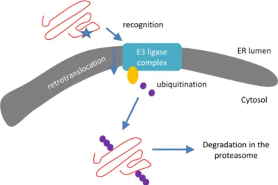
Next to UPR, the ER is also the site of a robust and continuously active protein degradation pathway that recognizes and destroys misfolded forms of both luminal and integral membrane proteins. This so-called ER-associated degradation (ERAD) directs unfolded proteins towards the cytosolic UPS (Section 3.3) and can be divided into three steps (Figure 3):
(1) Recognition of misfolded or mutated proteins in the ER. This process involves detection of substructures within proteins, such as exposed large hydrophobic regions, unpaired cysteine residues, and immature glycans.
(2) Retro-translocation of terminally misfolded proteins into the cytosol. The Hrd1E3 ubiquitin- protein ligase functions as a retrotranslocon (dislocon) to transport substrates to the cytosol. The direction of the transport is determined by the ubiquitin-binding factor Cdc48p in yeast and the valorin-containing protein (VCP/p97) in humans. The energy required for retro-translocation is provided by the ATPase activity of VCP/p97.
(3) Ubiquitin-dependent degradation by the proteasome. Misfolded polypeptides are ubiquitinated by a cascade of enzymatic reactions within the ER membrane, such as the ubiquitin ligases Hrd1 and Doa10 [92]. Next, the polyubiquitinated polypeptide is recognized by specific subunits of the 26S proteasome (and thus ERAD is attached to UPS; Section 3.3) and translocates to the Figure 2.Three signal pathways of ER-stress activate UPR and lead to either cell survival or apoptosis.
Under normal conditions, ER chaperon GRP78 binds all the three ER-stress sensors (PERK: protein kinase RNA like ER-kinase; IRE1α: inositol requiring enzyme 1α; ATF6: activating transcription factor 6).
Under ER-stress, GRP78 dissociates from the sensors. PERK and IRE1αbecome phosphorylated and form oligomers, and ATF6 translocates to the Golgi. ATF6, ATF4, and XBP1 activate UPR target genes to enhance the capacity of the ER to cope with unfolded proteins. Activation of Sig-1R inhibits the three branches of UPR. (Abbreviations: eIF2α: eukaryotic translation initiation factor 2α; XBP1: X-box binding protein 1 (spliced form); TRAF2: TNF-associated factor-2; ATF4: transcriptional activator factor-4;
MT: mitochondrion).
3.2. Endoplasmic Reticulum Associated Degradation
Next to UPR, the ER is also the site of a robust and continuously active protein degradation pathway that recognizes and destroys misfolded forms of both luminal and integral membrane proteins. This so-called ER-associated degradation (ERAD) directs unfolded proteins towards the cytosolic UPS (Section3.3) and can be divided into three steps (Figure3):
(1) Recognition of misfolded or mutated proteins in the ER. This process involves detection of substructures within proteins, such as exposed large hydrophobic regions, unpaired cysteine residues, and immature glycans.
(2) Retro-translocation of terminally misfolded proteins into the cytosol. The Hrd1E3 ubiquitin-protein ligase functions as a retrotranslocon (dislocon) to transport substrates to the cytosol. The direction of the transport is determined by the ubiquitin-binding factor Cdc48p in yeast and the valorin-containing protein (VCP/p97) in humans. The energy required for retro-translocation is provided by the ATPase activity of VCP/p97.
(3) Ubiquitin-dependent degradation by the proteasome. Misfolded polypeptides are ubiquitinated by a cascade of enzymatic reactions within the ER membrane, such as the ubiquitin ligases Hrd1 and Doa10 [92]. Next, the polyubiquitinated polypeptide is recognized by specific subunits of the 26S proteasome (and thus ERAD is attached to UPS; Section3.3) and translocates to the central chamber of the proteasome where the proteolytic active sites are located. ERAD has different branches for different misfolded domains [92].
Int. J. Mol. Sci. 2018, 19, x FOR PEER REVIEW 9 of 39
central chamber of the proteasome where the proteolytic active sites are located. ERAD has different branches for different misfolded domains [92].
Figure 3. Schematic representation of the ER-associated protein degradation (ERAD) pathway:
recognition, retro-translocation, and ubiquitination. Red line: protein/polypeptide chain; blue star:
misfolded domain; orange circle: Cdc48 ATPase; purple dots: ubiquitin molecules.
Indeed, ER-proteins with a misfolded domain in the cytoplasm (ERAD-C substrates) are degraded via the Doa10 complex, whereas proteins containing ER-luminal (ERAD-L) or intramembrane (ERAD-M) misfolded domains are degraded via the Hrd1 complex. Very recently, Zhu et al. [93] demonstrated that the ER-protein membralin is also an ERAD component which mediates degradation of ER-luminal and ER-membrane substrates (ERAD-L and -M). Interestingly, downregulation of membralin results in amyloid-pathology, synaptic deficits, and neuronal death.
Hence, membralin might play a critical role in AD pathogenesis.
3.3. Ubiquitination and UPS
Ubiquitination destines misfolded polypeptides for degradation through either UPS (Figure 3) or autophagy [94]. Although the ratios between the two protein degradation processes depend on physiological state and cell type, UPS is roughly responsible for approximately 80–90% of cellular proteolysis, whereas autophagy processes only manage around 10–20% [95–97]. Misfolded polypeptides are marked for degradation through the attachment of the 76 amino acid long ubiquitin protein. Next, polyubiquitination results in the attachment of additional ubiquitin units on any of the seven lysine residues of the original ubiquitin. This process is mediated by the cooperative action of three enzymes. First, the ubiquitin-activating enzyme E1 hydrolyses ATP and forms a thioester linkage with a cysteine residue of ubiquitin. This activated molecule is then passed on to the E2 ubiquitin- conjugating enzyme. Then, E3 ubiquitin-protein ligases bind to the misfolded proteins and the subsequent sequential ubiquitination of the first ubiquitin residue results in a polyubiquitinated misfolded polypeptide. Hence, ubiquitination generates linkage-specific degrons on substrates destined for destruction [94]. Subsequently, ubiquitin-based degrons are recognized by specific- adaptors with ubiquitin-binding domains (UBDs) [98]. The UBD adaptors RAD23 and UBQLVs deliver ubiquitinated substrates to the central chamber of the 26S proteasome for degradation, whereas the UBD adaptors p62 and NBR deliver ubiquitinated substrates to autophagic vacuoles, phagophores, and
Figure 3. Schematic representation of the ER-associated protein degradation (ERAD) pathway:
recognition, retro-translocation, and ubiquitination. Red line: protein/polypeptide chain; blue star:
misfolded domain; orange circle: Cdc48 ATPase; purple dots: ubiquitin molecules.
Indeed, ER-proteins with a misfolded domain in the cytoplasm (ERAD-C substrates) are degraded via the Doa10 complex, whereas proteins containing ER-luminal (ERAD-L) or intramembrane (ERAD-M) misfolded domains are degraded via the Hrd1 complex. Very recently, Zhu et al. [93]
demonstrated that the ER-protein membralin is also an ERAD component which mediates degradation of ER-luminal and ER-membrane substrates (ERAD-L and -M). Interestingly, downregulation of membralin results in amyloid-pathology, synaptic deficits, and neuronal death. Hence, membralin might play a critical role in AD pathogenesis.
3.3. Ubiquitination and UPS
Ubiquitination destines misfolded polypeptides for degradation through either UPS (Figure3) or autophagy [94]. Although the ratios between the two protein degradation processes depend on physiological state and cell type, UPS is roughly responsible for approximately 80–90% of cellular proteolysis, whereas autophagy processes only manage around 10–20% [95–97]. Misfolded polypeptides are marked for degradation through the attachment of the 76 amino acid long ubiquitin protein. Next, polyubiquitination results in the attachment of additional ubiquitin units on any of the seven lysine residues of the original ubiquitin. This process is mediated by the cooperative action of three enzymes. First, the ubiquitin-activating enzyme E1 hydrolyses ATP and forms a thioester linkage with a cysteine residue of ubiquitin. This activated molecule is then passed on to the E2 ubiquitin-conjugating enzyme. Then, E3 ubiquitin-protein ligases bind to the misfolded proteins and the subsequent sequential ubiquitination of the first ubiquitin residue results in a polyubiquitinated misfolded polypeptide. Hence, ubiquitination generates linkage-specific degrons on substrates destined for destruction [94]. Subsequently, ubiquitin-based degrons are recognized by specific-adaptors with ubiquitin-binding domains (UBDs) [98]. The UBD adaptors RAD23 and UBQLVs deliver ubiquitinated substrates to the central chamber of the 26S proteasome for degradation, whereas the UBD adaptors p62 and NBR deliver ubiquitinated substrates to autophagic vacuoles,
phagophores, and lysosomes. As a result, the interplay between polyubiquitination—the diverse ways to assemble ubiquitin chains—and the resulting complex “ubiquitin code” provide several means to modulate biological pathways by proteolytic and non-proteolytic processes [94]. Considering its central role in protein quality control, the UPS is a potential target for the management of AD [99].
3.4. Linkage between ERAD and UPR
UPR regulates a large battery of genes that are involved in nearly all aspects of ER protein production and delivery [80]. In fact, multiple genes required for ERAD and involved in the destruction of misfolded luminal and membrane proteins by the UPS pathway (for instance, hrd1/der3, hrd3, der1, and ubc7) are all UPR-regulated. Thus, UPR coordinates the pathways that mediate the two fates of misfolded ER-proteins: either folding and export, or retro-translocation (ERAD) and degradation.
Detailed examination of the link between UPR and ERAD places the degradation of proteins in the center of normal ER activity. ERAD is a continuous process and its loss results in an increase of unfolded proteins which are then recognized by the ER-scanning system UPR. However, knock out mouse mutants indicate that ERAD and UPR also function independently to some extent [80].
4. Endo-Lysosomal System and Autophagy
4.1. Endo-Lysosomal and Autophagy Dysfuntion in NDDs
The endo-lysosomal network contains a lot of communicating vesicular compartments with acidic pH ranging from 4.5 to 6.0. Material from outside the cell is taken up by endocytosis and passed on to the lysosomes, spherical vesicles containing more than 60 different hydrolytic enzymes able to break down proteins and many kinds of other biomolecules. As such, this network regulates cellular homeostasis and metabolism through degradation of cargo that is received by endocytosis from the plasma membrane. In addition, the autophagic pathways sequester cytosolic components, organelles, and toxic amyloids present in the cytoplasm, which are then digested by the lysosomes.
In AD, the endo-lysosomal network is involved in the generation of Aβthrough enzymatic cleavage of APP in the lysosomes byβ- and γ-secretases (the amyloidogenic pathway) [100,101].
In AD patients, the presence of amyloid plaques blocks the ability of lysosomes to travel within the axons and develop to mature lysosomes [102,103]. As such, these immature lysosomes are abnormally enriched inβ-secretase since these lysosomes are not able to degrade this APP-cleaving enzyme.
This represents a vicious circle since the increased number of immature lysosomes correlates with a higher local accumulation ofβ-secretase and a resulting higher amount of Aβ. Thus, accumulation of lysosomes even contributes to AD pathology. Next to lysosomes, Aβ is present in additional subcellular organelles of the degradative pathway such as endosomes, late endosomes/multivesicular bodies, autophagic vesicles, and lysosomes [104,105]. In addition to Aβ, other APP fragments (such as C83 and AICD) as well as aberrant post-translational APP modifications and dysregulation of APP processing contribute to the pathogenesis of AD [106–108]. Hence, perturbation of APP metabolism and subsequent Aβformation in the endo-lysosomal network is a central pathomechanism of AD [109].
Next to the lysosomal symptoms, AD is characterized by an endosomal pathology with increased endosomal size compared to healthy aged brains [104]. In fact, Aβaccumulation in the EL system increases lysosomal membrane permeability and causes the release of the lysosomal content (for instance cathepsins) into the cytoplasm ultimately resulting in apoptotic cell death [110].
In fact, one of the earliest events in Aβ-mediated neurotoxicity is just the release of lysosomal proteases. Interestingly, recent data indicate that the molecular chaperon Hsp70.1 acts as a lysosomal stabilizer [111,112]. The symptoms of the endocytic and lysosomal pathology are among the primary events of AD. In fact, AD patients exhibit endo-lysosomal network pathology in brain regions that do not yet show local Aβor tau-pathology [113]. Of note, these morphological changes were also observed in the brain of patients with Down syndrome [114].
Extracellular amyloid plaques of AD individuals accumulate endo-lysosomes and organelles of the autophagy network as these plaques are surrounded by dystrophic neurites that are enriched in lysosomal-like organelles [115]. A bidirectional relationship between the endo-lysosomal/autophagic network and AD might exist since the activity of certain lysosomal enzymes (e.g., cathepsin D) increases, whereas the activity of other enzymes decreases [116].
Interestingly, lysosomal storage disorders (LSDs) are also characterized by accumulated APP metabolites.
Consequently, AD and LSDs show common neuropathological features as lysosomal dysfunction in LSD leads to a kind of neurodegeneration and dementia similar to AD. Hence, endo-lysosomal dysfunction should be accepted as a potential risk factor for AD [116] and might be a converging pathomechanism in NDDs [117,118].
4.2. Autophagy
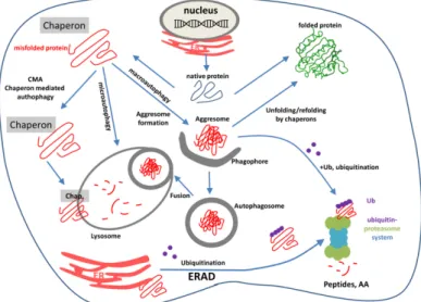
Autophagy or autophagocytosis refers to the mechanism which allows the cell to destroy and degrade unnecessary and/or dysfunctional components in the lysosome [119]. However, autophagy is a double edged sword. In disease, autophagy pathways represent an adaptive response to stress which promotes survival, whereas in other cases, autophagy promotes cell death. The molecular definitions of autophagy and its related processes were recently reviewed [120]. Three main forms of autophagy are commonly described: chaperon-mediated-autophagy (CMA), macro-, and microautophagy.
CMA is a very complex and specific multistep process. In CMA, soluble cytosolic proteins are selected in a chaperon-dependent manner, targeted to lysosomes, and directly translocated across the lysosomal membrane for degradation. Of note, CMA directly shuttles proteins across the lysosomal membrane without formation of additional vesicles. In addition, CMA differs from other types of autophagy as it is extremely selective towards the proteins which cross the lysosomal barrier.
In CMA, chaperon-bound autophagy substrates bind to the protein-translocation factor LAMP2A monomer on the cytosolic side of the lysosome forming an oligomeric LAMP2A translocation complex [121]. Through the LAMP2A complex, CMA substrates are translocated into the lysosomal lumen while they are unfolding and dissociating from the chaperons. During this process, LAMP2A complexes are stabilized by the lysosomal chaperon Hsp90AA1 and the cytosolic pool of glial acidic fibrillary protein (GFAP) [121]. CMA is responsible for the selective removal of damaged and unnecessary proteins. By facilitating the recycling of amino acids of the degraded (misfolded) proteins, CMA plays an important role in the regulation of the cellular metabolism. Of note, CMA is a continuous process which is active in different tissues and organs, such as brain, liver, and kidney.
Macroautophagy is the main, multistep pathway for degradation of damaged cell organelles and unused proteins with participation of the lysosome (Figure 4). The first step is the formation of a crescent-shaped double membrane (phagophore or isolation membrane) derived from multiple sources, such as cytoplasmic membrane, mitochondria, or ER- and Golgi apparatus [96,122]. During the following steps, the double-membrane phagophore grows in size and closes to form a complete autophagosome.
Next, the membrane of the autophagosome fuses with lysosomes to form autolysosomes. Finally, the cargoes (e.g., misfolded polypeptides) are degraded by lysosomal hydrolases.
In microautophagy, cytoplasmic entities destined for degradation are directly taken up by lysosomes via direct membrane invagination, without formation of protein complexes [123]. Although microautophagy is not well studied, two forms of this pathway were recently defined: microautophagy and endosomal microautophagy. The non-selective microautophagy begins with invagination of the cytosolic substrate. It is a tubular process by which the autophagic tube is formed. Endosomal microautophagy relies on multiple endosomal sorting complexes required for transport (ESCRT) systems [124]. In mammalian cells, this mechanism involves mainly late endosomes. Endosomal microautophagy degrades cytosolic proteins. Selectivity of this autophagy requires chaperon interaction, for instance only proteins containing a Lys-Phe-Glu-Arg-Gln (KFERQ-like) motif are recognized by Hsp78 [124].
Int. J. Mol. Sci. 2018, 19, x FOR PEER REVIEW 12 of 39
Figure 4. Macroautophagy is a multistep process: initiation, elongation, completion, fusion, and lysosomal degradation. Red coil: misfolded protein; red lines: oligopeptide and amine acids; blue dots: lysosomal enzymes.
In microautophagy, cytoplasmic entities destined for degradation are directly taken up by lysosomes via direct membrane invagination, without formation of protein complexes [123]. Although microautophagy is not well studied, two forms of this pathway were recently defined: microautophagy and endosomal microautophagy. The non-selective microautophagy begins with invagination of the cytosolic substrate. It is a tubular process by which the autophagic tube is formed. Endosomal microautophagy relies on multiple endosomal sorting complexes required for transport (ESCRT) systems [124]. In mammalian cells, this mechanism involves mainly late endosomes. Endosomal microautophagy degrades cytosolic proteins. Selectivity of this autophagy requires chaperon interaction, for instance only proteins containing a Lys-Phe-Glu-Arg-Gln (KFERQ-like) motif are recognized by Hsp78 [124].
The three types of autophagy are regulated by similar signaling pathways induced by starvation, nitrogen deprivation, rapamycin treatment, etc. Autophagy is executed by autophagy-related (Atg) genes. Recent studies focusing on the relationship between aging and autophagy gave interesting results: senescence of cells was associated with decreased autophagy and decreased expression of autophagy genes [125,126]. It occurred as the result of the increased methylation of autophagy genes by the DNA methyl transferase DNMT23. The autophagy gene ATG5 was one of the highly methylated autophagy genes. In mammals, Atg1 complex is based on the kinases ULK1 or ULK2, and plays an essential role in regulating autophagy [127]. ULK is part of a larger protein complex containing autophagy-related proteins mAtg13, Atg101, and FIP200. The ULK complex is regulated by a series of phosphorylations on ULK1/2, mAtg13, and FIP200. The ULK1/2 function (and autophagy) is regulated by mTOR complex1 and 5′-AMP-activated protein kinase (AMPK) through a complicated network of phosphorylation events on a large number of phospho-sites of ULKs Interestingly, nutrient starvation prolongs lifespan in C. elegans through the induction of caloric restriction in parallel with a high level of autophagy, an effect which can be simulated by resveratrol [128]. Figure 5 schematically represents the cellular fate of misfolded polypeptides indicating ERAD, UPS, and the three different autophagy pathways.
Figure 4. Macroautophagy is a multistep process: initiation, elongation, completion, fusion, and lysosomal degradation. Red coil: misfolded protein; red lines: oligopeptide and amine acids;
blue dots: lysosomal enzymes.
The three types of autophagy are regulated by similar signaling pathways induced by starvation, nitrogen deprivation, rapamycin treatment, etc. Autophagy is executed by autophagy-related (Atg) genes. Recent studies focusing on the relationship between aging and autophagy gave interesting results:
senescence of cells was associated with decreased autophagy and decreased expression of autophagy genes [125,126]. It occurred as the result of the increased methylation of autophagy genes by the DNA methyl transferase DNMT23. The autophagy gene ATG5 was one of the highly methylated autophagy genes. In mammals, Atg1 complex is based on the kinases ULK1 or ULK2, and plays an essential role in regulating autophagy [127]. ULK is part of a larger protein complex containing autophagy-related proteins mAtg13, Atg101, and FIP200. The ULK complex is regulated by a series of phosphorylations on ULK1/2, mAtg13, and FIP200. The ULK1/2 function (and autophagy) is regulated by mTOR complex1 and 50-AMP-activated protein kinase (AMPK) through a complicated network of phosphorylation events on a large number of phospho-sites of ULKs Interestingly, nutrient starvation prolongs lifespan inC. elegans through the induction of caloric restriction in parallel with a high level of autophagy, an effect which can be simulated by resveratrol [128]. Figure5schematically represents the cellular fate of misfolded polypeptides indicating ERAD, UPS, and the three different autophagy pathways.Int. J. Mol. Sci. 2018, 19, x FOR PEER REVIEW 13 of 39
Figure 5. The most important pathways of intracellular protein degradation: endoplasmic reticulum associated-degradation (ERAD), the ubiquitin-proteasome system (UPS), and autophagy pathways.
Purple dots: ubiquitin molecules; red coil: misfolded aggregated proteins; red lines: oligopeptides or amino acids.
5. Prevention and Treatment of NDDs
5.1. Heat Shock Proteins in Neurodegenerative Disorders
Many neurodegenerative diseases are characterized by the accumulation of misfolded proteins in different forms: intra- and extracellular aggregates, plaques, inclusion bodies, and proteinaceous fibrillary structures (Table 1).
The formation of extracellular aggregates is mainly a tool for sequestration of toxic, pathogenic proteins since, in case of Aβ, the soluble oligomers and protofibrils are the most cytotoxic forms [129,130]. In fact, according to the intracellular Aβ hypothesis, intracellular Aβ oligomers initiate the disease by interacting with cytoplasmic proteins and membranes of cell organelles (mitochondria, ER), thereby triggering apoptosis [131]. In addition, polyQ proteins interact with transcription factors, proteasomal subunits, and cytoskeletal proteins eventually leading to repression of transcription, impairment of the protein degradation system, and alteration of the neurofilament network [132]. Although Aβ plaques do not appear to be as neurotoxic as the soluble oligomers, they do provoke local inflammatory responses. Ultimately, this leads to uncontrolled activation of microglia and release of inflammatory cytokines and thus chronic inflammation in the brain [133].
Due to a different and neuron-specific protein quality control system compared to glial cells, ubiquitously expressed amyloids are selectively deposited in neurons [134]. Hence, compared to neurons, glial cells are much less affected in neurodegenerative diseases. In fact, neurons have a lower basal level and a weaker heat-inducibility of HspA1 compared to astrocytes, whereas astrocytes have higher CHIP activity and degrade toxic, aggregation-prone proteins faster than neurons [53].
In parallel with the increasing level of misfolded polypeptides, Hsp expression levels are usually upregulated in the affected brain regions. For instance, the levels of HspB1 and HspB5 are highly elevated in the cerebral cortex of AD patients [135,136], in the reactive astrocytes in PD [135], and in
Figure 5.The most important pathways of intracellular protein degradation: endoplasmic reticulum associated-degradation (ERAD), the ubiquitin-proteasome system (UPS), and autophagy pathways.
Purple dots: ubiquitin molecules; red coil: misfolded aggregated proteins; red lines: oligopeptides or amino acids.
5. Prevention and Treatment of NDDs
5.1. Heat Shock Proteins in Neurodegenerative Disorders
Many neurodegenerative diseases are characterized by the accumulation of misfolded proteins in different forms: intra- and extracellular aggregates, plaques, inclusion bodies, and proteinaceous fibrillary structures (Table1).
The formation of extracellular aggregates is mainly a tool for sequestration of toxic, pathogenic proteins since, in case of Aβ, the soluble oligomers and protofibrils are the most cytotoxic forms [129,130].
In fact, according to the intracellular Aβhypothesis, intracellular Aβoligomers initiate the disease by interacting with cytoplasmic proteins and membranes of cell organelles (mitochondria, ER), thereby triggering apoptosis [131]. In addition, polyQ proteins interact with transcription factors, proteasomal subunits, and cytoskeletal proteins eventually leading to repression of transcription, impairment of the protein degradation system, and alteration of the neurofilament network [132]. Although Aβplaques do not appear to be as neurotoxic as the soluble oligomers, they do provoke local inflammatory responses.
Ultimately, this leads to uncontrolled activation of microglia and release of inflammatory cytokines and thus chronic inflammation in the brain [133].
Due to a different and neuron-specific protein quality control system compared to glial cells, ubiquitously expressed amyloids are selectively deposited in neurons [134]. Hence, compared to neurons, glial cells are much less affected in neurodegenerative diseases. In fact, neurons have a lower basal level and a weaker heat-inducibility of HspA1 compared to astrocytes, whereas astrocytes have higher CHIP activity and degrade toxic, aggregation-prone proteins faster than neurons [53].
In parallel with the increasing level of misfolded polypeptides, Hsp expression levels are usually upregulated in the affected brain regions. For instance, the levels of HspB1 and HspB5 are highly elevated in the cerebral cortex of AD patients [135,136], in the reactive astrocytes in PD [135], and in the spinal cord in a mouse model of ALS [28]. Thus, the levels of several Hsps are constitutively increased in neurodegenerative diseases as a compensatory mechanism.
In addition, Hsps co-localize with the abnormal protein aggregates (reviewed by [26,137]).In fact, several members of the small Hsp family, such as HspB1, HspB2, HspB5, and HspB6 associate with senile plaques in the AD brain [138]. In addition, HspB5 is mainly localized in astrocytes, microglia, and oligodendrocytes, while HspB1 was found in degenerating neurons [136,139]. HspB5 localizes to the Lewy bodies [140] and is present in the spinal cord of ALS patients [141]. In addition, HspA1 co-localizes with Aβpeptides andα-synuclein, whereas HspA8 was found in intracellular inclusions in ALS [142].
In certain cases, aggregation-prone polypeptides evade the protein quality control systems. In fact, due to the increasing rate of aggregate formation, Hsps get trapped within the aggregates what reduces their availability. For instance, an increased level of aggregate formation correlates with a decreased ability of small Hsps to preventα-synuclein fibril formation in vitro [143].
In parallel, the inducibility and chaperon activity of Hsps are impaired during aging (reviewed in [55]) which is accompanied by a reduced activity of the protein degradation systems as well [144]. Hence, the combined effect of the weakening of the capacity of the chaperons and the protein degradation complex as well as an increased formation of misfolded proteins in aged organisms finally leads to the overload of the system what further accelerates the development of NDDs [55].
Therefore, restoration of the heat shock response and the increase of chaperon activity of Hsps represent useful therapeutic strategies. In fact, each neurodegenerative disorder is characterized by a specific subset of Hsps able to ameliorate the specific symptoms [145]. Consequently, the protective effect of elevated levels of Hsps in different animal models of NDDs has been shown. For instance, HspB1 overexpression delays the decline of motor strength and improves the survival of the spinal motor neurons in a transgenic mouse model of ALS [146]. In addition, we showed that HspB1 overexpression rescues the impaired learning abilities, decreased the number of amyloid plaques, and normalized synaptic abnormalities, such as increased excitability and impaired long-term potentiation, in a mouse model of AD [147]. Overexpression of HspA1 preventsα-synuclein-induced
dopaminergic neuronal loss [148] and polyQ-induced neurodegeneration [149] inDrosophilaand in SCA1 transgenic mice [150]. In addition, transgenic overexpression of HspA1A reduces Aβplaque formation, neuronal loss, and cognitive deficits in a mouse model of AD [151]. These results indicate that overexpression of Hsps ameliorates certain symptoms of neurodegenerative diseases.
The protective function of Hsps in NDDs lies in their capacity to suppress pathological protein aggregation via their chaperon-like activity. This potent anti-aggregation property of Hsps is well documented by in vitro studies, both in experiments performed with solutions of aggregation-prone proteins and in cell culture experiments [152].
In fact, several studies demonstrate that even individual Hsps can prevent protein aggregation.
For instance, HspB1, HspB5, and HspB8 bindα-synuclein and inhibit mature fibril formation [153].
Mechanistically, HspB1 and HspB5 prevent aggregation through transient interactions with α-synuclein monomers as such stabilizing their structure and inhibiting the formation of oligomeric nuclei [143]. Similarly, by binding to fibril seeds rather than forming a stable complex with the monomeric amyloid peptides, HspB5 prevents the induction ofβ-sheet structure and the amyloid fibril growth of Aβ1-40 [154]. In addition, HspB1 inhibits Aβ1-42 amyloidogenesis in vitro [152].
Most of the in vitro studies show that addition of individual Hsps to the reaction after the completion of aggregation has no effect on the size of aggregates, which suggests that Hsps bind to monomeric and/or prefibrillar forms of aggregation prone proteins as such keeping them in a soluble form which prevents fibril growth.
Several cell culture models provide evidence that Hsps prevent abnormal protein aggregation.
For instance, overexpression of DNAJ protein family members in cerebrovascular cells suppresses the nuclear aggregation of mutant ataxin-1 and ataxin-3 [129,155], whereas HspB1, HspB5, and HspB6 are able to bind to Aβinhibiting its fibril formation [156]. In addition, HspB1 and HspB5 expression can significantly reduceα-synuclein aggregation [157]. Of note, the capacity of small Hsps to reduce α-synuclein inclusion formation is retained even in the presence of inhibitors of autophagy or proteasomal degradation [143]. Hence, by preventing initiation of aggregation, Hsps are potent inhibitors of protein aggregation in living cells. Interestingly, in most of these studies, the reduced aggregation was accompanied by a decreased toxicity of the aggregation-prone polypeptides.
Small Hsps family members have several additional non-chaperon functions that could contribute to their neuroprotective function. In fact, small Hsps are potent inhibitors of the apoptotic cascade.
In addition, the increased level of Hsps can prevent apoptotic cell death induced by aggregation-prone proteins, such asα-synuclein or Aβ[158,159]. Hence, since neurological disorders such as AD, PD, HD, and ALS are characterized by enhanced apoptotic cell death, the neuroprotective function of small Hsps might lie in their capacity to prevent apoptosis [160–162].
Cellular oxidative stress increases during aging and in different NDDs. For instance, HspB1 has been shown to decrease polyQ toxicity without suppressing protein aggregation by protecting cells against oxidative stress indicating that Hsps can regulate cellular redox homeostasis and have protective effects against oxidative stress independently from their anti-aggregation effect [163]. Similarly, Hsp70.1 has dual roles: it acts as a molecular chaperon for damaged proteins while simultaneously it is a guardian of lysosomal integrity. It is known, that in the acidic environment of the lysosome lumen, Hsp70.1 interacts with bis(monoacylglycero)phosphate (BMP), an anionic phospholipid bound to the inner lysosomal membrane. BMP is a cofactor for the enzyme acid sphingomyelinase (ASM).
The Hsp70.1-BMP interaction enhances the association of BMP with ASM, activating the enzyme so that it breaks down sphingomyelin to form ceramide. Thus, the increased production of ceramide in lysosomes protects lysosomal membranes from rupturing [40]. According to the “calpain-cathepsin hypothesis” of AD [111], both age-dependent oxidative stress and decreasing Hsp70.1 levels might be responsible for elevated lysosomal membrane permeability and cell death. Specific membrane lipids containing linoleic and arachidonic acids are vulnerable to cumulative oxidative stresses, especially because their toxic peroxidation product 4-hydroxy-2-nonenal (HNE) preferentially carbonylates Hsp70.1, and, in the next step, calpain cleaves and inactivates this carbonylated Hsp70.1 pool.
This event ultimately causes elevated lysosomal permeabilization and rupture, with the release of cathepsins and finally cell death [112].
While essential for the normal neuronal function, the dendritic network is usually altered in NDDs.
For instance, in AD patients, dendritic degeneration is remarkable characterized by a significant loss of total dendritic length and the density of dendritic spines [164,165]. However, during neuronal stress conditions, the upregulated HspB5 plays an important role in the preservation of dendritic complexity and neuronal connectivity by stimulating dendritic branching [166].
5.2. Targeting Hsps for Treatment of NDDs
Misbalanced Hsp expression levels are central in the pathogenesis of multiple prevalent disorders.
Hence, compounds able to restore Hsp expression levels have high therapeutic potential. It has been demonstrated that overexpression of HspA1A and HspB1 proteins in a transgenic mouse models of AD (APPxPS1 mouse) can ameliorate the major symptoms of AD by improving synaptic and cognitive functions, decreasing the number of amyloid plaques, and increasing neuronal survival [147,151].
These results indicate that chaperon molecules are potential targets in AD therapy and subsequently have drawn attention to search for chaperon inducer and co-inducer molecules, both natural plant extracts as well as synthetic small molecules. However, only diseased tissues or cells should be targeted as a general increase in Hsp expression levels correlates with carcinogenesis. As a therapeutic class, Hsp co-inducers cannot amplify Hsp expression without a concomitant stress. However, they further elevate Hsp levels in the presence of cellular stress underlying the phenotype of several pathological conditions. Here, we describe some selected compounds that exert neuroprotective properties via chaperon induction/co-induction. A comprehensive review on the protective role of plant biophenols in mechanisms of AD was recently published [167].
5.2.1. Therapeutic Potential of Small Molecule Hsp Co-Inducers
Hydroximic acid derivatives including bimoclomol, arimoclomol, NG-094, and BGP-15 are nontoxic small molecules based on the structure of the beta-blocker propranolol [59].
Vigh et al. were the first to demonstrate the Hsp co-inducing capacity of hydroximic derivatives by showing that the first generation derivative bimoclomol, a hydroxylamine derivative ((2-hydroxy-3-(1-piperidinyl) propoxy)-3-pyridinecarboximidoil-chloride maleate), synergistically enhances heat-induced Hsp60, Hsp70, and Hsp90 levels [168]. The compound facilitates the formation of chaperon molecules in eukaryotic cells by inducing or amplifying expression of heat-shock genes.
The cytoprotective effects observed under several experimental conditions, including a murine model of ischemia and wound healing in the diabetic rat, are likely mediated by the coordinate expression of all major Hsps. Consequently, potential therapeutic value for bimoclomol was established in diabetes [169,170] and cardiovascular disease [171–173], pathologies characterized by an imbalance in Hsp expression.
Next, arimoclomol((3Z)-N-((2R)-2-hydroxy-3-piperidin-1-ylpropoxy)-1-oxidopyridin-1-ium-3- carboximidoyl chloride) reduces lysosomal accumulation in primary fibroblasts from patients with lysosomal storage diseases, a pathology associated with severe systemic and central nervous system symptoms [174]. In addition, in a rhodopsin transgenic rat models, a model for retinitis pigmentosa—a group of inherited diseases that cause blindness due to the progressive death of rod and cone photoreceptors in the retina, pharmacological potentiation of the stress response with arimoclomol improved electroretinogram responses and prolonged photoreceptor survival [175]. In a mouse model of spinal and bulbar muscular atrophy, an adult-onset hereditary neurodegenerative disorder caused by an expansion of polyQ repeats in the first exon in the androgen receptor gene, arimoclomol significantly improved hindlimb muscle force and contractile characteristics, rescued motor units and improved motor neuron survival while upregulating the expression of the vascular endothelial growth factor which possess neurotrophic activity [176]. In a mouse model of ALS, an incurable neurodegenerative disorder characterized by progressive degeneration of motor neurons