Journal of Experimental Botany, Vol. 70, No. 18 pp. 4903–4917, 2019 doi:10.1093/jxb/erz217 Advance Access Publication May 14, 2019
This paper is available online free of all access charges (see https://academic.oup.com/jxb/pages/openaccess for further details)
© The Author(s) 2019. Published by Oxford University Press on behalf of the Society for Experimental Biology.
This is an Open Access article distributed under the terms of the Creative Commons Attribution Non-Commercial License (http://creativecommons.org/licenses/
by-nc/4.0/), which permits non-commercial re-use, distribution, and reproduction in any medium, provided the original work is properly cited. For commercial re-use, please contact journals.permissions@oup.com
RESEARCH PAPER
The mitogen-activated protein kinase 4-phosphorylated heat shock factor A4A regulates responses to combined salt and heat stresses
Norbert Andrási1, , Gábor Rigó1,2, Laura Zsigmond1, Imma Pérez-Salamó3, Csaba Papdi3, Eva Klement1, Aladár Pettkó-Szandtner1, Abu Imran Baba1, Ferhan Ayaydin1, Ramakrishna Dasari1,4, Ágnes Cséplő1 and László Szabados1,*,
1 Biological Research Centre, Temesvári krt 62, H-6726 Szeged, Hungary
2 Department of Plant Biology, University of Szeged, 6726 Szeged, Hungary
3 School of Biological Sciences, Royal Holloway, University of London, Egham Hill, Surrey, TW20 0EX, UK
4 Department of Biotechnology, Kakatiya University, Warangal-506009, India
*Correspondence: szabados.laszlo@brc.mta.hu
Received 22 December 2018; Editorial decision 30 April 2019; Accepted 4 May 2019 Editor: Christine Foyer, University of Birmingham, UK
Abstract
Heat shock factors regulate responses to high temperature, salinity, water deprivation, or heavy metals. Their function in combinations of stresses is, however, not known. Arabidopsis HEAT SHOCK FACTOR A4A (HSFA4A) was previously reported to regulate responses to salt and oxidative stresses. Here we show, that the HSFA4A gene is induced by salt, elevated temperature, and a combination of these conditions. Fast translocation of HSFA4A tagged with yellow fluorescent protein from cytosol to nuclei takes place in salt-treated cells. HSFA4A can be phosphorylated not only by mitogen-activated protein (MAP) kinases MPK3 and MPK6 but also by MPK4, and Ser309 is the dominant MAP kinase phosphorylation site. In vivo data suggest that HSFA4A can be the substrate of other kinases as well. Changing Ser309 to Asp or Ala alters intramolecular multimerization. Chromatin immunoprecipitation assays confirmed binding of HSFA4A to promoters of target genes encoding the small heat shock protein HSP17.6A and transcription factors WRKY30 and ZAT12. HSFA4A overexpression enhanced tolerance to individually and simultaneously applied heat and salt stresses through reduction of oxidative damage. Our results suggest that this heat shock factor is a component of a complex stress regulatory pathway, connecting upstream signals mediated by MAP kinases MPK3/6 and MPK4 with transcription regulation of a set of stress-induced target genes.
Keywords: Arabidopsis, combined stress, heat, heat shock factor A4A, MAP kinases, phosphorylation, promoter binding, salinity, transcription regulation.
Introduction
In nature, a number of simultaneously acting environmental effects determine plant growth and development. Extreme conditions require the capability of particular adaptations to face challenges of several, often simultaneous abiotic stresses.
Combinations of adverse conditions have a more serious
impact on plants than separately imposed stresses and physio- logical consequences are more dramatic than effects of indi- vidual stresses (Rizhsky et al., 2004b; Suzuki et al., 2014). System biology analysis has revealed that combinations of stresses lead to novel transcript and metabolome profiles, which are not HeadA=HeadB=HeadA=HeadB/HeadA
HeadB=HeadC=HeadB=HeadC/HeadB
HeadA=Materials_HeadB=HeadA=Materials_HeadB/HeadA
Materials_HeadB=Materials_HeadC=Materials_HeadB=Materials_HeadC/HeadB Extract2=HeadB=Extract=HeadB
Extract2=HeadA=Extract=HeadA Extract3=HeadA=Extract1=HeadA Extract3=HeadB=Extract1=HeadB BList1=SubBList1=BList1=SubBList BList1=SubBList3=BList1=SubBList2
SubBList1=SubSubBList3=SubBList1=SubSubBList2 SubSubBList3=SubBList=SubSubBList=SubBList SubSubBList2=SubBList=SubSubBList=SubBList SubBList2=BList=SubBList=BList
Keywords=Keywords=Keywords_First=Keywords HeadA=HeadB=HeadA=HeadB/HeadA
HeadB=HeadC=HeadB=HeadC/HeadB HeadC=HeadD=HeadC=HeadD/HeadC Extract3=HeadA=Extract1=HeadA
REV_HeadA=REV_HeadB=REV_HeadA=REV_HeadB/HeadA REV_HeadB=REV_HeadC=REV_HeadB=REV_HeadC/HeadB REV_HeadC=REV_HeadD=REV_HeadC=REV_HeadD/HeadC REV_Extract3=REV_HeadA=REV_Extract1=REV_HeadA EDI_HeadA=EDI_HeadB=EDI_HeadA=EDI_HeadB/HeadA EDI_HeadB=EDI_HeadC=EDI_HeadB=EDI_HeadC/HeadB EDI_HeadC=EDI_HeadD=EDI_HeadC=EDI_HeadD/HeadC EDI_Extract3=EDI_HeadA=EDI_Extract1=EDI_HeadA
CORI_HeadA=CORI_HeadB=CORI_HeadA=CORI_HeadB/HeadA CORI_HeadB=CORI_HeadC=CORI_HeadB=CORI_HeadC/HeadB CORI_HeadC=CORI_HeadD=CORI_HeadC=CORI_HeadD/HeadC CORI_Extract3=CORI_HeadA=CORI_Extract1=CORI_HeadA ERR_HeadA=ERR_HeadB=ERR_HeadA=ERR_HeadB/HeadA ERR_HeadB=ERR_HeadC=ERR_HeadB=ERR_HeadC/HeadB ERR_HeadC=ERR_HeadD=ERR_HeadC=ERR_HeadD/HeadC ERR_Extract3=ERR_HeadA=ERR_Extract1=ERR_HeadA
EDI_Affiliation_Last=EDI_Affiliation_rLast=EDI_Affiliation=EDI_Affiliation_Last Affiliation_Last=OPI_Affiliation_Last=Affiliation=OPI_Affiliation
Affiliation=OPI_Affiliation=Affiliation_Last=OPI_Affiliation
CORI_Text_First=CORI_Text=CORI_Text_First=CORI_TextInd Box_Head=Box_AHead=Box_Head=Box_AHead/Head
CopyrightLine_rule=CopyrightLine_rule=CopyrightLine_rule=CopyrightLine Contributor=Towhom=Contributor=Towhom1
Towhom1=Towhom=Towhom1=Towhom1
Downloaded from https://academic.oup.com/jxb/article-abstract/70/18/4903/5489060 by Biological Research Centre of the Hungarian Academy of Sciences user on 19 November 2019
just the sum of responses present in plants under individual stresses (Rasmussen et al., 2013; Sewelam et al., 2014; Georgii et al., 2017). A combination of salinity and heat resulted in specific metabolite profiles that included modulation of ion balance, water status, and photosynthetic activity, along with production of protective compounds, which are not typical for salinity or heat alone (Rivero et al., 2014). Experiments with combined salt, mannitol, and heat stresses revealed that heat- and salt-induced genes have usually higher, while osmotically induced genes have lower, expression levels (Sewelam et al., 2014). More than half of the transcriptome changes in com- bined treatments could not be predicted from single stresses (Rasmussen et al., 2013).
Production of reactive oxygen species (ROS) is a conse- quence of aerobic metabolism that is enhanced by most adverse conditions, generating oxidative damage as a secondary stress.
Maintaining ROS homeostasis in drought and salt stress is es- sential for plant survival and acclimation, especially in combin- ation with heat or high light (Miller et al., 2010). Besides being damaging, ROS and in particular hydrogen peroxide (H2O2) are important components of stress signaling that are essential to coordinate responses to a number of single or combined biotic and abiotic stress conditions (Volkov et al., 2006; Baxter et al., 2014; Choudhury et al., 2017).
Heat shock factors (HSFs) are essential regulators of responses to heat and a number of other adverse conditions. In the ab- sence of stress, HSFs form complexes with heat shock proteins (mainly HSP90) in cytosol, which are dissolved during stress when HSFs relocalize to nuclei. HSFs activate target genes in trimeric form, recognizing heat shock elements (HSEs) com- prising palindromic transcription factor (TF) binding domains (5′-AGAAnnTTCT-3′) (Akerfelt et al., 2010; Anckar and Sistonen, 2011). While in yeast and animals one or a few genes encode HSFs, plants have large HSF gene families, which are composed of 21 genes in Arabidopsis (Nover et al., 2001), 25 genes in rice (Chauhan et al., 2011), and 64 genes in rapeseed (Zhu et al., 2017), representing broad functional diversification.
Plant HSFs regulate responses not only to high temperatures but also to diverse environmental stresses (Scharf et al., 2012; Albihlal et al., 2018). Genome-wide transcript profiling experiments have revealed that plant HSFs not only regulate the expression of heat shock protein (HSP) and chaperon genes but control genes that are implicated in transcriptional regulation, protein biosynthesis, metabolism, development, transport, and signal transduction (Busch et al., 2005). A recent study identified almost a thousand target genes of HSFA1b factor in Arabidopsis and established a hierarchical network of 27 HSFA1b-controlled TFs, which regulate the activity of 1780 genes (Albihlal et al., 2018). Earlier we showed that ROS-controlled HSFA4A (AT4G18880) of Arabidopsis promotes the expression of a wide set of defense genes including ZnF, MYB and WRKY-type transcription fac- tors (TFs), and regulates tolerance to salinity (Pérez-Salamó et al., 2014). HSFA4A-type factors were shown to confer cadmium tolerance to rice (Shim et al., 2009) and desiccation tolerance to sunflower (Carranco et al., 2017) and rapeseed (Lang et al., 2017). Rapeseed HSFA4A is upregulated by both drought and heat, confirming that it is implicated in multiple stress responses (Zhu et al., 2017).
HSFs can undergo multiple post-translational modifications, such as phosphorylation, acetylation, or sumoylation, which can modulate their activity and stability (Akerfelt et al., 2010).
Phosphorylation of human HSF1 by the mitogen-activated protein (MAP) kinase extracellular signal-regulated kinase 1 (ERK1) and glycogen synthase kinase 3 (GSK3) was shown to inhibit HSF1’s function and repress the heat shock response in non-stress conditions (Chu et al., 1996). Such inhibitory phosphorylation can promote interaction with HSP90 and is required for sumoylation, which represses HSF1-dependent activation of target genes (Hietakangas et al., 2003; Wang et al., 2006). In contrast to ERK1 or GSK3, hyperphosphorylation of the human HSF1 on particular Ser residues by calcium/
calmodulin (CaM)-dependent protein kinase II (CaMKII) or polo-like kinase 1 (PLK1) has a stimulatory effect as it enhances nuclear translocation and promotes transcription (Holmberg et al., 2001; Kim et al., 2005). Yeast HSF1 is hyperphosphorylated upon heat shock, which is required for recognition of HSEs of target promoters and is a prerequisite for the activation (Hashikawa and Sakurai, 2004). In plants, HSF phosphoryl- ation is more complex due to the large gene families of heat shock factors and protein kinases. In Arabidopsis the cyclin- dependent CDC2s kinase was shown to phosphorylate HSF1, which prevented binding to target DNA (Reindl et al., 1997).
An example of positive regulation is the calcium-dependent heat activated MAP kinase of tomato that phosphorylates and activates HSFA3 (Link et al., 2002). CaM-binding protein kinase 3 (AtCBK3) can interact with and phosphorylate AtHSFA1 of Arabidopsis, promoting binding to HSEs and transcription of HSP genes (Liu et al., 2008). Heat stress can activate MAP kinase 6 (MPK6) in Arabidopsis, which forms a complex with and phosphorylates HSFA2 (Evrard et al., 2013). MPK6 and the closely related MPK3 are key components of cellular de- fenses regulating resistance against various pathogens, oxida- tive stress responses and ethylene or ABA signaling (Rasmussen et al., 2012; Su et al., 2017; Sun et al., 2018; Bigeard and Hirt, 2018). MPK3 and MPK6 can phosphorylate and control the activity of various TFs including ZAT10, which controls oxi- dative stress responses (Nguyen et al., 2012b), MYB44 involved in ABA signaling (Nguyen et al., 2012a), and pathogen-related WRKY33 or ERF6 (Mao et al., 2011; Meng et al., 2013) and ICE1 implicated in cold tolerance (Li et al., 2017). Earlier we reported that the Arabidopsis HSFA4A is a substrate of MAP kinases MPK3 and MPK6 and determined phosphorylation sites by mass spectrometry (Pérez-Salamó et al., 2014). The bio- logical or molecular function of such MAP kinase-mediated phosphorylation was, however, not studied. Here we describe that HSFA4A is phosphorylated by MPK4, another key stress- related MAP kinase in Arabidopsis, and show that HSFA4A is implicated in responses not only to salinity but also to com- bined salt and heat stresses.
Materials and methods
Plant material and growth conditions
Arabidopsis Col-0 ecotype was used in all experiment. HSFA4A cDNA was overexpressed under the control of estradiol-inducible promoter of pER8GW vector (Papdi et al., 2008). In pHSFA4A::HSFA4A-YFP lines,
Downloaded from https://academic.oup.com/jxb/article-abstract/70/18/4903/5489060 by Biological Research Centre of the Hungarian Academy of Sciences user on 19 November 2019
expression of yellow fluorescent protein (YFP)-tagged HSFA4A cDNA was controlled by the 2 kb HSFA4A promoter.
Arabidopsis plants were grown in sterile conditions on half-strength Murashige and Skoog (½MS) medium in growth chambers with an 8 h light–16 h dark light cycle at 22 °C and 100 µE m−2 s−1 light inten- sity (control condition). To induce the transcription, 5 µM estradiol was added to the culture medium 24 h before and during stress treatments (Pérez-Salamó et al., 2014).
Stress tolerance was evaluated in vitro. In one test system, seeds were germinated and plantlets were grown on agar-solidified ½MS medium supplemented by 50–150 mM NaCl and 5 µM estradiol. Heat stress was imposed by incubating 10-day-old plants in high temperatures: 37 °C in light and 30 °C in dark for 2–8 d. In the second test system seeds were germinated and plants were grown on nylon mesh (SEFAR 07-20/13) on agar-solidified standard ½MS medium for 10 d and then transferred to the surface of liquid culture medium (10 ml medium in a 13 cm diam- eter Petri dish). Salt stress was imposed by supplementing liquid medium with 100 or 150 mM NaCl. Heat stress was 37 °C in light and 30 °C in dark for 2–4 d. Stress combinations were implemented by simultaneous application of salt and heat treatments. Plants were subsequently removed and transferred to standard ½MS culture medium for recovery. Plant sur- vival and growth was evaluated 10 d later. Transgenic Arabidopsis lines were generated by the floral dip method as described (Clough and Bent, 1998). Agrobacterium-mediated transformation of cell suspension culture was made as described (Pérez-Salamó et al., 2014).
Stress treatments for transcript and western analysis, phosphorylation studies, and chromatin immunoprecipitation (ChIP) assays were per- formed on 10-day-old in vitro-grown seedlings using the sterile hydro- ponic system described above. Unless otherwise stated, salt stress was imposed with 150 mM NaCl and heat stress was generated with 37 °C in light and 30 °C in dark (8/16 h of light/dark cycle). Stress treatments were initiated 2 h after the start of light period (time 0). Transcript pro- filing and western analysis were performed with samples collected after 2, 6 and 24 h of stress, while in vivo phosphorylation and chromatin binding (ChIP assay) were tested on plants treated for 6 h.
Gene cloning
To generate pHSFA4A promoter and HSFA4A–YFP gene fusion, 2kb-long promoter fragment of the HSFA4A gene (AT4G18880) was cloned and fused to the YFP-tagged HSFA4A of the pPCV- HSFA4A-YFP construct (Pérez-Salamó et al., 2014). After sequencing, the pHSFA4A::HSFA4A-YFP fragment was moved in the pMDC99 binary Gateway vector by a Gateway LR clonase reaction (Curtis and Grossniklaus, 2003).
To generate a phosphorylation-mimicking version of HSFA4A, the Ser309 residue was replaced by Asp309 as described (Pérez-Salamó et al., 2014). The sequenced S309D-HSFA4A cDNA was cloned into the pENTR2b Gateway vector using BamHI and XhoI restriction sites, and moved into the binary destination vector pER8GW (Papdi et al., 2008) with Gateway LR clonase reaction, producing the estradiol-inducible pER8-HSFA4A-S309D.
To generate mutant gene constructs for a bimolecular fluorescence complementation (BiFC) assay, the HSFA4A-S309A (Pérez-Salamó et al., 2014) and the newly generated HSFA4A-S309D fragments were PCR amplified from pENTR2b-HSFA4A-S309A and pENTR2b-HSFA4A- S309D plasmids using the HSFA4A-HindIII and HSFA4A-SmaI-NoStop primers. The PCR fragments were subsequently cloned into the HindIII and SmaI sites of pSAT1A-nEYFP-N1a and pSAT1A-cEYFP-N1 BiFC vectors (http://www.bio.purdue.edu/people/faculty/gelvin/nsf/index.
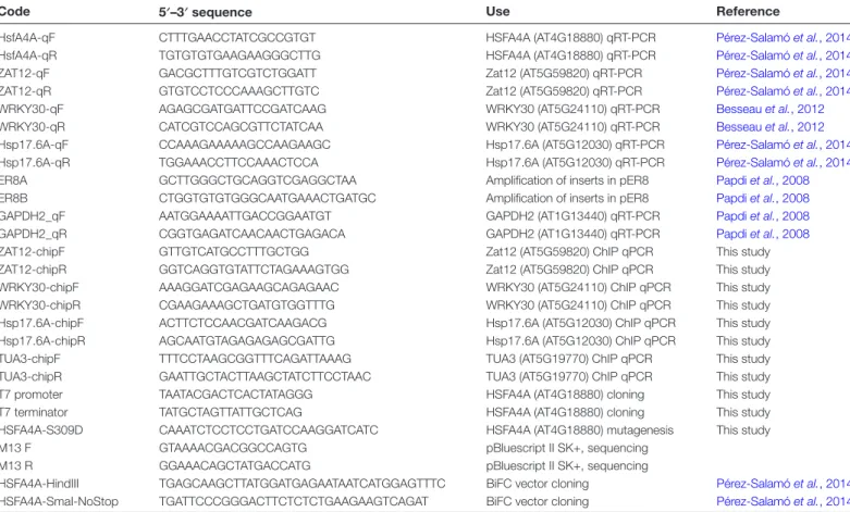
htm). Oligonucleotides used in this study are listed in Table 1.
Physiological parameters
Lipid peroxidation was determined in 10-day-old plants exposed to single or combined stresses for 48 h, measuring the malondialdehyde (MDA) content, using a thiobarbituric acid (TBA) test (Hodges et al., 1999). The amount of MDA–TBA complex was calculated according to the fol- lowing equation: X (%)=100×(OD532−OD600) (Zsigmond et al., 2012).
Microscopic techniques
To study intracellular localization of HSFA4A, 5-day-old seedlings ex- pressing the pHSFA4A::HSFA4A-YFP construct were imaged using an Olympus FV1000 confocal laser scanning microscope. To follow changes in intracellular localization, the YFP signal was recorded in roots by taking pictures of the same cells in every 5 min, up to 30 min. Plantlets were placed on slides and immersed either in standard ½MS culture me- dium or medium supplemented by 100 mM NaCl. BiFC assays were per- formed as described (Pérez-Salamó et al., 2014). Fluorescence intensities were analysed in nuclear regions and cytoplasm using ImageJ2 (https://
imagej.net/ImageJ2).
Chromatin immunoprecipitation assay
A ChIP assay was used to verify the interaction of HSFA4A protein with three selected gene promoters: ZAT12 (AT5G59820), HSP17.6A (AT5G12030), and WRKY30 (AT5G24110). The chromatin was iso- lated from stress-treated and control pHSFA4A::HSFA4A-YFP ex- pressing plants, following the Abcam ChIP protocol (Haring et al., 2007).
The immunoprecipitation was carried out with anti-green fluorescent protein (GFP) antibody (Roche) and Dynabeads Protein G (Invitrogen/
Thermo Fisher Scientific). Control ChIP experiment was carried out with Dynabeads Protein G without anti-GFP antibody. Reverse cross- linking and DNA purification were carried out by the Abcam ChIP protocol. Fragments of immunoprecipitated DNA were amplified by quantitative PCR using specific primers for the HSE-containing pro- moter regions of ZAT12, HSP17.6A, and WRKY30 genes. For refer- ence, the tubulin α-3 (TUA3, AT5G19770) promoter fragment was used, which has no HSE. qPCR data were normalized with values obtained on TUA3. TF binding was calculated as fold enrichment over the control (Aleksza et al., 2017).
SDS-PAGE, non-denaturing PAGE and western blot
Total protein was isolated from 100 mg seedlings with 50 mM Tris–HCl pH 7.5, 150 mM NaCl, 1% Triton X-100 and 1× Protease Inhibitor Cocktail (Sigma-Aldrich/Merck). For the multimerization studies, DTT was added to the 100 µg total protein extract in different final concentra- tions (0, 25, 50, 75 mM) and incubated for 10 min in room temperature.
The samples were size separated on 7% non-denaturating SDS-PAGE and transferred to Immobilon polyvinylidene difluoride (PVDF) membrane (Merck Millipore) for western detection. For denaturing electrophoresis, 25 µg total protein was size separated on 8% SDS-PAGE, transferred onto Immobilon PVDF membrane. For western hybridization membranes were incubated in 1×TBST blocking buffer (50 mM Tris–HCl pH 8.0, 150 mM NaCl, 0.05% Tween-20, 5% dry skimmed milk) for 1 h and with anti-GFP antibody (Roche, 1:2000 dilution) in blocking buffer for 1.5 h.
After washing with 1× TBST three times for 10 min, the membranes were incubated for 1.5 h with an anti-mouse-POD secondary antibody (Pierce, dilution 1:5000), washed with 1× TBST as before. The membrane was subsequently overlaid with Immobilon Western Chemiluminescent HRP Substrate (Merck Millipore) and chemiluminescence was detected with a Fusion FX5 camera system (Vilber Lourmat).
RNA isolation and RT-PCR (qPCR)
Total RNA was isolated from 100 mg Arabidopsis seedlings, using a Nucleospin Plant RNA Kit (Macherey-Nagel). Isolated RNA was DNase treated with TURBO DNA-free™ Kit (Invitrogen/Thermo Fisher Scientific), and first-strand cDNA synthesis of 1 µg of total RNA was carried out with a High Capacity cDNA Reverse Transcription Kit (Applied Biosystems/Thermo Fisher Scientific), using random hexamers.
Real-time PCR was carried out with the ABI 7900 Fast Real Time System (Applied Biosystems) with the following protocol: 40 cycles at 95 °C for 15 s, 60 °C for 60 s, using Maxima SYBR Green qPCR Master Mix (Thermo Fisher Scientific). GAPDH2 (AT1G13440) was used as reference gene. The normalized relative transcript levels were calculated with the 2−ΔΔCt method (Czechowski et al., 2005).
Downloaded from https://academic.oup.com/jxb/article-abstract/70/18/4903/5489060 by Biological Research Centre of the Hungarian Academy of Sciences user on 19 November 2019
Protein phosphorylation
All gene constructs used in phosphorylation experiments (His6- MPK3, maltose-binding protein (MBP)–HSFA4A, MBP, glutathione S-transferase (GST)–MPK4) were expressed in BL21DE39 Rosetta cells (Novagen). The GST–MPK4 construct was obtained from Robert Dóczi (Martonvásár, Hungary). Proteins were purified by affinity chro- matography, following the manufacturer’s instructions (Dóczi et al., 2007).
Proteins were dialysed overnight in 50 mM NaCl, 10% glycerin, 2 mM β-mercaptoethanol, 10 mM Tris–HCl pH 7.5. Protein integrity was checked by SDS-PAGE. In vitro phosphorylation was performed with purified proteins as described (Pérez-Salamó et al., 2014); 1–2 µg of pro- teins were used in each phosphorylation reaction, which was performed in 20 mM Tris–HCl (pH 8.0), 5 mM MgCl2, 1 mM DTT, containing 185 kBq [γ-32P]ATP) at room temperature (23–25 °C) for 1 h. Proteins were separated on 12% SDS-PAGE and the polyacrylamide gel was stained with Coomassie Blue (Thermo Fisher Scientific). Autoradiography was performed with AGFA Medical X-ray Blue Film (AGFA, Germany).
Identification of MPK4 phosphorylation sites in HSFA4A
The phosphorylation reaction for mass spectrometry was iden- tical to that described above, except that non-radiolabeled 1 µM ATP (Thermo Fisher Scientific) was used. After the reaction, sam- ples were separated by SDS-PAGE, and protein bands were visual- ized by Coomassie Blue staining. The protein band corresponding to MBP–HSFA4A was excised and analysed by mass spectrometry in the Laboratory of Proteomics Research of the Biological Research Centre (www.szbk.u-szeged.hu/services_proteomics_research.php).
Proteins were in-gel digested by trypsin at 37 °C for 6 h as described (https://msf.ucsf.edu/protocols.html). The digest was split and half of the sample subjected to Fe(III)-immobilized metal affinity chro- matography phosphopeptide enrichment (Ficarro et al., 2009). The digest with and without enrichment was analysed on an Orbitrap
Elite (Thermo Scientific) mass spectrometer, online coupled to a Waters nanoAcquity HPLC. Mass spectrometry (MS) data acquisition was performed in a data-dependent fashion; in each survey scan the five most abundant multiply charged precursor ions were selected for high-energy collision dissociation fragmentation at 30% normalized collision energy. Both, MS and tandem mass spectrometry (MS/MS) spectra were acquired in the orbitrap at a resolution of 60 000 and 15 000, respectively. Dynamic exclusion was 30 s.
Data were processed by Protein Discoverer (v1.3), and the gen- erated peak lists were subjected to a database search using Protein Prospector (v5.22.0) against the Uniprot 1.11.2017 database, consid- ering also the protein sequence of the expressed MBP–HSFA4A and the MPK4 kinase. Monoisotopic masses were used with a precursor mass tolerance of ±5 ppm and a fragment mass tolerance of ±20 ppm.
Carbamidomethylation of Cys residues was set as fixed modification;
acetylation of protein N-termini, cyclization of peptide N-terminal Gln residues, Met oxidation and phosphorylation of Ser, Thr, and Tyr residues were selected as variable modifications and a maximum two variable modifications were permitted per peptide. Search results were accepted at a false discovery rate of 5% at the protein level and 1% at the peptide level. Modification site assignments with a minimum site localization in peptide (SLIP) score of six were accepted (Baker et al., 2011). MS/MS spectra were manually inspected.
Relative quantification of the phosphorylation at a given site was performed at the MS1 level from the liquid chromatography–MS ana- lysis of the non-enriched tryptic digest. The peak areas of the non- phosphorylated and phosphorylated peptides were calculated by Pinnacle (v1.0.83.0) using the isotopic cluster of the corresponding precursor ions:
PP signal % = PP peak area
PP peak area + non−PP peak area×100 where PP is phosphopeptide.
Table 1. List of oligonucleotides used in this study
Code 5′–3′ sequence Use Reference
HsfA4A-qF CTTTGAACCTATCGCCGTGT HSFA4A (AT4G18880) qRT-PCR Pérez-Salamó et al., 2014
HsfA4A-qR TGTGTGTGAAGAAGGGCTTG HSFA4A (AT4G18880) qRT-PCR Pérez-Salamó et al., 2014
ZAT12-qF GACGCTTTGTCGTCTGGATT Zat12 (AT5G59820) qRT-PCR Pérez-Salamó et al., 2014
ZAT12-qR GTGTCCTCCCAAAGCTTGTC Zat12 (AT5G59820) qRT-PCR Pérez-Salamó et al., 2014
WRKY30-qF AGAGCGATGATTCCGATCAAG WRKY30 (AT5G24110) qRT-PCR Besseau et al., 2012
WRKY30-qR CATCGTCCAGCGTTCTATCAA WRKY30 (AT5G24110) qRT-PCR Besseau et al., 2012
Hsp17.6A-qF CCAAAGAAAAAGCCAAGAAGC Hsp17.6A (AT5G12030) qRT-PCR Pérez-Salamó et al., 2014 Hsp17.6A-qR TGGAAACCTTCCAAACTCCA Hsp17.6A (AT5G12030) qRT-PCR Pérez-Salamó et al., 2014 ER8A GCTTGGGCTGCAGGTCGAGGCTAA Amplification of inserts in pER8 Papdi et al., 2008 ER8B CTGGTGTGTGGGCAATGAAACTGATGC Amplification of inserts in pER8 Papdi et al., 2008
GAPDH2_qF AATGGAAAATTGACCGGAATGT GAPDH2 (AT1G13440) qRT-PCR Papdi et al., 2008
GAPDH2_qR CGGTGAGATCAACAACTGAGACA GAPDH2 (AT1G13440) qRT-PCR Papdi et al., 2008
ZAT12-chipF GTTGTCATGCCTTTGCTGG Zat12 (AT5G59820) ChIP qPCR This study
ZAT12-chipR GGTCAGGTGTATTCTAGAAAGTGG Zat12 (AT5G59820) ChIP qPCR This study
WRKY30-chipF AAAGGATCGAGAAGCAGAGAAC WRKY30 (AT5G24110) ChIP qPCR This study
WRKY30-chipR CGAAGAAAGCTGATGTGGTTTG WRKY30 (AT5G24110) ChIP qPCR This study
Hsp17.6A-chipF ACTTCTCCAACGATCAAGACG Hsp17.6A (AT5G12030) ChIP qPCR This study Hsp17.6A-chipR AGCAATGTAGAGAGAGCGATTG Hsp17.6A (AT5G12030) ChIP qPCR This study
TUA3-chipF TTTCCTAAGCGGTTTCAGATTAAAG TUA3 (AT5G19770) ChIP qPCR This study
TUA3-chipR GAATTGCTACTTAAGCTATCTTCCTAAC TUA3 (AT5G19770) ChIP qPCR This study
T7 promoter TAATACGACTCACTATAGGG HSFA4A (AT4G18880) cloning This study
T7 terminator TATGCTAGTTATTGCTCAG HSFA4A (AT4G18880) cloning This study
HSFA4A-S309D CAAATCTCCTCCTGATCCAAGGATCATC HSFA4A (AT4G18880) mutagenesis This study
M13 F GTAAAACGACGGCCAGTG pBluescript II SK+, sequencing
M13 R GGAAACAGCTATGACCATG pBluescript II SK+, sequencing
HSFA4A-HindIII TGAGCAAGCTTATGGATGAGAATAATCATGGAGTTTC BiFC vector cloning Pérez-Salamó et al., 2014 HSFA4A-SmaI-NoStop TGATTCCCGGGACTTCTCTCTGAAGAAGTCAGAT BiFC vector cloning Pérez-Salamó et al., 2014
Downloaded from https://academic.oup.com/jxb/article-abstract/70/18/4903/5489060 by Biological Research Centre of the Hungarian Academy of Sciences user on 19 November 2019
Identification of phosphorylation sites in vivo
To test HSFA4A phosphorylation in vivo, protein extracts were iso- lated from 10-day-old salt-treated (150 mM NaCl, 6 h) and control pHSFA4A::HSFA4A-YFP expressing plants. Seedlings (500 mg) were harvested, frozen in liquid nitrogen and ground with a TissueLyser at 30 Hz. Total proteins were extracted using the manufacturer’s Lysis buffer supplemented with 1 mM DTT, 1 mM phenylmethylsulfonyl fluoride, 1× Sigma protease inhibitor cocktail, 3 mM p-nitrophenyl phos- phate, 1 µM MG132. Protein extracts (3 mg/immunoprecipitate) were immunopurified using anti-GFP antibody-coupled very small (50 nm) magnetic beads (MACS® Technology, Miltenyi), digested in-column with trypsin, and analysed by MS as described (Horvath et al., 2017).
Data analysis, bioinformatics
Protein sequences were obtained from Phytozome (https://phytozome.
jgi.doe.gov/pz/portal.html). Multiple sequence alignment was performed with Clustal Omega (https://www.ebi.ac.uk/Tools/msa/clustalo/).
Transcription factor binding sites were identified with AthaMap (http://
www.athamap.de). DAP-seq data were compiled manually from on- line data of the Neomorph database: http://neomorph.salk.edu/
PlantCistromeDB.
Results
Regulation of HSFA4A
The HSFA4A gene was shown to be induced by a number of abiotic and biotic stresses (Pérez-Salamó et al., 2014; see eFP Browser: http://bar.utoronto.ca/efp/cgi-bin/efpWeb.cgi?pri maryGene=AT4G18880&modeInput=Absolute). Expression of this factor with a stress combination was, however, not studied. When wild-type Arabidopsis plants were treated by high salinity, high temperature, and a combination of these conditions, HSFA4A expression changed dramatically, but induction followed different kinetics. Transcription in con- trol plants was slightly and temporally enhanced, probably as consequence of lid opening and touching these plants during transfer to fresh medium. Salt (150 mM NaCl) induced tran- scription in 2 h and remained 2- to 3-fold elevated for up to 24 h. Heat (37 °C) and the combination of heat and salt stress had no effect on transcript levels in the first hours but enhanced HSFA4A transcription after 24 h (Fig. 1A). These results sug- gest that heat and salinity regulate HSFA4A through different signaling pathways. To study the HSFA4A protein in vivo, a transgenic Arabidopsis line was generated that expresses the YFP-tagged HSFA4A under the control of its own 2 kb-long promoter (pHSFA4A::HSFA4A-YFP, Fig. 1B). Western hy- bridization confirmed the presence of the HSFA4A–YFP chi- meric protein in transgenic plants, which was more abundant in salt, heat, and combined salt and heat stressed plants cor- relating with stress-dependent induction of the endogenous HSFA4A gene (Fig. 1; Supplementary Fig. S1 at JXB online).
Confocal microscopic observations revealed weak HSFA4A–
YFP-derived fluorescence in green parts of the plants (not shown) while it was well detectable in roots: the signal was strong in cells of root caps, elongation and differentiation zones, and root hairs. In root cells and root hairs HSFA4A–YFP was present in both cytoplasm and nuclei. Parallel with enhanced western signal, several hours of salt treatment led to stronger fluorescence in root cells, which became particularly strong
in nuclei (Fig. 2A). Several hours of stress enhanced content of HSFA4–YFP protein, which could be detected either by western assay or visualization with confocal microscopy (Figs 1B, 2A; Supplementary Fig. S1).
Heat shock factors are shuttling proteins with predominant cytoplasmic localization in non-stressed conditions and nuclear accumulation upon heat and other stress (Scharf et al., 1998;
Heerklotz et al., 2001; Akerfelt et al., 2010). To study intra- cellular shifts of HSFA4A during stress, pHSFA4A::HSFA4A- YFP expressing roots were treated with salt (100 mM NaCl) and change of YFP-derived fluorescence was monitored in the same root cells. Salt stress led to a fast and temporal accumu- lation of HSFA4A–YFP in nuclei while the fluorescence pat- tern did not change significantly in non-treated control cells (Fig. 2B, C). In nuclei, YFP-derived fluorescence reached its maximum 20 min after the initiation of salt treatment, which was followed by a gradual decrease (Fig. 2C). In cytosol only
Fig. 1. Regulation of HSFA4A. (A) Transcriptional regulation of HSFA4A gene in wild-type Arabidopsis plants treated with salt (150 mM NaCl), heat stress (37 °C in light and 30 °C in dark), and their combination for 2, 6, and 24 h. Relative expression is shown where 1 corresponds to transcript level at 0 h. Error bars indicate standard error; asterisks indicate significant differences from control: *P<0.05 and **P<0.01 (Student’s t-test). (B) Schematic map of the pHSFA4A::HSFA4A-YFP gene construct.
(C) Detection of HSFA4A–YFP fusion protein in 10-day-old control and salt-stressed plants (150 mM NaCl, 0–24 h) transformed with the pHSFA4A::HSFA4A-YFP gene construct. Salt treatment led to enhanced HSFA4A–YFP specific western signal. (This figure is available in color at JXB online.)
Downloaded from https://academic.oup.com/jxb/article-abstract/70/18/4903/5489060 by Biological Research Centre of the Hungarian Academy of Sciences user on 19 November 2019
minimal change in YFP-derived fluorescence could be ob- served. These results suggest rapid transfer of HSFA4A to nu- clei that starts within minutes upon onset of salt stress and most probably takes place before gene activation and de novo protein biosynthesis.
Binding of HSFA4A to promoter elements of target genes
Whole-genome transcript profiling has identified genes that were upregulated by HSFA4A overexpression (Pérez-Salamó
et al., 2014). These genes can be direct targets of this heat shock factor, binding to their cis-acting HSEs, or can indirectly be induced by TFs, which are themselves regulated by HSFA4A.
Three HSFA4A-induced genes were selected to test promoter binding: HSP17.6A, ZAT12, and WRKY30, encoding a small heat shock protein, a zinc finger and a WRKY-type TF, re- spectively (Pérez-Salamó et al., 2014). Promoter regions of these genes contain several HSE motifs, suggesting that they can be direct targets of HSFs (Fig. 3A). Promoter binding was tested in vivo by ChIP assays, using transgenic plants expressing the pHSFA4A::HSFA4A-YFP gene construct, treated with salt (150 mM NaCl), high temperature (37 °C), or both stresses for 6 h before chromatin extraction. The ChIP assay revealed specific enrichment of HSE-containing promoter regions of all three tested genes (Fig. 3B). Heat treatment significantly enhanced enrichment on all three promoters, while salt pro- moted HSFA4A binding to ZAT12 and WRKY30 promoters but only slightly influenced binding to HSP17.6A. The com- bination of both stresses was additive on promoter binding on ZAT12 and WRKY30 genes, while on the HSP17.6A pro- moter it was similar to heat treatment (Fig. 3B). Binding to the TUA3 promoter, which lacked HSE motifs, was not altered and was used as a non-specific control. These results demon- strate that the HSFA4A factor can directly bind to the pro- moters of the three target genes, which is enhanced depending on the type of stress.
HSFA4A is phosphorylated by MAP kinase 4
Earlier we showed that MAP kinases MPK3 and MPK6 can interact with and phosphorylate HSFA4A. Five phosphoryl- ated amino acid residues were identified by MS, and Ser309 was found to be the dominant MPK3 and MPK6 phosphor- ylation site (Pérez-Salamó et al., 2014). An alternative stress- related MAP kinase signaling pathway is controlled by MPK4, which is implicated in pathogen responses and ROS homeo- stasis (Pitzschke et al., 2009; Dóczi and Bögre, 2018). To reveal possible involvement of HSFA4A in MPK4-controlled stress signaling, phosphorylation of HSFA4A by MPK4 was tested in vitro. We found that HSFA4A can be phosphorylated not only by MPK3 but also by MPK4 (Fig. 4A). Subsequent analysis by mass spectrometry identified six amino acid residues of HSFA4A which were phosphorylated by MPK4: Thr124, Ser198, Ser239, Ser309, Thr396, and Ser397 (Fig. 4B; Supplementary Dataset S1). Four of the identified sites coincided with amino acid res- idues phosphorylated also by MPK3 (Ser198, Ser239, Ser309, Thr396) (Pérez-Salamó et al., 2014). The two phospho-isoforms modified at Thr396 and Ser397 could be distinguished based on differences in retention time and fragmentation pattern.
Calculation of phosphopeptide signal frequencies showed that 80% of the Ser309 residues can be phosphorylated by MPK4, in contrast to the other Ser or Thr residues, which were phos- phorylated with much lower frequencies (0.1–8%, Fig. 4B), suggesting that Ser309 is the primary phosphorylation site for MPK4. Ser309 is the dominant phosphorylation site also for MPK3 and MPK6 (Pérez-Salamó et al., 2014), suggesting that it is the primary target of MAP kinases.
To test phosphorylation of HSFA4A in vivo, YFP-tagged HSFA4A was purified from control and salt-treated transgenic
Fig. 2. Intracellular localization and transfer of HSFA4A. (A) Confocal microscopic detection of the HSFA4A–YFP fusion protein in different segments of roots. Root hair is stained with propidium iodide to
demonstrate nuclear localization of the YFP signal. Segments of elongation zone are shown with and without salt treatment (100 mM NaCl, 2 h).
(B) HSFA4A is transported into nuclei during salt stress. Roots were treated with 100 mM NaCl, and HSFA4A–YFP-derived fluorescence was monitored in individual cells at regular intervals. Arrow indicate position of a nucleus. (C) Quantitative evaluation of YFP fluorescence in cytosol and nuclei. Relative fluorescence is shown, where 1 corresponds to intensity measured in cytosol at time 0. YFP-derived fluorescence was rapidly enhanced in nuclei of salt-treated cells, while it did not change in control cells. Scale bar on images indicates 20 µm. Error bars indicate standard error; asterisks indicate significant differences from time 0: *P<0.05 and
**P<0.01 (Student’s t-test).
Downloaded from https://academic.oup.com/jxb/article-abstract/70/18/4903/5489060 by Biological Research Centre of the Hungarian Academy of Sciences user on 19 November 2019
plants expressing the pHSFA4A::HSFA4A-YFP gene con- struct (Fig. 1B, C), and phosphopeptides were identified by mass spectrometry (Fig. 4C). Phosphorylation of Ser239 and Ser309 was observed in both in vivo phosphorylation detec- tion assays and in vitro MPK3 and MPK4 phosphorylation ex- periments (Fig. 4D; Supplementary Table S1) confirming that these amino acids are indeed in vivo targets of MAP kinases.
Phosphorylation of Ser112 and Ser306 was also revealed in vivo; they were not phosphorylated by MAP kinases (Fig. 4C).
Computer prediction suggested that these amino acid res- idues can be phosphorylated by protein kinases such as protein kinase A (PKA), cyclin dependent kinase (CDK), casein kinase
1 (CK1), casein kinase 2 (CK2), and GSK3 (Supplementary Table S1). These results suggest that HSFA4A is under complex post-translational control as it can be phosphorylated not only by MAP kinases but also by other classes of protein kinases.
Multiple alignment of amino acid sequences of 33 HSFA4- type TFs from 27 plant species revealed that the identi- fied phosphorylation sites can be assigned to three groups (Supplementary Dataset S2). Ser112 is present in all plant HSFA4 proteins, while Thr124, Ser306, and Thr396 are con- served in most of the proteins. Ser198, Ser239, and Ser309 are present only in HSFA4A-type proteins of closely related plant species that belong to the Brassicaceae family (Arabidopsis,
Fig. 3. Binding of HSFA4A on target gene promoters. (A) Schematic map of ZAT12, HSP17.6A, and WRKY30 promoters according to AthaMap.
Promoter regions between −1000 and +200 bp are shown. Black line indicates promoter, dark grey corresponds to 5′-UTR and exon while light grey is intron sequence. HSE motifs are indicated by grey boxes and sequences connected to the amplified regions are shown above the target region. Dashed arrows indicates transcription initiation. Amplified target sequences by qPCR are indicated by black double arrows. (B) ChIP assay with YFP-tagged HSFA4A using transgenic plant expressing the pHSFA4A::HSFA4A-YFP gene construct (see Fig. 1B, C). Plants were treated by salt (150 mM NaCl, 6 h), heat stress (37 °C, 6 h), and their combination before ChIP assay. ChIP results are shown as relative enrichment by qPCR, where reference (value 1) is the qPCR value of the TUA3 promoter, which lacks any HSE motif, at control conditions. Note enrichments on different promoter regions, which can be enhanced by salt or heat treatments. Error bars indicate standard error; asterisks indicate significant differences from ChIP values of TUA3: *P<0.05 and
**P<0.01 (Student’s t-test).
Downloaded from https://academic.oup.com/jxb/article-abstract/70/18/4903/5489060 by Biological Research Centre of the Hungarian Academy of Sciences user on 19 November 2019
Arabis halleri, Capsella rubella, Brassica rapa, and Eutrema salsugineum). On the other hand, it is intriguing that Thr396 was missing in the Arabidopsis HSFA4C and five proteins most related to this HSF in Brassicaceae species (Supplementary Dataset S2; Supplementary Fig. S2). A conserved MAP kinase docking domain is present in all HSFA4-type kinases (Fig.
4D; Supplementary Dataset S2). These results suggest that the most conserved phosphorylation sites are present in all plant HSFA4-type TFs, but MPK3-, MPK4-, and MPK6-mediated phosphorylation is characteristic only of HSFA4 factors of the Brassicaceae species.
Phosphorylation affects intramolecular interactions of HSFA4A
Recognition of heat shock elements and transcriptional activa- tion of target genes requires trimerization of heat shock factors (Miller and Mittler, 2006; Akerfelt et al., 2010). Intramolecular dimerization of HSFA4A has previously been demonstrated, and conserved Cys residues were shown to be essential to stabilize such interactions (Pérez-Salamó et al., 2014). To study multimer formation of HSFA4A in vivo, protein ex- tracts from HSFA4A–YFP-expressing plants were separated on non-denaturing gels and the YFP-tagged TF was detected by western hybridization using anti-GFP antibody. In crude extracts a high molecular mass complex was detected with molecular mass of approximately 200 kDa. DTT treatment reduced the abundance of the high molecular mass complex while a 70–80 kDa band appeared, suggesting that in reducing conditions the HSFA4A-containing complex is disassembled and the monomer HSFA4A is released (Fig. 5A). Similar results were obtained with protein extracts isolated from HSFA4A–
YFP-expressing cell suspension cultures (Supplementary Fig.
S3A). In plant cells most HSFA4A protein seem to exist in high molecular mass complexes, which can be monomerized in a reducing environment. In vitro assay showed that oxida- tive conditions favor HSFA4A multimerization while in a re- ducing environment monomers are predominantly formed (Supplementary Fig. S3B).
To test the effect of MAP kinase-mediated phosphoryl- ation on HSFA4A multimerization in vivo, protein–protein interactions of wild-type and point mutants of HSFA4A pro- teins were compared in BiFC assays using a protoplast-based transient expression system. The dominant phosphorylation site Ser309 was replaced by either Ala (non-phosphorylating, S309A) or Asp (phosphorylation mimicking, S309D) amino acids. Dimerization of wild-type and mutant HSFA4A could be confirmed by detecting YFP-derived fluorescence in all three versions of HSFA4A, while the BiFC signal was missing in the control samples transformed with empty vectors alone or in combination with one of the HSFA4A–YFP partners (Fig. 5B). When BiFC fluorescence intensities with wild-type and mutant HSFA4A were compared, S309A displayed 30%
weaker, while the S309D mutant had 50% stronger, fluores- cence than the wild-type TF (Fig. 5C). Western detection of
Fig. 4. Phosphorylation of HSFA4A. (A) In vitro phosphorylation of HSFA4A by MAP kinases MPK3 and MPK4. MBP-tagged HSFA4A was phosphorylated in vitro by His-MPK3 or GST–MPK4. (B) List of phosphopeptides identified by MS. Phosphorylated amino acids are indicated with bold letters (pT, pS). MBP-tagged HSFA4A was phosphorylated in vitro by MPK4, in-gel digested by trypsin, and analysed by mass spectrometry. The modified sites within the detected tryptic peptides were determined from MS/MS spectra acquired following ferric nitrilotriacetate chelate (Fe(III)-NTA) phosphopeptide enrichment (Supplemental Dataset S1). Phosphopeptide signal% was calculated from MS signal areas of the unmodified and phosphorylated peptides detected in the tryptic digest without phosphopeptide enrichment.
Note that these values are not absolute phosphorylation ratios. (C) Detection of phosphopeptides in vivo. HSFA4A–YFP fusion protein was immunoprecipitated from transgenic plants, and phosphopeptides were detected by mass spectrometry. Bold letters indicate phosphorylated amino acids. (D) Amino acid sequence of HSFA4A. Amino acids phosphorylated by MPK3 (Pérez-Salamó et al., 2014) and MPK4 (this study) or detected in immunoprecipitated samples are shown with bold and underlined letters. Boxed letters indicate amino acids that were detected in both in vitro and in vivo phosphorylation assays. Underlined letters in italics indicate predicted MAPK docking motif (RKRRFPR).
Conserved DNA binding domain is underlined. GST, glutathione S-transferase; His, polyhistidine tag; MBP, maltose binding protein.
Downloaded from https://academic.oup.com/jxb/article-abstract/70/18/4903/5489060 by Biological Research Centre of the Hungarian Academy of Sciences user on 19 November 2019
Fig. 5. Multimerization of HSFA4A. (A) Detection of HSFA4A–YFP multimers in Arabidopsis plants transformed with the pHSFA4A::HSFA4A-YFP gene construct. Protein extracts were treated with or without DTT and separated on non-denaturing polyacrylamide gels. HSFA4A–YFP was detected by western hybridization with anti-GFP antibody. Separated and membrane-blotted proteins were stained with Ponceau Red. (B) BiFC assay of wild- type HSFA4A (HSFA4A-wt), and mutants in which Ser309 was changed to Ala (HSF-S309A) or Asp (HSF-S309D). nYFP and cYFP indicates N- and C-terminal half of YFP protein. Controls include polyethylene glycol-treated protoplasts without plasmids, protoplasts transformed with plasmids having nYFP and cYFP fragments, or protoplasts expressing HSFA4A-cYFP in combination with the empty nYFP plasmid (upper row). Typical BiFC images are shown. (C) Quantitative evaluation of fluorescence signals in YFP-expressing transformed protoplasts in BiFC experiments. Relative fluorescence intensities are shown, where 1 equals signals of protoplasts expressing the wild-type HSFA4A constructs (HSFA4A-wt) while HSF-S309A and HSF- S309D indicate S309A and S309D mutants, respectively. (D) Western detection of HSFA4A–YFP fusions in BiFC experiment. Anti-GFP antibody was used to detect the proteins in transformed protoplasts. Note that comparable amount of HSFA4A–YFP was produced in each BiFC samples. In fact, slightly lower amount of wild-type and higher amount of S309A version of HSFA4A was produced. Error bars indicate standard error; asterisks indicate significant differences from HSFA4A-wt: *P<0.05 and **P<0.01 (Student’s t-test). Scale bar on images indicates 10 μm.
Downloaded from https://academic.oup.com/jxb/article-abstract/70/18/4903/5489060 by Biological Research Centre of the Hungarian Academy of Sciences user on 19 November 2019
YFP with anti-GFP antibody confirmed the efficient pro- duction of all HSFA4A–YFP protein forms in our transient expression system (Fig. 5D). These data suggest that MAP kinase-mediated phosphorylation is not essential for HSFA4A dimerization but has a positive influence on it.
HSFA4A can enhance tolerance to combined salt and heat stresses
Overexpression of HSFA4A in Arabidopsis could enhance tolerance to salt, heavy metal, and oxidative agents while the knockout mutant showed hypersensitivity to salt (Pérez-Salamó et al., 2014; Faragó et al., 2018). Whether this TF could modu- late responses to combined stresses is, however, not known. To evaluate responses to combined salt and heat stresses, toler- ance of wild-type and HSFA4A-overexpressing Arabidopsis lines were tested in two experimental systems. In the first trials, seedlings were germinated and grown on culture media con- taining 0, 50, 75, 100, 125, and 150 mM NaCl, and 10-day-old plantlets were treated by 4 or 8 d of heat stress (37 °C in light and 30 °C in dark). Survival rates of HSFA4A-overexpressing plants were higher than wild-type plants on salt-containing media with or without heat stress (Supplementary Fig. S4).
In the second set of experiments, wild-type seeds (Col-0) and transgenic lines overexpressing wild-type and S309D mutant versions of HSFA4A were germinated on standard culture medium and 10-day-old plantlets were exposed to different doses of salt, heat, and combined stresses followed by transfer to standard culture medium to allow recovery. Heat stress had only a minor effect on plant viability in these conditions, while salinity affected plants in a concentration-dependent manner.
Damage was clearly alleviated by overexpression of both forms of HSFA4A, especially when higher salt doses (150 mM NaCl) were used. Around 50% of HSFA4A-overexpressing and 15%
of wild-type plants recovered completely, while 15–20% of HSFA4A-overexpressing and 40% of wild-type plants died after exposure to 150 mM NaCl for 2 d (Fig. 6A, B). When 100 mM NaCl was combined with high temperature for 2 d, 15% of the transgenic but none of the wild-type plants re- covered completely. After 4 d of combined stress, 10% of the wild-type and 30–40% of transgenic plants survived (Fig.
6A, B). In these conditions both HSFA4A forms alleviated damage to a similar extent. Combination of higher doses of salt (150 mM NaCl) with heat led to complete lethality (not shown). These results indicate that overexpression of HSFA4A not only improved tolerance to salt but could increase viability under simultaneous heat and salt stresses.
Reactive oxygen species (ROS) are generated by many ad- verse environmental conditions imposing oxidative damage to stressed plants. One of the deleterious effect of ROS is lipid peroxidation, which damages membranes and is a good indi- cator of oxidative damage. To assess the effect of HSFA4A on ROS-triggered damage, lipid peroxidation rates were compared in wild-type and HSFA4A-overexpressing plants. All individual or combined stress treatments enhanced lipid peroxidation in wild-type plants and the damage was proportional to the se- verity of the stress (Fig. 6C). Overexpression of both forms of HSFA4A reduced lipid peroxidation when 150 mM NaCl or
combined 100 mM NaCl and heat stress was imposed. In mild conditions (heat or 100 mM NaCl alone) the S309D mutant was slightly more efficient in reducing lipid peroxidation (Fig.
6C). These results indicate, that HSFA4A can reduce oxidative damage imposed not only by individual salt or heat stresses, but also by stress combinations.
Discussion
Heat shock factors in plants are components of complex regulatory networks with various levels of control including transcription regulation, posttranslational modifications, intra- cellular transport, intra- and intermolecular interactions, and homo- and heteromeric trimer formation (Akerfelt et al., 2010;
Scharf et al., 2012). HSFA4A responds to various stress con- ditions including salinity, heavy metals, oxidative agents, high temperatures, and treatments that generate protein misfolding (Pérez-Salamó et al., 2014; Lin et al., 2018). Our results show that salt and heat induction of HSFA4A follow a different pat- tern: expression is upregulated by salt in 2 h while heat alone or in combination with salt stress promotes expression only after 24 h (Fig. 1). Change in protein abundance roughly correlated with alterations in transcript levels (Fig. 1; Supplementary Fig.
S1). Information on expression control of plant HSFs is scarce and no TF has been described that regulates the HSFA4A gene. Binding of different classes of TFs to the promoter and 5′-untranslated region (UTR) of HSFA4A could be revealed by online data mining of genome-wide DNA affinity puri- fication sequencing (DAP-seq) (O’Malley et al., 2016) or ChIP-seq (Albihlal et al., 2018) studies. ChIP-seq data suggest HSFA1B binding, while DAP-seq revealed binding of bZIP, WRKY, C2H2, MYB, and HSF-type TFs to HSFA4A pro- moter, which has conserved recognition motifs for such factors (Supplementary Figs S5, S6). These TFs can regulate HSFA4A expression in different conditions.
Regulation at the post-translational level includes various types of protein modifications such as phosphorylation, sumoylation, or modulation of intracellular localization, which can profoundly affect the activity of a regulatory pro- tein. These features were studied with YFP-tagged HSFA4A, expressed in transgenic Arabidopsis plants under the control of its own promoter. HSFA4A–YFP accumulated in a salt- and heat-dependent manner (Fig. 1; Supplementary Fig. S1), confirming that transcriptional activation by several stresses in fact leads to enhanced HSFA4A protein levels. Subcellular distribution of HSFs is regulated by the balance between nu- clear import/export processes (Scharf et al., 2012). In non- stressed roots cells, HSFA4A–YFP could be detected in cytosol and nuclei while salt treatment led to fast nuclear ac- cumulation (Fig. 2). These observations correlate with earlier reports describing the cytoplasmic localization of inactive HSFs, which move to nuclei in stress conditions allowing HSE recognition and activation of target genes (Scharf et al., 1998, 2012; Heerklotz et al., 2001). Nuclear localiza- tion signals are among the most conserved sequence elem- ents of plant HSFs, composed by basic amino acid residues (RKRRF/LPR, Supplementary Dataset S2). While precise
Downloaded from https://academic.oup.com/jxb/article-abstract/70/18/4903/5489060 by Biological Research Centre of the Hungarian Academy of Sciences user on 19 November 2019
Fig. 6. HSFA4A overexpression enhances tolerance to heat and salt stresses. Ten-day-old in vitro-grown plantlets were treated by salt (100 mM, 150 mM NaCl), heat (37 °C in light, 30 °C in dark) or their combination for 2 or 4 d. Rates of surviving healthy (vigorous growth with several new green leaves), damaged (small plants with retarded growth and/or chlorotic leaves), and dead plants (completely chlorotic with no green leaves) were scored 10 d after recovery. Similar results were obtained with independent transgenic lines of both constructs and one representative transgenic line was used for each construct in this experiments. (A) Growth of wild-type (Col-0) and transgenic plants overexpressing the wild-type (HSFox-wt) and S309D mutant (HSFox-m) forms of HSFA4A after heat, 150 mM NaCl, and combined 100 mM NaCl and heat treatments. (B) Frequencies of healthy, damaged, and dead plants after heat, salt, and combined heat and salt stresses applied for 2 or 4 d. Survival frequencies of control, non-stressed plants (all survived and healthy) and plants treated by 150 mM NaCl and heat (all dead) are not shown. (C) Lipid peroxidation rates of wild-type and HSFA4A-overexpressing lines. Values are normalized to control, non-treated plants. Error bars indicate standard deviation; asterisks indicate significant differences to Col-0 wild-type plants: *P<0.05 and **P<0.01 (Student’s t-test).
Downloaded from https://academic.oup.com/jxb/article-abstract/70/18/4903/5489060 by Biological Research Centre of the Hungarian Academy of Sciences user on 19 November 2019
transport of plant HSFs is not known, animal models suggest, that nuclear import is mediated by special transport proteins (Wang and Lindquist, 1998). Interaction of tomato A2- and A1-type HSFs was shown to be important for efficient nu- clear import (Scharf et al., 1998). In plant cells HSFA4A–
YFP protein was predominantly detected in high molecular mass complexes. Such complexes could be disrupted with reducing treatments generating monomer proteins (Fig. 5A;
Supplementary Fig. S3). Sensitivity of HSFs to redox changes has been reported, suggesting that ROS accumulation during stress conditions, particularly H2O2, can stabilize the tran- scriptionally active trimers (Miller and Mittler, 2006; von Koskull-Döring et al., 2007). Conserved Cys residues were suggested to stabilize intramolecular interactions of HSFA4A through redox-sensitive disulfide bonds (Pérez-Salamó et al., 2014). Our data suggest that the high molecular mass com- plex formation of HSFA4A depends on the redox environ- ment, and reducing conditions favor monomerization (Fig. 5;
Supplementary Fig. S3).
ROS accumulation is an important physiological con- sequence of environmental stress, which generates add- itional oxidative damage (Apel and Hirt, 2004; Choudhury et al., 2017). Maintenance of ROS homeostasis is essential in adverse conditions and plants have a sophisticated ROS sensing, signaling, and detoxification system to avoid or re- duce ROS-generated damage (Miller et al., 2010). H2O2 is a common secondary signaling messenger, which can activate MAP kinase phosphorylation cascades, namely the MKK4/5–
MPK3/6 and the MKK1/2–MPK4 modules, which are im- portant stress signaling pathways in Arabidopsis (Nakagami et al., 2006; Colcombet and Hirt, 2008; Smékalová et al., 2014).
Our results suggest, that ROS signals converge on HSFA4A through MPK4 and MPK3/6-mediated phosphorylation (Fig. 4; Pérez-Salamó et al., 2014). It is intriguing that the Ser309 residue is the dominant phosphorylation site for both MPK3/6 and MPK4 (Fig. 4; Pérez-Salamó et al., 2014). A re- cent phosphoproteomic analysis identified sets of common and specific targets for MPK3, MPK4, and MPK6 kinases and suggested that substrate specificities depend on particular Ser/
Thr phosphorylation sites on the target proteins (Rayapuram et al., 2018). In HSFA4A and in the related heat shock fac- tors of the Brassicaceae family, the conserved Ser198, Ser239, and Ser309 residues are followed by Pro, forming an authentic (S/T)P phosphorylation site for all three MAP kinases. It is interesting that the other phosphorylated amino acids iden- tified by MS/MS analysis (Thr124, Thr238, Thr396, Ser397) are not followed by Pro in HSFA4A, nor in closely related HSFs (Fig. 4; Supplementary Dataset S2). Sequence analysis identified a single MAP kinase docking domain that does not overlap with the phosphorylation sites of HSFA4A (Fig. 4;
Pitzschke, 2015; Dóczi and Bögre, 2018). The DNA binding domain (DBD), and MAP kinase docking domains are the most conserved motifs in the HSFA4-type factors (Supplementary Dataset S2) suggesting that MAP kinase-mediated phosphor- ylation of HSFA4-type TFs is evolutionarily conserved in both monocots and dicots. MAP kinase substrates are phosphoryl- ated either by MPK3/6 or MPK4 kinases but not both (Dóczi and Bögre, 2018). Rare exceptions include WRKY33 and
HSFA4A, which are substrates of both MPK4 and MPK3/6 (Fig. 4; Pérez-Salamó et al., 2014; Leissing et al., 2016). These TFs seem to be controlled by both the MKK4/5–MPK3/6 and the MKK1/2–MPK4 regulatory modules (Colcombet and Hirt, 2008; Smékalová et al., 2014; Xu and Zhang, 2015; Dóczi and Bögre, 2018). While MPK3 and MPK6 are predominantly positive regulators of plant defenses, MPK4 has multiple func- tions and can play a negative role in stress signaling (Pitzschke et al., 2009). Both MAP kinase modules coordinate defenses against different plant pathogens and abiotic stresses such as high salinity, osmotic and oxidative stresses, and extreme tem- peratures (Colcombet and Hirt, 2008; Rasmussen et al., 2012;
Smékalová et al., 2014; Dóczi and Bögre, 2018). HSFs can be phosphorylated by different kinases, exerting either negative or positive effects on their activity (Chu et al., 1996; Holmberg et al., 2001; Hietakangas et al., 2003; Evrard et al., 2013).
Mass spectrometry identified novel phosphorylation sites of HSFA4A, which are not substrates of the studied MAP kinases (Fig. 4; Supplementary Table S1). HSFA4A can therefore be regulated by several classes of kinases through multiple post- translational modifications. Identity and function of these kin- ases remains to be determined by further studies.
HSFA4-type TFs are implicated in responses to salinity, heavy metals, or desiccation, which generate ROS and cause oxidative damage in plant cells (Shim et al., 2009; Pérez-Salamó et al., 2014; Lang et al., 2017; Li et al., 2018). Oxidative stress can be more damaging when plants are exposed to a combin- ation of adverse conditions, and therefore ROS control is par- ticularly important in extreme environments. Overexpression of HSFA4A improved plant survival and reduced lipid peroxidation in individual and combined heat and salt stresses suggesting that this TF is important to reduce oxidative stress (Fig. 6). HSFA4A can alleviate cellular damage either by dir- ectly activating effector genes with protective functions such as chaperones and ROS scavengers or enhance expression of other TFs that regulate the expression of different target genes (Fig. 7). HSFA4A directly binds to the promoters of a set of stress-induced target genes including HSP17.6A, ZAT12, and WRKY30 (Fig. 3). HSP17.6A belongs to the molecular chaperones, which are critical to maintain protein homeostasis during stress conditions (Wang et al., 2004). Overproduction of HSP17.6A in Arabidopsis increased salt and drought tolerance (Sun et al., 2001) and reduced ABA sensitivity in germinating seeds (Papdi et al., 2008). ZAT12 and WRKY30 are im- portant TFs in stress signaling. ZAT12 controls ROS signaling and promotes transcription of genes involved in redox con- trol such as ROS scavenging ascorbate peroxidase 1 (APX1) (Rizhsky et al., 2004a; Davletova et al., 2005). ZAT12 reduces iron uptake by responding to peroxide signals thereby allevi- ating Fe-promoted oxidative stress (Le et al., 2016). In tomato ZAT12 can reduce heat-derived oxidative stress by activating various defense genes encoding small HSPs and antioxidant enzymes (Shah et al., 2013). WRKY-type TFs are principal regulators of defenses against pathogen attack (Eulgem et al., 2000). WRKY30 is induced by salt and oxidative conditions and upon overexpression enhances tolerance to salt and oxi- dative stress (Scarpeci et al., 2013) and controls leaf senescence through salicylic acid-dependent signals (Besseau et al., 2012).
Downloaded from https://academic.oup.com/jxb/article-abstract/70/18/4903/5489060 by Biological Research Centre of the Hungarian Academy of Sciences user on 19 November 2019