Characterization of a Selenium-resistance-enhancing Homocysteine S-methyltransferase from Aegilops tauschii
L.J. Wu1,2, Y. Shang 1,2, T. Liu1,2, W.J. Chen1,3, B.L. Liu1,3, L.Q. Zhang4, D.C. Liu4, B. Zhang1,3* and h.g. Zhang1,3*
1Key Laboratory of Adaptation and Evolution of Plateau Biota (AEPB), Northwest Institute of Plateau Biology, Chinese Academy of Sciences, Xining 810008, China
2University of Chinese Academy of Sciences, Beijing 100049, China
3Qinghai Province Key Laboratory of Crop Molecular Breeding, Xining 810008, China
4Triticeae Research Institute, Sichuan Agricultural University, Chengdu 611130, China (Received 13 May 2017; Accepted 15 August 2017;
Communicated by A. Börner)
In this study, the cDNA of homocysteine S-methyltransferase was isolated from Aegilops tauschii Coss., with the gene accordingly designated as AetHMT1. Similar to other methyl- transferases, AetHMT1 contains a GGCCR consensus sequence for a possible zinc-binding motif near the C-terminal and a conserved cysteine residue upstream of the zinc-binding motif. Analysis of AetHMT1 uncovered no obvious chloroplast or mitochondrial targeting sequences. We functionally expressed AetHMT1 in Escherichia coli and confirmed its bio- logical activity, as evidenced by a positive HMT enzyme activity of 164.516 ± 17.378 nmol min−1 mg−1 protein when catalyzing the transformation of L-homocysteine. Compared with the bacterium containing the empty vector, E. coli harboring the recombinant AetHMT1 plasmid showed much higher tolerance to selenate and selenite. AetHMT1 transcript amounts in different organs were increased by Na2SeO4 treatment, with roots accumulating higher amounts than stems, old leaves and new leaves. We have therefore successfully iso- lated HMT1 from Ae. tauschii and characterized the biochemical and physiological functions of the corresponding protein.
Keywords: homocysteine S-methyltransferase, Aegilops tauschii, selenium
Abbreviations: HMT: homocysteine S-methyltransferase, Bp: base pair, RT-PCR:
reverse transcription polymerase chain reaction, SMT: selenocysteine methyltransferase
Introduction
Selenium (Se) is an essential micronutrient in both animals and humans, with several specific proteins requiring Se-cysteine in their active positions (White and Brown 2010;
Hu et al. 2016). Se deficiency-related diseases caused by local low availability in food are well documented (Chen et al. 1980; Peng and Yang 1991). A daily dietary supplement of Se is one of the most effective Se-enrichment strategies. The main food sources of Se are wheat (Guo et al. 2013; Szira et al. 2014), red meat and seafood (Ebert and Jakob 2007),
*Corresponding author; E-mails: zhangbo@nwipb.cas.cn; hgzhang@nwipb.ac.cn
while plants can serve as an effective buffer to prevent accidental intake of excessive Se by humans (Schiavon and Pilon-Smits 2017), which is sometimes caused by direct sup- plementation (Hartikainen 2005). The breeding or selection of crops with greater Se con- centrations in their edible tissues is an effective approach to increase the dietary Se intake of animals and humans (White 2016).
A precondition of higher Se accumulation ability in plants is toleration of high Se con- centrations in large tissues (Brown and Shrift 1982). Se tolerance is primarily related to the ability to divert Se away from the production of selenocysteine and selenomethionine, which might be incorporated into non-functional proteins, through the synthesis of less toxic Se metabolites (White 2016). Selenocysteine methyltransferase (SMT) is thought to be the main enzyme specifically catalyzing methylate selenocysteine (SeCys) to produce the non-protein amino acid methylselenocysteine, thereby reducing intracellular concen- trations of SeCys and selenomethionine (SeMet) (Neuhierl and Böck 1996). The SMT protein has been characterized as having over 40% identity to yagD from Escherichia coli, with the latter protein having been shown to catalyze the methylation of homocyst- eine, selenohomocysteine and SeCys (Neuhierl et al. 1999). Similarly, comparison of the SMT amino acid sequence from Astragalus bisulcatus against various databases has re- vealed that this sequence is 70% and 65% identical to homocysteine S-methyltransferase (HMT, EC 2.1.1.10) proteins from Arabidopsis thaliana and Oryza sativa, respectively.
In fact, the cDNAs encoding HMT from Arabidopsis thaliana were cloned through func- tional complementation of yagD E. coli mutations (Ranocha et al. 2000). The HMT en- zyme catalyzes the synthesis of methionine from homocysteine using SMM as a methyl donor. These results collectively suggest that SMT and HMT have similar structures and functions, with both possessing the ability to methylate cysteine and homocysteine, as well as their Se isologs, with varying specificities (Sors et al. 2005).
Se in wheat grains has high bioavailability (Thomson 2004). Bread wheat (Triticum aestivum L., genome AABBDD), a major staple crop worldwide, is humanity’s main food source. Bread wheat is an allohexaploid that originated from hybridization between culti- vated tetraploid wheat (T. turgidum, BBAA) and the diploid wheat relative Aegilops tauschii (DD). A previous study has indicated that Ae. tauschii has a higher Se concentra- tion than bread wheat (Lyons et al. 2005).
In the present study, we isolated and characterized a novel gene that encodes the HMT enzyme from Ae. tauschii (AetHMT1). To functionally analyze AetHMT1, we expressed this gene in E. coli exposed to high Se concentrations. We investigated the potential activ- ity of the enzyme encoded by AetHMT1 and also analyzed temporal and spatial patterns of AetHMT1 expression in response to Se treatments.
Materials and Methods Plant materials and growth conditions
Aegilops tauschii. ssp. strangulata accession AS2407 was obtained from Sichuan Agri- cultural University and used in this study. One-week-old seedlings germinated in dishes
were transferred to pots containing a 1:3 (v/v) mixture of natural soil and artificial sub- strate (Klasmann, Germany) and were grown in a greenhouse at 24 °C under 16-h day- length conditions for 8 weeks. Young leaf samples from at least three individual plants were then combined, frozen in liquid nitrogen, and stored at −80 °C for RNA extraction.
For RT-PCR, AS2407 seedlings with consistent growth were selected, transplanted into a hydroponic growth system, and aerated hourly with Hoagland’s solution for 10 min. When plants were at the two-leaf and one-center stage, sodium selenate (Na2SeO4) at a concentration of 10 μM was added to half of the plants, with the non-treated plants serving as a control. Various organs, including new leaves, old leaves, stems and roots, were harvested after 72 h of treatment, frozen in liquid nitrogen, and stored at −80 °C for RNA extraction.
Isolation of HMT1 from Ae. tauschii
Total mRNA was extracted from young leaf samples using an RNAprep Pure kit (Tian- gen) and stored at −80 °C. The extracted mRNA was reverse transcribed into cDNA using a RevertAid First Strand cDNA Synthesis kit (Thermo Scientific) according to the manu- facturer’s instructions, detected by electrophoresis, and stored at −20 °C. The homocyst- eine S-methyltransferase 1 gene from Aegilops tauschii (AetHMT1) was cloned using HMT1-specific primers HMT1F and HMT1R (Table S1*) designed according to http://
www.ncbi.nlm.nih.gov/ protein/EMT27714.1. PCR amplifications were performed with Phusion High-Fidelity DNA polymerase (Thermo). The resulting products were purified using an EasyPure Quick Gel Extraction kit (Tran), sub-cloned into a pGEM-T Easy vec- tor, transformed into E. coli strain DH5α, and sequenced (Ausubel et al. 1993).
Construction of a prokaryotic expression vector
The AetHMT1 coding sequence was amplified from the original cDNA clones with Phu- sion High-Fidelity DNA polymerase using primers BH1F and BH1R (Table S1). RT-PCR products of the AetHMT1 gene were gel-purified and cloned into a pGEM-T Easy vector to yield pGEM-AetHMT1. The sequenced recombinant pGEM-AetHMT1 plasmid was digested with BamH1 and ligated into a pET-30a-c (+) expression vector. The recombi- nant plasmid was verified by PCR and restriction enzyme digestion. The correct recom- binant plasmid was designated as pET-AetHMT1 and transformed into competent cells of E. coli strain BL21 (DE3).
Overproduction of the AetHMT1 gene in E. coli and preparation of a crude extract Escherichia coli strain BL21 (DE3) containing the pET-AetHMT1 plasmid was cultured in liquid Luria–Bertani medium containing kanamycin (50 U ml/1) at 37 °C with rotation at 250 rpm until an OD600 of 0.6 was obtained. IPTG was then added to a final concentra-
*Further details about the Electronic Supplementary Material (ESM) can be found at the end of the article.
tion of 0.01 mM. After incubating the culture at 17 °C for 20 h, 1-ml aliquots were col- lected. All collected samples were analyzed by sodium dodecyl sulfate-polyacrylamide gel electrophoresis (SDS-PAGE).
Bacterial cells (50 ml) cultured above were collected by centrifugation at 12,000 rpm for 5 min. The supernatant was discarded, and the cell pellets were resuspended in 10 ml PBS buffer containing 1 mM phenylmethanesulfonyl fluoride and 1 mM DL-dithiothrei- tol. The prepared samples were then sonicated in a JY96-II sonifier cell disrupter (Sci- ente) on ice. The sonicated cells were collected by centrifugation at 4 °C and 12,000 rpm for 15 min. To check for AetHMT1 expression, the supernatant and precipitate were ana- lyzed by SDS-PAGE. The supernatant containing AetHMT1 protein was sterilized by filtration with a 0.22-μm filter unit (Millex GP). The expressed protein was isolated with a His GravTrap and His Buffer kit (GE Healthcare) according to the manufacturer’s in- structions and stored in a −80 °C freezer.
SDS-PAGE
SDS-PAGE was performed according to Laemmli (1970). All samples were denatured in boiling water for 10 min after adding protein denaturation liquid and then centrifuged at 12,000 rpm for 1 min. Following SDS-PAGE, gels were stained overnight with Coomas- sie brilliant blue, decolorized with glacial acetic acid:alcohol:water (1:3:6, v/v/v), and visualized.
Enzyme separation and activity assay
AetHMT1 enzyme activity was assayed by measuring the reduction of the substrate L-homocysteine according to previous methods (Liu et al. 2014; Lozada-Ramirez et al.
2008) with slight modification. A 2-ml centrifuge tube containing 20 mM HEPES-KOH buffer (pH 7.2), 200 μl enzyme extract, 0.2 mM L-homocysteine and 200 μM S-adenosyl- L-methionine was incubated at 37 °C for 25 min. A standard curve was prepared for L-homocysteine (0.00, 0.05, 0.10, 0.15, 0.20 and 0.25 mM). After the addition of 200 μl of 100 mΜ 5,5'-dithiobis-(2-nitrobenzoic acid (DTNB) for visualization, samples were incubated for 10 min in 96-well plates (EIA/RIA 1×8 Stripwell Plant, Costar). Following the reaction, 100 μl of solution remaining on the 96-well plates was incubated with an equal volume of DTNB for 10 min. The concentration of L-homocysteine was then measured by reading the absorbance value at 412 nm on a Multimode Plate Reader (PerkinElmer EnSpire). The concentration of AetHMT1 protein was determined accord- ing to the Bradford method (Bradford 1976).
Bacterial Se tolerance analysis
To test clone functionality in the BL21 (DE3) cells, an individual colony from each con- struct was grown overnight in M9 medium (Sambrook et al. 1989) containing 0.8% gly- cine, 100 mM L-methionine and 100 μg ml−1 kanamycin (Ranocha et al. 2000). The
overnight culture was reinoculated in 3 ml of the same medium, collected at an OD600 of approximately 0.4, and washed twice with 1 ml of 0.9% NaCl. The pretreated cells were adjusted to an OD600 of 0.05 and grown in the presence or absence of 100 μM of selenate or selenite at 37 °C for 16 h on a shaker (Shaker). Cell density (OD600) was then measured (Lyi et al. 2005).
AetHMT1 gene expression pattern after Se treatment
To analyze AetHMT1 gene expression patterns, different organs (new leaves, old leaves, stems and roots) of AS2407 were harvested after Se treatment. HMT1F and HMT1R primers (Table S1) were used for semi-quantitative RT-PCR, with the tubulin gene chosen as an internal control.
Results
Isolation and characterization of a cDNA encoding AetHMT1 from Ae. tauschii
A full-length sequence with close homology to HMT was obtained by a search against the NCBI GenBank database. A primer pair based on this sequence was then designed for RT-PCR amplification of the open reading frame of the HMT1 gene of Ae. tauschii. ssp.
strangulata AS2407. Sequencing of the PCR product indicated a size of 975 bp, exactly the same as the sequence identified in GenBank (accession number KD510783.1). The deduced AetHMT1 sequence was predicted to encode 324 amino acid residues, with a calculated molecular mass of approximately 35.15 kDa and an isoelectric point of 5.74.
Blast searches revealed that AetHMT1 shared high sequence similarity with several methyltransferases. Some HMT or SMT genes have been cloned: for example, SMT from Astragalus bisulcatus (AbSMT) (Ari et al. 2010), HMT-1 from Arabidopsis thaliana (AtHMT-1) (Ranocha et al. 2000), SMT from broccoli (Brassica oleracea var. italica;
BoSMT) (Lyi et al. 2007), and SMT from Camellia sinensis (CsSMT) (Zhu et al. 2007).
Like other related methyltransferases (Ranocha et al. 2000; Lyi et al. 2005), AetHMT1 contained a GGCCR consensus sequence near the C-terminal and three conserved cysteine residues, Cys236, Cys303 and Cys304, considered to play a crucial role in creat- ing a ternary structure for the zinc-binding motif (Millian and Garrow 1998; Peariso et al.
1998; Koutmos et al. 2008). The deduced amino acid sequence of AetHMT1 was found to share the following identities: 51.92% with AbSMT, 52.02% with BoSMT, 91.09%
with TuHMT1 and 65.96% with GaHMT (Fig. S1). AetHMT1 was found to share 65.96%
identity with AtHMT-1 and 86.73% with ZmHMT1, and contained no obvious chloro- plast or mitochondrial targeting sequences.
The high sequence similarity between SMT and HMT is responsible for their common function of catalyzing cysteine into methylcysteine, but with different affinity capabilities (Sors et al. 2009). This similarity suggests that SMT and HMT are derived from a com- mon ancestor and are functionally related. Different conserved amino acids may be re- sponsible for the different enzyme catalytic capabilities between HMT and SMT (Zhao et al. 2015).
Phylogenetic analysis placed AetHMT1 in a clade together with HMT-1 from Arabi- dopsis thaliana and HMTs from T. urartu and Zea mays, which was separate from SMTs of Astragalus bisulcatus and C. sinensis (Fig. S2).
Expression and purification of recombinant protein
To confirm the identity of the protein encoded by AetHMT1, the gene was cloned into plasmid pET-30a-c (+) to be expressed in E. coli strain BL21 (DE3). The pET-30a-c (+) vector is designed for cloning and high-level expression of sequences fused with a His tag. Cloning sites (BamHI) are available for producing proteins. Overexpression in E. coli BL21 (DE3) containing AetHMT1 recombinant plasmid generated a gene product (Fig. 1). The molecular mass of AetHMT1 was approximately 40 kDa according to SDS- PAGE (Fig. 1), which is basically in agreement with the expected size of 40.58 kDa (35.15 kDa derived from the gene sequence and 5.44 kDa from the pET-30a-c (+) expres- sion vector) as calculated using the tool at http://web.expasy.org/peptide_mass/. In addi- tion, the empty pET-30a-c (+) vector and non-IPTG-induced pET-AetHMT1 did not pro- duce AetHMT1 protein in E. coli BL21 (DE3).
Figure 1. SDS-PAGE analysis of recombinant AetHMT1 protein expressed in E. coli. The AetHMT1 enzyme was purified by using 20 mM imidazole to elute other proteins. M, standard protein molecule weight marker;
1, crude cell extracts before induction; 2 and 3, crude cell extracts after induction; 4, supernatants from induced E. coli; 5, in precipitation; 6, affinity-purified recombinant AetHMT1 eluted by 20 mM immidazole
AetHMT1 enzyme activity assays
Enzyme extracts from E. coli cells expressing AetHMT1 and the empty vector control were prepared and assayed for HMT activity, with S-adenosyl-L-methionine (SAM) used as the methyl donor and L-homocysteine as the methyl acceptor at saturating concentra- tions (Ranocha et al. 2000). Extracts from the E. coli cells possessing AetHMT1 clones were found to catalyze methyl transfer from SAM to homocysteine, thus demonstrating HMT activity, with testing to reduce the amount of homocysteine also conducted (Loza- da-Ramirez et al. 2008). Homocysteine and SAM concentrations for optimal AetHMT1 activity were determined to be 200 μM and 200 μM, respectively. The apparent HMT activity of the AetHMT1 extracts was 164.516 ± 17.378 nmol min−1 mg−1 protein. No enzyme activity was detected in the empty vector control.
Increased Se tolerance of AetHMT1-expressing E. coli
To examine if AetHMT1 improves the Se tolerance of E. coli, the growth of BL21 (DE3) cells expressing AetHMT1 and the empty vector were examined in the presence or ab- sence of 100 μM selenate or selenite. After 16 h of incubation with 100 μM of selenate, the growth of empty vector control cells was significantly reduced by 69.96%. In contrast, cells containing AetHMT1 showed Se tolerance, with only a 26.76% reduction in growth (Fig. 2). Similarly, enhanced Se tolerance was also observed when cells expressing Ae- tHMT1 were exposed to 100 μM selenite. Compared with selenate, selenite appeared to be more toxic to bacterial cell growth (Fig. 2; pET-30a-c (+) control). In contrast to cells containing AetHMT1, which showed Se tolerance with only a 75.56% reduction in growth, the growth of empty vector control cells was significantly reduced (by 83.91%).
Figure 2. The effects of AetHMT1 expression on bacterial tolerance to selenate and selenite. Bacterial strain BL21 (DE3) transformed with empty pET-30a-c (+) vector and AetHMT1 were grown overnight and the cell density in the absence and presence of 100 μM Na2SeO4 or 100μM Na2SeO3, then samples were measured as
detailed in “Materials and Methods”. The experiments were repeated with three replications
Transcriptional expression analysis of AetHMT1 in Ae. tauschii
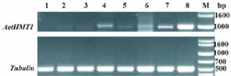
The expression of AetHMT1 in new leaves, old leaves, stems and roots was examined by semi-quantitative RT-PCR using gene-specific HMT1F and HMT1R primers, with the constitutively expressed tubulin gene as a reference (Fig. 3). AetHMT1 transcripts were relatively abundant in all tissues. AetHMT1 expression was dramatically upregulated in all tissues after 72-h treatment with 10 mM Na2SeO4. Interestingly, roots accumulated higher transcript amounts than stems or leaves, which implies that AetHMT1 may play a crucial role in Se uptake in Ae. tauschii.
Discussion
Plants are known for their unique ability to synthesize methionine from homocysteine using HMT (Lyi et al. 2007). Three HMT cDNAs have been identified from Arabidopsis thaliana on the basis of homology to E. coli YagD (Ranocha et al. 2000; Ranocha et al.
2001). The HMT gene encodes cytosolic enzymes that have zinc-binding motifs and share significant protein sequence similarity with SMT from Astragalus bisulcatus, a Se hyper- accumulator (Neuhierl et al. 1999). Although HMT and SMT enzymes may play different roles in plants (Ranocha et al. 2001), both HMT and SMT genes are thought to confer Se tolerance. Because Ae. tauschii is an important genetic resource and the D-genome donor for common wheat, the characterization of the HMT1 protein from Ae. tauschii can indi- rectly contribute to the development of Se-rich wheat. In this study, the complete cDNA of AetHMT1 was isolated from Ae. tauschii for the first time. The cDNA was cloned into a prokaryotic expression vector, pET-30a (+), and expressed in E. coli BL21 (DE3) after induction with IPTG. According to SDS-PAGE, AetHMT1 was successfully expressed in the pET prokaryotic expression system.
Figure 3. Semi-quantitative RT-PCR analysis of AetHMT1 gene expression in new leaves, old leaves, stems and roots after treated by selenium for 72 hours. The As2407 seedlings were cultivated under 0 mM Na2SeO4 con- ditions and 10 mM Na2SeO4 conditions as described in the Materials and methods. Levels of mRNA were analyzed by semi-quantitative RT-PCR using gene specific primers for AetHMT1. And Tubulin was used as internal control. 1, new leaves treated by 0 mM Na2SeO4; 2, new leaves treated by 10 mM Na2SeO4; 3, old leaves treated by 0 mM Na2SeO4; 4, old leaves treated by 10 mM Na2SeO4;5, stems treated by 0 mM Na2SeO4; 6, stems treated by 10 mM Na2SeO4; 7, roots treated by 0 mM Na2SeO4; 8, roots treated by 10 mM Na2SeO4;
M, Marker
Sequence alignment and phylogenetic analysis of the deduced AetHMT1 protein with other HMT and SMT proteins grouped AetHMT1 in the same clade with HMTs, which is separate from SMTs (Fig. S2). The deduced amino acid sequence of AetHMT1 shares high sequence identity with TuHMT1 from T. urartu (Ling et al. 2013), ZmHMT1 from Z. mays (Ranocha et al. 2001) and BoSMT from B. oleracea var. italica (Lyi et al. 2005).
AetHMT1 contains a zinc-binding domain as well as an upstream conserved cysteine residue, characteristics typical of Zn2+ dependent thiol and selenol methyltransferases (Zhu et al. 2007; Lyi et al. 2007; Lyi et al. 2005). The presence of these features suggests that this enzyme requires a zinc cofactor for binding and/or activating the thiol group of homocysteine (Millian and Garrow 1998; Ranocha et al. 2000), as the GGCCR binding motif may be important for methyltransferase activity (Lyi et al. 2005). AetHMT1 has no obvious chloroplast or mitochondrial targeting sequences and appears to be a cytosolic enzyme, which is consistent with previous results (Lyi et al. 2005; Zhu et al. 2007; Lyi et al. 2007). Methylation of selenocysteine or selenomethionine, as well as metabolism in- volving the synthesis of S-methylmethionine, most likely takes place in the cytosol (Bourgis et al. 1999). According to our SDS-PAGE results, the AetHMT1 protein had the same mass as conjectured (Jia et al. 2013). The biochemical identity of purified AetHMT1 protein was confirmed, with its positive HMT enzyme activity in catalyzing the transfor- mation of L-homocysteine measured as 164.516 ± 17.378 nmol min−1 mg−1 protein.
The Se tolerance of E. coli was enhanced by the expression of recombinant AetHMT1.
AetHMT1 conferred better protection against selenite than selenate, as the AetHMT1- transformed cells had a higher ratio of growth on selenite vs. selenate compared with the empty vector. This result is consistent with observations of previous studies (Lyi et al.
2005; Zhu et al. 2007). A precondition of higher Se accumulation ability is tolerance to high Se concentrations in large tissues (Brown and Shrift 1982). Our results demonstrated that AetHMT1 can improve bacterial Se endurance.
AetHMT1 is inferred to play an active role in Se absorption, as AetHMT1 expression was dramatically upregulated in all tissues, especially roots, after treatment with 10 mM Na2SeO4. The expression pattern of AetHMT1 after exposure to selenate is similar to that of BoSMT (Lyi et al. 2007). AetHMT1 expression amount was highest in roots, followed by stems, old leaves and then new leaves. This order may be similar to the pattern of Se delivery in plants. This observation indicates that the effect of Na2SeO4 on AetHMT1 is likely a consequence of the role of this gene in Se metabolism.
In summary, cDNA of the HMT gene from Ae. tauschii, designated as AetHMT1, was isolated for the first time. The AetHMT1 protein was successfully expressed using the pET prokaryotic expression system. The biochemical activity of AetHMT1 was con- firmed, with a positive HMT enzyme activity in catalyzing the transformation of L-homo- cysteine measured as 164.516 ± 17.378 nmol min−1 mg−1 protein. AetHMT1 can improve bacterial Se endurance. Our research suggests the possibility of developing Se-rich bread wheat by selecting new Ae. tauschii AetHMT1 haplotypes with strong Se endurance and Se accumulation ability.
Acknowledgements
This research was funded by National Natural Science Foundation of China (31101140), CAS “Light of West China” Program, the Project from Qinghai Province (2011-Z-716, 2016-HZ-808), the Strategic Priority Research Program of Chinese Academy of Sciences (No. XDA08030106), the Project of State Key Laboratory Breeding Base – Key Labora- tory for Spring Rapeseed Genetic Improvement of Qinghai Province (2017-07) and the Program of Science and Technology Innovation Platform of Qinghai Province (2017-ZJ- Y14).
References
Ari, Ş., Çakır, Ö., Turgut-Kara, N. 2010. Selenium tolerance in Astragalus chrysochlorus: identification of a cDNA fragment encoding a putative Selenocysteine methyltransferase. Acta Physiol. Plant. 32:1085–1092.
Ausubel, F.M., Glazebrook, J., Greenberg, J., Katagiri, F., Mindrinos, M., Yu, G.L. 1993. Analysis of the Arabidopsis Defense Response to Pseudomonas Pathogens. J. Cell Biol. 14:393–403.
Bourgis, F., Roje. S., Nuccio, M.L., Fisher, D.B., Tarczynski, M.C., Li, C.J., Herschbach, C., Rennenberg, H., Pimenta, M.J., Shen, T.L., Gage, D.A., Hanson, A.D. 1999. S-methylmethionin plays a major role in phloem sulfur transport and is synthesized by a novel type of methyltransferase. Plant Cell 11:1485–1497.
Bradford, M.M. 1976. Rapid and Sensitive Method for Quantitation of Microgram Quantities of Protein Utilizing Principle of Protein-Dye Binding. Anal. Biochem. 72:248–254.
Brown, T. A., Shrift, A. 1982. Selenium: Toxicity and Tolerance in Higher Plants. Biol. Rev. 57: 59–84.
Chen, X., Yang, G., Chen, J., Chen, X., Wen, Z., Ge, K. 1980. Studies on the relations of selenium and Keshan disease. Biol. Trace Elem. Res. 2:91–107.
Ebert, R., Jakob, F. 2007. Selenium deficiency as a putative risk factor for osteoporosis. International Congress Series 1297:158–164.
Guo, Z.F., Zhang, Z.B., Xu P., Guo, Y.N. 2013. Analysis of Nutrient Composition of Purple Wheat, Cereal Res.
Commun. 41:293–303.
Hartikainen, H. 2005. Biogeochemistry of selenium and its impact on food chain quality and human health. J.
Trace Elem. Med. Bio. 18:309–318.
Hu, B. L., Huang, D.R., Xiao Y.Q., Fan Y.Y., Chen D.Z., Zhuang J.Y. 2016. Mapping QTLs for Mineral Element Contents in Brown and Milled Rice Using an Oryza sativa ×O. rufpogon Backcross Inbred Line Population. Cereal Res. Commun. 44:57–68.
Jia, J., Zhao, S., Kong, X., Li, Y., Zhao, G., He, W., Appels, R., Pfeifer, M., Tao, Y., Zhang, X., Jing, R., Zhang, C., Ma, Y., Gao, L., Gao, C., Spannagl, M., Mayer, K.F., Li, D., Pan, S., Zheng, F., Hu, Q., Xia, X., Li, J., Liang, Q., Chen, J., Wicker, T., Gou, C., Kuang, H., He, G., Luo, Y., Keller, B., Xia, Q., Lu, P., Wang, J., Zou, H., Zhang, R., Xu, J., Gao, J., Middleton, C., Quan, Z., Liu, G., Wang, J., Yang, H., Liu, X., He, Z., Mao, L., Wang, J. 2013. Aegilops tauschii draft genome sequence reveals a gene repertoire for wheat adap- tation. Nature 496:91–95.
Koutmos, M., Pejchal, R., Bomer, T.M., Matthews, R.G., Smith, J.L., Ludwig, M.L. 2008. Metal active site elasticity linked to activation of homocysteine in methionine synthases. Proc. Natl. Acad. Sci. U.S.A.
105:3286–3291.
Laemmli, U.K. 1970. Cleavage of structural proteins during the assembly of the head of bacteriophage T4.
Nature 227:680–685.
Ling, H.Q., Zhao, S., Liu, D., Wang, J., Sun, H., Zhang, C., Fan, H., Li, D., Dong, L., Tao, Y., Gao, C., Wu, H., Li, Y., Cui, Y., Guo, X., Zheng, S., Wang, B., Yu, K., Liang, Q., Yang, W., Lou, X., Chen, J., Feng, M., Jian, J., Zhang, X., Luo, G., Jiang, Y., Liu, J., Wang, Z., Sha, Y., Zhang, B., Wu, H., Tang, D., Shen, Q., Xue, P., Zou, S., Wang, X., Liu, X., Wang, F., Yang, Y., An, X., Dong, Z., Zhang, K., Zhang, X., Luo, M.C., Dvorak, J., Tong, Y., Wang, J., Yang, H., Li, Z., Wang, D., Zhang, A., Wang, J. 2013. Draft genome of the wheat A-genome progenitor Triticum urartu. Nature 496:87–90.
Lozada-Ramirez, J.D., Martinez-Martinez, I., Garcia-Carmona, F., Sanchez-Ferrer, A. 2008. Cloning, overex- pression, purification, and characterization of S-adenosylhomocysteine hydrolase from Corynebacterium efficiens YS-314. Biotechnol. Progr. 24:120–127.
Lyi, S.M., Heller, L.I., Rutzke, M., Welch, R.M., Kochian, L.V., Li, L. 2005. Molecular and biochemical char- acterization of the selenocysteine Se-methyltransferase gene and Se-methylselenocysteine synthesis in broccoli. Plant Physiol. 138:409–420.
Lyi, S.M., Zhou, X., Kochian, L.V., Li, L. 2007. Biochemical and molecular characterization of the homocyst- eine S-methyltransferase from broccoli (Brassica oleracea var. italica). Phytochem. 68:1112–1119.
Lyons, G.H., Judson, G.J., Ortiz-Monasterio, I., Genc, Y., Stangoulis, J.C., Graham, R.D. 2005. Selenium in Australia: selenium status and biofortification of wheat for better health. J. Trace Elem. Med. Bio. 19:75–82.
Millian, N.S., Garrow, T.A. 1998. Human betaine-homocysteine methyltransferase is a zinc metalloenzyme.
Arch. Biochem. Biophys. 356:93–98.
Neuhierl, B., Böck, A. 1996. On the mechanism of selenium tolerance in selenium-accumulating plants.
Purification and characterization of a specific selenocysteine methyltransferase from cultured cells of Astragalus bisculatus. Eur. J. Biochem. 239:235–238.
Neuhierl, B., Thanbichler, M., Lottspeich, F., Böck, A. 1999. A family of S-methylmethionine-dependent thiol/
selenol methyltransferases – Role in selenium tolerance and evolutionary relation. J. Biolog. Chem.
274:5407–5414.
Peariso, K., Goulding, C.W., Huang, S., Matthews, R.G., Penner-Hahn, J.E. 1998. Characterization of the zinc binding site in methionine synthase enzymes of Escherichia coli: The role of zinc in the methylation of homocysteine. J. Am. Chem. Soc. 120:8410–8416.
Liu, P., Zhou, J.P., Xu, X.L., Hao, Q.Q., Wen, X.Y., Wang, L.,Tian, Y.P. 2014. Activity assay and preservation of S-homocysteine methyltransferas. International Journal of Laboratory Medicine 35:1526–1528.
Peng, A., and Yang, C.L. 1991. Examination of the roles of selenium in the Kaschin-Beck disease. Biol. Trace Elem. Res. 28:1–9.
Ranocha, P., Bourgis, F., Ziemak, M.J., Rhodes, D., Gage, D.A., Hanson, A.D. 2000. Characterization and functional expression of cDNAs encoding methionine-sensitive and insensitive homocysteine S-methyltransferases from Arabidopsis. J. Biolog. Chem. 275:15962–15968.
Ranocha, P., McNeil, S.D., Ziemak, M.J., Li, C., Tarczynski, M.C., Hanson, A.D. 2001. The S-methylmethionine cycle in angiosperms: ubiquity, antiquity and activity. Plant J. 25:575–584.
Sambrook, J., Fritsch, E.F., Maniatis, T. 1989. Molecular cloning: A laboratory manual: 2nd ed. Immunology 49:895–909.
Schiavon, M., Pilon-Smits, E.A. 2017. Selenium Biofortification and Phytoremediation Phytotechnologies:
A Review. J. Environ. Qual. 46:10–19.
Sors, T.G., Ellis, D.R., Salt, D.E. 2005. Selenium uptake, translocation, assimilation and metabolic fate in plants. Photosynth. Res. 86:373–389.
Sors, T.G., Martin, C.P., Salt, D.E. 2009. Characterization of selenocysteine methyltransferases from Astragalus species with contrasting selenium accumulation capacity. Plant J. 59:110–122.
Szira, F., Monostori, I., Galiba,G., Rakszegi, M., Bálint, A.F. 2014. Micronutrient Contents and Nutritional Values of Commercial Wheat Flours and Flours of Field-grown Wheat Varieties – A Survey in Hungary.
Cereal Res. Commun. 42:293–302.
Thomson, C.D. 2004. Assessment of requirements for selenium and adequacy of selenium status: a review. Eur.
J. Clin. Nutr. 58:391–402.
White, P.J. 2016. Selenium accumulation by plants. Ann. Bot. 117:217–235.
White, P.J., Brown, P.H. 2010. Plant nutrition for sustainable development and global health. Ann. Bot.
105:1073–1080.
Zhao, D.Y., Sun, F.L., Zhang, B., Zhang, Z.Q., Yin, L.Q. 2015. Systematic Comparisons of Orthologous Selenocysteine Methyltransferase and Homocysteine Methyltransferase Genes from Seven Monocots Species. Notulae Scientia Biologicae 7:210–216.
Zhu, L., Jiang, C.J., Deng, W.W., Gao, X., Wang, R.J., Wan, X.C. 2007. Cloning and expression of selenocyst- eine methyltransferase cDNA from Camellia sinensis. Act. Physiol. Plant. 30:167–174.
Electronic Supplementary Material (ESM)
Electronic Supplementary Material (ESM) associated with this article can be found at the website of CRC at http://www.akademiai.com/content/120427/
Electronic Supplementary Table S1. DNA primers used for amplification of cloning, expressing protein in E. coli and semi-quantitative RT-PCR for AetHMT1
Electronic Supplementary Figure S1. Sequence alignment of the deduced amino acids for AetHMT1 (EMT27714.1) with those of related proteins. Conserved amino acids residues are shaded in black. Dash depict gaps were added for optimum alignment. The arrowhead indicated the third conserved Cys residue. The left- right arrow shows the possible zinc-binding motif that is typical for the structural family of Zn2+-dependent thiol and selenol methyltransferases is underlined. TuHMT1, Triticum urartu HMT1(EMS47619.1); ZmHMT1, Zea mays HMT1(AAG22537.1); AtHMT-1, Arabidopsis thaliana HMT1(NP_189219.1); GaHMT1, Gossypium arboretum HMT1(KHG14282.1); BoSMT, Brassica oleracea var. italica SMT(AAX20123.1); AbSMT, A. bisulcatus SMT (CAA10368); AtHMT-1, Arabidopsis thaliana HMT-1(AAF23821.1); CsSMT, Camellia
sinensis SMT(ABF47292.1)
Electronic Supplementary Figure S2. Phylogenetic analysis of AetHMT1 and the aligned protein sequences.
The analysis was conducted using the maximum likelihood method and a single unrooted phylogram tree was identified. Numbers presented at each branch point represent bootstrap values from 1000 replicates. Branch
lengths on the tree are proportional to the amount of change as shown by the bar