Mechanisms of
Mitochondrial Ion Transport
Cyril L. Moore
I. Introduction 5 7 3
II. Monovalent Cation Transport 574
A. Noninduced Transport 5 7 4
B. Mitochondrial H20 5 7 9
C. Induced Transport ( M+) 5 8 2
III. Divalent Cation Transport 5 9 6
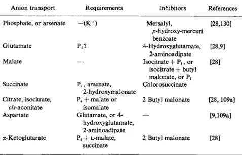
IV. Anion Transport 605 A. Inorganic Anions 605
V. Nucleotide Transport 6 1 0
VI. Nonelectrolytes 6 1 3
VII. Energetics of Ion Transport 613 VIII. The Mechanisms of Ion Transport 617
References 622
I. INTRODUCTION
Over the years the mitochondrion has been isolated, fed electrons, filled with water and ions, tapped of its energy, punctured with elec- trodes, inhibited, activated, fragmented (by sonication, by enzymatic digestion or detergency) separated into outer and inner membranes, and yet it has refused to truly divulge its innermost secret, i.e., how it transfers energy from electron transport (substrate oxidation) to ATP synthesis. This is not to say that progress has not been made in this dynamic field, it merely indicates the inherent difficulty with which it is beset.
One of the areas in which some progress has been made is that of ion transport and the transduction of energy from substrate oxidation or ATP to support this phenomenon.
Here we review the subject of ion transport by presenting as much up-to-date information as possible with as little partiality as possible.
573
Active transport can be defined as a process in which ions are trans
located contra the existing electrochemical gradient and is thus reliant upon metabolic energy. In order to maintain electrical neutrality (if this is indeed physiologically desirable), an ion of opposite charge could accompany the migrating ion, or a counter ion of similar charge could move in the opposite direction to the migrating ion, or both of these might operate to satisfy electrical neutrality.
The data of several investigators [7,11,40,121] indicate that mito
chondria have the capacity to accumulate monovalent cations in an energy-dependent reaction. It was also noted that water was accumu
lated during K+ uptake, and that there was a requirement for specific anions [11].
Slater and Cleland [120] observed the binding of C a2 + to heart mitochondria and considered it to be an energy-independent process.
Subsequent studies by several investigators [26,72,112,118] opened the Pandora's Box of mitochondrial ion transport.
The problem is discussed as it has evolved since then.
II. M O N O V A L E NT CATION TRANSPORT
A. Noninduced Transport
Stanbury and Mudge [122] were among the first to relate aerobic phosphorylation to the accumulation of K+ by retinal and kidney slices. Prompted by the findings in Lardy's laboratory [105] that mitochondria contained substantial amounts of K+, they examined rat liver mitochondria and reported:
(a) A stable fraction of mitochondrial K+ and an easily extractable one
(b) K+ accumulation at the expense of oxidizable substrate and oxygen
(c) A DNP-induced discharge of K+
(d) Significant decreases in intramitochondrial K+ caused by low concentrations of inorganic phosphate, as observed by others.
Their experiments showed that mitochondria had the capacity to accumulate K+ in a reaction requiring an oxidizable substrate and oxygen. They also showed that DNP affected mitochondrial K+ as a function of the DNP concentration as shown in Fig. 1. At concentra
tions below 10"5 Μ most of the K+ was discharged from the mito
chondria, leaving behind a small stable fraction. At 10"3 Μ DNP the
I ι ι ι ι I
6 5 4 3
- LOG Μ [DNP]
FIG. 1. The effect of D N P on mitochondrial K+. The ratio of specific activity of K+
inside (Kt) to K+ outside ( K0) . The mitochondria is on the left y axis. The dashed line represents the internal K+ ( K , ) and follows the right y axis. Note that at approximately 1 0 "3 Μ DNP, the Kt remains stable. Modified from the data of Stanbury and Mudge [122].
rate of exchange of K+ was increased without further loss of K+. These findings, shown in Table I, were corroborated by those of Spector [121]. Slater [40] showed that, at 23° in 0.25 Μ sucrose, rat liver mitochondria lost 95% of their K+ within 4 hours. There was, however, no significant loss of N a+. Gamble also found that DNP (5 χ 10"5 M) and Pf (3 χ 10"3 Μ) potentiated this loss even in the presence of 10 mM KC1. The Pf promoted loss of K+ was accompanied by mitochondrial swelling. The explanation for this swelling was not given but could be related to the phosphate-induced swelling reported more recently by Azzoni [8]. Nonetheless, Bartley and Amoore [10]
demonstrated that K+ accumulation from sucrose solutions was accompanied by swelling. It is possible that the K+ loss during the Ρ rinduced swelling observed by Gamble was intimately coupled to the uptake of divalent cations such as C a2 + known to be lost from mitochondria during periods of anaerobiosis [83,27a].
This phosphate-induced swelling apparently involves parameters other than inhibition or reversal of K+ uptake. More recently the studies of Azziand Azzone [9] have indicated a metabolism-dependent shrinkage
TABLE I
CONCENTRATION" OF K+ IN RAT LIVER MITOCHONDRIA AFTER 2 0 MINUTES AT 3 7 ° IN 0 . 2 5 Μ SUCROSE
Anion, 0.01 Μ D N P labile K+ D N P stable K+
Chloride 2 1 5 0 2 1 5
Sulfate 1 4 0 5 1 3 5
Phosphate 7 5 0 7 5
Glutamate* 6 8 5 5 9 5 9 0
Citrate 8 5 0 5 5 5 2 9 5
Bicarbonate 5 8 5 5 4 5 4 0
a Micromoles per gram of protein.
b Adapted from data of Spector [121].
of mitochondria induced by succinate in the presence of valinomycin and a potentiation in the presence of 50 mM KC1. Lehninger [67,68]
has shown that ATP promotes mitochondrial shrinkage in salt-free solutions. The presence of 25 mM tris HC1 in the medium could be contributory to this shrinkage especially in view of the more recent data of Gear and Lehninger [43] on the H+ ion extrusion from mito
chondria in the absence of energy.
If water is accumulated in the absence of energy [25,92], then the prob
lem involves the integrity of the osmotic barrier of the mitochondrion. It was shown that ATP and sulfhydryl compounds prevented or reversed the energy-deficient swelling of mitochondria [93]. Increasing the per
meability of mitochondria by valinomycin in the absence of energy [9] exaggerates the swelling far beyond that which would be expected in the valinomycin-induced swelling which accompanies K+ uptake.
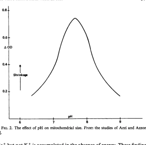
Thus, with an energy source, two events occur: (1) shrinkage due to the alteration of the membrane and (2) swelling due to the accumulation of cations. The magnitude of the overall expression is most likely a function of the cation concentration [101a] and the pH of the medium as judged from the studies of Azzi and Azzone [9] and shown in Fig. 2.
It is fair to agree with statements of the complexity of the process of swelling and shrinking cycles, since it involves the type of cation, the pH of the system, the age of the preparation, the substrate, and indeed the availability of energy as well as the type of anion and other cations which are present in the medium. In regard to the anionic species, heart mitochondria swell spontaneously in a medium of isotonic acetate salts of K+ and N a+, but not in chloride solutions. This swelling has been shown by Brierley and co-workers [16] to be an index of an energy requiring M+ uptake as shown in Table II. According to these authors,
0 . 4 4 -
0 . 2 1 0 . 6 F
Shrinkage
6 7 8 9
FIG. 2 . The effect of pH on mitochondrial size. From the studies of Azzi and Azzone
N a+ but not K+ is accumulated in the absence of energy. These findings are contrary to those of several investigators [10,28,40,121], working with liver mitochondria, but agree with those of Lynn and Brown [73]. The difficulty here is that one has to consider the role of the anion (acetate) in this system; when this is not done, the events may take on different characteristics and dimensions. It is becoming increasingly obvious, however, that monovalent cations may be accumulated via mechanisms which are far from unitarian.
One interesting thought which we may interject here is that the low intramitochondrial Κ f: N a+ ratio of approximately 20-30 observed by Brierley and co-workers [16] could be explained on the basis that N a+ transport is outwardly directed in an energy-requiring reaction, and in the absence of such energy it is accumulated in an osmotic or cation- specific reaction, which is a function of the chemistry of the membrane.
The opposite vectorial relationship could be postulated for K+. The extent of these reactions again could be a function of the source and age of the mitochondrial preparation.
Thus, according to the studies of Christie and co-workers [32], mitochondria prepared from sheep kidney cortex and stored at 0° lost
TABLE I I
SWELLING RATES OF HEART MITOCHONDRIA SUSPENDED IN ISOTONIC SALT SOLUTIONS"·*
Endogenous respiration dzrotenone
± — + Succinate Acetate salts
Li 0.05 0.20 0.16
N a+ 0.12 0.29 0.38
K+ 0.00 0.07 0.20
M g2 + 0.01 0.03 0.08
N H4 + 0.80 0.80 0.80
Choline 0.05 0.09 0.19
Chloride salts
N a+ 0.00 0.00 0.00
N H4 + 0.00 0.00 0.00
a System: 1.25 mg mitochondria in 3 ml of the listed salt solu
tions: 120 m M M + , 80 m M M2 + pH 6.8-7.2; 1.25 imxmoles of rotenone used.
b From Brierley et al. [16].
their ability to maintain K+ and N a+ against adverse concentration gradients. C a2 + ions were found to promote the release of K+ at 0°
with no affect on N a+. At 25°, N a+ uptake was increased in the presence of C a2 + while M g2 + promoted its release. These findings have been confirmed by Lehninger and co-workers [69].
It would appear that, if energy is preferentially utilized for Ca2 + uptake, then it would be derived from the processes of N a+ expulsion and K+ uptake, and in a way this is an expression of the findings of Christie and co-workers [32]. M g2 + ions which are involved in the coupling of electron transport to ATP synthesis, as documented for brain mitochondria by Moore and Jobsis [86], and also in reactions utilizing ATP, promote release of N a+. This would be expected if the energetics of the mitochondria are well controlled. Other experiments demonstrating the almost total loss of K+ and 7% loss of N a+ at 37°
in 0.25 Μ sucrose [40] would lend credence to the preceding theory.
This 7% loss of N a+ was prevented by addition of the sodium salt of glutamic acid, with no net sodium uptake. ATP and MgCl2 reversed the K+ depletion and promoted its accumulation against a concentration
gradient. The K+ accumulation, a function of the anion present, was also found to be accompanied by little or no change in total water.
The reaction of mercury salts (HgCl2) in promoting K+ release with little eifect on N a+ [40,115] also speaks for the vectorial and energy requirements of K+ and N a+ transport by mitochondria. As will be seen later, this theorem may be true only in the absence of ionophoresis induced by cationophiles.
B. Mitochondrial H20
Most investigators in the field of mitochondrial ion transport have grown used to the idea of equating mitochondrial swelling with the accompaniment of ion accumulation by osmotic changes. Whether or not the parallelism always exists is quite dubious from the discussions above, as well as from the fact that swelling occurs during the energy- requiring histone-induced cation loss from mitochondria [52]. In con
sidering any movement of water, however, the dynamism of this pheno
menon must be carefully evaluated. Accordingly, the studies of several laboratories [33,75,114,126-128] indicate that mitochondria behave to a high degree like osmometers. This osmometric characteristic is not related to the total mitochondrial water, mostly to the sucrose-inaccess
ible space and to a lesser degree to the sucrose accessible space [30].
Rapid changes in mitochondrial volume brought about by varied osmolarity of the suspending medium are reversible and involve 60%
of the total mitochondrial water. This leaves approximately 40%
osmotically dead space [128]. This is not to say that all mitochondria have the same water content under identical conditions, as was dramatic
ally demonstrated by Amoore and Bartley [7]. They found that light and heavy fractions of mitochondrial preparations differed in their water content, the lighter fraction containing more water than the heavier.
The methods used for measuring mitochondrial water (swelling) include (a) wet minus dry weight [13,32,74]; (b) light scattering (90°) [27b] and/or turbidimetric methods (% transmission at 0°) [128] at 546 or 520 πιμ; (c) method (a) has been supplemented by the 1 4C-carb- oxypolyglucose method to account for extra mitochondrial water;
(d) the mitocrit method which measures packed wet volume.
Mitochondrial swelling has been found to occur under a variety of conditions; for example, in 0.15 Μ alkali metal acetate salts, in the presence of histones, of cationophiles, of aging, or in the absence of
energy. The reversal of some of these swelling phases is also of signifi- cance.
Blondin and Green [13] described the swelling in alkali metal acetate salts and showed that KCN substantially decreased but did not eliminate the swelling. The degree of swelling in acetate salts is appar- ently a function of the size of the hydrated alkali metal ion as shown in Table III. The ratio 8 : 1 : 2 of accumulation of ions R b+ : N a+ : K+
TABLE I I I
HYDRATED ALKALI METAL IONS"
A L i+ 3.07 N a+ 2.25 K+ 1.63 R b+ 1.57 C s+ 1.56
a From Bockris (1949).
is not a reflection of the degree of swelling, since Rb+-induced swelling was smaller in magnitude than that observed with N a+ or K+. Instead it must be related to the possible extent of alkali metal hydration.
Swelling supported by electron flow is also observed in the presence of other cations (as well as anions such as phosphate), in a manner apparently competitive with ADP. Packer and co-workers [96] have observed that oligomycin inhibits the shrinkage caused by ADP. Harris and Van Dam [48] have pointed out that, in the presence of rotenone, mitochondrial water increased with increasing osmolarity of the sus- pending medium. Since the sucrose-inaccessible space decreased, the actual volume increase must be related to the sucrose-accessible space.
This overall swelling is, to say the least, complex. One would like to suggest that the shrinkage of the sucrose-inaccessible space is osmotic and that the swelling of the accessible space involves sequestering of sucrose plus the water from both the inacessible space and the extra- mitochondrial compartment in an attempt to create from a dynamic system a stable equilibrium. Oscillations are therefore to be expected and the time of assay is critical.
Several investigators [30,48,64-66] have shown that ATP leads to a loss of water from the sucrose-accessible space. Since this is not the strictly osmotically active space, it could be presumed that conforma- tional changes possibly involved in physicomechanical work (e.g.,
syneresis) are occurring. More recently the role of ATP in- causing shrinkage of mitochondria has become demonstrable as a change in conformation of the inner membrane, since the magnitude of such a change is enormous enough to be visualized via electron microscopy [46]. Caplan and Greenawalt [22] have also shown that ATP causes shrinkage of crude ghost fractions obtained by water lysis of rat liver mitochondria. Thus it becomes difficult not to agree with Lehninger, [64-67] as well as Blondin and Green [13], that the ATP-dependent functions of water uptake and extrusion belie the possibility of strictly osmometric function for mitochondria. The osmometric nature of the inner membrane compartment (i.e., sucrose-inaccessible space) is impure, and the lack of osmometric behavior of the outer compartment is clouded by some of the experimental observations. The true picture may thus be described as follows. The sucrose-available space is under very little osmotic control, and any control of volume change may be related to a functional or alterable outer membrane. The sucrose-inac- cessible space is under a high degree of osmotic control; nonetheless, there is within this compartment controllable nonosmotic swelling.
The control of this type of swelling resides in the energy-dependent functionality of the inner membrane. This dependence is sensitive to aging and could be lost during preparative procedures as foreseen in the light and heavy fractions of Amoore and Bartley [7]; the inner mitochondrial compartment undergoes osmotic swelling and shrinking different from that observed during active ion uptake and release [13]. The presence of nondiffusible large molecules within the matrix space leads to the imbibition of water from hypotonic media, and thus the osmotic-type volume changes are of the Donnan type.
ATP reversible swelling may also be induced by surfactive agents such as fatty acids or CC14 [21,44,47,67,75]. Indeed, electron micro- graphs of muscle from different hyperactive muscle diseases show mitochondrial abnormalities and the presence of fatty deposits in their vicinity [Wisniewski and Raine, personal communication].
The studies cited indicate that mitochondria upon aging at various temperatures lose their ability to accumulate K+ or maintain high intramitochondrial K+. It was also noted that ATP and MgCl2 could protect against the loss due to aging. The suggestions that mitochondria contain (a) an ion complex carrier for K+, which could be part of the ATP-synthesizing apparatus [122] and (b) carrier molecules capable of maintaining an exchange diffusion across their membrane [11] find support in the recent findings by several groups of investigators inspired by the discovery of the philicity of valinomycin for K+, Cs+, R b+ and the ionophorous property of this depsipeptide in inducing the transport of monovalent cations into mitochondria [88].
C. Induced Transport (M + )
The apparent uncoupling of oxidative phosphorylation by depsipep- tide antibiotics (e.g., valinomycin, dianemycin) was first observed by Lardy and co-workers (1961), and since then this laboratory has done a magnificent job of documenting several antibiotics not only with uncoupling activity, but also with cationophilic and ionophoretic properties. The induction of monovalent cation transport by mito
chondria is a phenomenon first described by Moore and Pressman [88]; some of these studies are described in Fig. 3.
:t
0.1 mM
1 MIN
FIG. 3. The effect of phosphate (permeant anion) on respiration and K+ uptake during valinomycin induction. The uptake of K+ and release of Η + in the presence of valinomycin.
The solid lines indicate the absence of phosphate, and the dashed lines its presence. Note also only slight respiratory stimulation in the absence of phosphate. Open circles represent H+; open triangles, K+. (AA) 2/xg antimycin A; (ATP) 5 mM tris ATP. The medium consisted of 220 mM sucrose, 20 mM choline chloride, 2 mM tris HC1 (pH 7.4) ± 5 mM tris phosphate; 2.5 mg mitochondrial protein per milliliter was used.
While isolated mitochondria were found to accumulate only small amounts of monovalent cations, addition of valinomycin to a suspen
sion of mitochondria, in a 0.25 Μ sucrose medium containing oxidiz
able substrates, oxygen, K+, and a permeant anion, resulted in a stimulation of oxygen uptake.
Of special interest is the question of the mechanism of this transport.
In order to examine this adequately, one should have some general knowledge of the chemistry of the cationophiles, which are separable into the following categories.
1. TYPES OF CATIONOPHILIC COMPOUNDS
Type 1. Nonneutral compounds
(a) Noncyclic acidic—Nigericin (Fig. 4), Dianemycin, the monesins (Fig. 5)
(b) Acidic cyclic peptide alamethicin (1 free COOH) (c) Basic polyhydroxy compound—monazomycin (1 dis
sociable amino group)
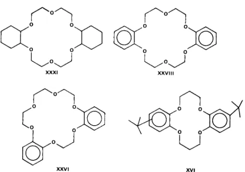
Type 2. Macrotetralides—nactin series (Fig. 6) Type 3. Cyclic crown polyethers of Pedersen (Fig. 7)
Type 4. Linear Oligopeptides—gramicidins (other than the cyclic gramicidin S)
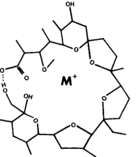
Type 5. Cyclic depsipeptides—valinomycin, enniatin (Figs. 8, 9)
FIG. 4. Monensin-M+ complex according FIG. 5. Nigericin-M+ complex according to the model of Sleinrauf et al. [120a]. to the model of Sleinrauf et al. [120a].
FIG. 6. (a) Nonactin (Pauling model), (b) Nonactin-K+ complex viewed down through a crystal axis to demonstrate the enclosure of th K+ within the sphere presented as an outline, and with models. Open circles represent C; closed circles 5 oxygen, (c) Nonactin-K+ comple viewed down the b-crystal axis to demonstrate the hugging of the K+ in both model and outline forms. Data according to Kilbourn et al. [54]
X X VI xvi
FIG. 7. Synthetic polyethers of Pedersen. XXXI: dicyclohexyl-18-crown 6; (XXVIII):
dibenzo-18-crown-6; (XXVI): asymetric dibenzo-18-crown 6; XVI: di-terf-butylbenzo- 14-crown-4.
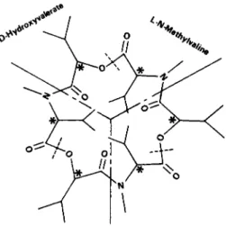
D-Hydroxy-
FIG. 8. Valinomycin.
FIG. 9 . Enniatin.
2. BEHAVIOR OF CATIONOPHILE
a. Philicity. The incomplete structures of nigericin and monensin are given in Figs. 4 and 5. These compounds are quite similar and both have high philicity for monovalent cations. X-ray crystallographic data are indicative of a replacement of the shell of hydration of the cation M+ by the ring oxygens of monensin and nigericin as depicted in Figs.
4 and 5. This is not meant to indicate complete understanding of the nature of the complexes but instead to rationalize their possible nature.
As with Type 1(a) cationophiles, the possibility of displacement of the hydration shell of the cation can be visualized if Pauling models for enniatin B - K+ complex of the Type 5 cationophile are constructed.
If indeed the hydration shell is removed, the strain necessary for conformation of the cationophile to the metal ion would predict its ion specificity. This is reflected in the greater K+ to N a+ selectivity displayed by nonactin (Fig. 6a). The K+ ion complexes of the macrotetralide actins (Type 2) are exemplified by the nonactin-K+ (N-K+) complex (Figs. 6b and c) where the K+ is apparently trapped within the shell of the nonactin molecule. There is no specific interaction between the positively charged N - K+ complex and anions such as NCS" which according to the crystallographic studies of Kilbourn and co-workers (1967) occupy spherical cavities between the complexed nonactin molecules.
The K+ forms a symmetrical coordination to the 4 carbonyl oxygens of the four subunits of nonactin. This according to the studies of
Prestegard and Chan (1969) gives rise to salt-induced proton-magnetic shifts. Resonance shifts are greatest for the protons in close (geometric) proximity to the centrally directed carbonyl groups. K+ ions are bound to the nonactin without water of hydration. If the ions are hydrated before entering the central aperture of the nonactin molecule, a confor
mational change occurs with the loss of water, and the resultant coord
ination of the cation. The members of the nactin family of cationophiles with their 40-membered rings differ by substitution of methyl groups.
In the case of trinactin (i.e., nonactin substituted with three methyl side groups), the philicity for K+ decreases substantially, while that for N a+ is slightly altered. The spacing of the molecule becomes less ac
comodating to K+ and its binding characteristics now approach those of N a+. The substitutions as stated above alter the ion selectivity, but do not abolish it. The ring size apparently does not hinder the col
lapse of these molecules around the cations, and thus the alteration and not the abolition of ion selectivity is observed.
The synthetic cyclic polyethers (Type 3, Fig. 7) form complexes with alkaline earth metals, and in some cases with the following groups of the periodic table: 1A, IB, most IIA, and some IIB, a few IIIA and IIIB and IVB. Pedersen [97] has shown that Type 3 cationophiles, the polyethers, have some degree of selectivity among the group I cations.
Dibenzo-14-crown-4 cyclic ether (compound XVI) with 14 ring atoms including 4 ether oxygens, has a high philicity for Li+ and N a+ but very little for the other members of their group. Dicyclohexyl-21- crown-7 (compound XXXIV) has a high affinity for K+ and R b+.
Pedersen has also shown that the mono- and dibenzo-12-crown-4 ethers (compounds VI and XXXIV) with pore sizes large enough to accomodate Li+ and N a+, the two largest hydrates (cf Table III) do not, however, complex these ions because of lack of coplanarity of the ether oxygens. Compound XXVIII the symmetrical dibenzo-18-crown-6 is quite active in complexing cations, whereas the asymmetric dibenzo- 18-crown-6 is highly unreactive. The lack of activity of some of the polyethers could be blamed on steric hindrance and/or rigidity of the molecules. In a general manner it may be stated that, as the ring size of the cyclic polyethers increases, the philicity for monovalent and even polyvalent cations decreases.
In the case of Type 5 cationophiles (Figs. 8, 9) (valinomycin and its analogs of varying ring sizes), the complexing of K+ by enniatinB, would suggest that this size of ring (18 members) is adequate for Κ+binding;
however, the high degree of philicity for M+ might be responsible for the decreased ionophoretic properties of the enniatin (as well as the 18 crown ethers). The 36-membered ring of valinomycin must undergo
a conformational change to accommodate K+, and observations are indicative of a change in conformation of valinomycin in the presence of K+ [89a]. The K+ philicity present in valinomycin is lost in the synthetic valinomycin analogs with 24 or 48 atoms in the ring [116].
In both cases it would appear that conformational changes become unwieldy. In the case of the 48-membered ring there is too much freedom of motion to contain the M+ firmly, and in the 24-membered ring there is too much restriction to allow the necessary conformational change.
The enantiomorphs of valinomycin, like the parent compound, are excellent cationophiles [116].
The phenomenon of cationophilicity is multivariable; the variables include (a) ring size; (b) atomic and functional group composition of the ring; (c) steric effects of functional groups; (d) the ability to form a ring if none exists; and (e) the rigidity or flexibility of the molecule and its ability to undergo the proper conformational change to accommo
date the cation and give the resultant ion selectivity.
These variables are also important to the ionophoretic (cation transport) properties of the cationophiles.
The linear gramicidins have the advantage of chemical adequacy.
They possess the correct functional groups and, structurally, are cap
able of wrapping themselves around the cations. Thus they show a low degree of ion selectivity and will bind all of the alkali metal ions with a high degree of philicity.
b. Ionophoresis. Ionophoresis may be defined for mitochondrial systems as the ability of cationophiles to induce the uptake or release (transport) of cations through their direct interaction with the cation and mitochondria. This transport is exemplified in Figs. 3, 10, and 11 where valinomycin and gramicidin, respectively, interact with rat liver or brain mitochondria, and promote the accumulation of N a+ and/or K+ [83]. In these figures the exchange of Η+ for Μ+ as well as the increase in 02 uptake during the accumulation are also presented.
In general, most cationophiles have ionophoretic properties, char
acteristic of their structure. It would have been comforting to speculate that the 18-membered ring size of enniatin Β as well as of the crown 18 cyclic polyethers might be the exact fit for cation binding. However, in valinomycin we find a 36-membered ring, with a high degree of philicity and greater ionophoretic potency than the 18-membered ring compounds. This lower ionophoric behavior could be due to just such a tight fit without the requirement for conformational changebility.
Yet it must be pointed out that the binding constants of the cationo- phile-cation complex parallel (more often than not) ionophoretic capabilities.
ο
2MIN
I 1 [ 02 = 0 ] FIG. 10. Energy-linked gramicidin-induced ion transport in rat liver mitochondria.
Note uptake of both N a+ and K+ (downward deflection), release of H+ (upward deflec
tion), and increased rate of 02 uptake upon addition of gramicidin. Antimycin inhibition of respiration, and reversal of ion movements are counteracted by energy derived from ascorbate (ASC)-TMPD oxidation, and by ATP in the CN"-inhibited system. The system consisted of 220 mM sucrose, 15 mM choline chloride, 5 mM tris HC1, 10 mM each of glutamate and malate. 16 mg mitochondria were used in a total volume of 2.5 ml.
The chamber is fitted with a Clark oxygen electrode, Beckman cation, and N a+ and H+
electrodes. The traces are identified by the labels in the figure.
Although this relationship has been found to be true for valinomycin and for enniatin A and B, enniatin C does not conform to this simple criterion. Enniatin C has three isobutyl side groups in place of the secondary butyl and isopropyl groups of enniatins A and Β respectively.
This substitution does not allow the enniatin C-K+ complex to undergo in alcohol a conformation change different from that observed in the absence of K+. On the other hand, enniatin A and Β undergo a con
formational change in the presence of K+ and are ionophoretic, where
as enniatin C is practically inactive.
This ability to undergo conformational changes in a lipophilic environment could be projected to membrane systems, where, in the lipophilic milieu, valinomycin and enniatins A and B, having formed strong clathrate-type complexes with the cations, now migrate to a polar region of the membrane. A conformational change in the ionic
DINACTIN X X XI
Τ \
50 μΜ \
ν X X V I II
\
^\ X X V I II
/ \ /
D I N A C T IN \ /
\y
Ϊ
1.0 mM
1
(a)
1 1 MIN |
— o2
Ϊ
1.0 mM
1
(a) (b)
FIG. 11. (a) Dinactin-induced K+ uptake and stimulation of respiration, and the reversal bydibenzo-18-crown6(XXVIII). (b) Reversal of dicyclohexyl-18-crown-6 (XXXI)- induced K+ uptake and inhibition of stimulated respiration by XXVIII. Dinactin on the other hand increased both respiration and K+ uptake.
and dipolar region of the clathrate coupled with the hydrophobic nature of the cationophile and the ability of the hydrophobic side chains to undergo conformational change all contribute to the release and capture of the cations.
In this case the cationophile could remain in the hydrophobic regions of the membrane, and in the absence of cation it could then either move back to the outer surface of the mitochondria or, if you will, be anchored at some site available to both hydrophilic and hydrophophobic regions of the membrane.
If, as several investigators have shown, cations can bind to mito
chondria in a surface active fashion, then it would not be desirable for the cationophile to leave the hydrophobic regions of the membrane where its solubility is greatest, since this would not facilitate optimum ion transport. With enniatin C, the lack of significant conformational change (as described for the alcohol system) would promote retention
of the cation with a high degree of philicity, low turnover, and low ionophoresis.
The side chains of the crown ethers (Type 3), like those of Type 5 cationophiles (the enniatins), are determinants in their ionophoretic activity. The difference in behavior of the cyclic polyethers is clear from the ability of dieyclohexyl-18-erown-6 (XXXI) to induce K+ accumulations by mitochondria and the absence of this property in its aromatic counterpart dibenzo-18-crown-6 (XXVIII), Fig. 11. Compound XXVIII causes a reversal of mitochondrial K+ uptake induced by din- actin (Figs. 11, 12) or valinomycin [60]. Alone, however, XXVIII shows no effect on K+ movement, 02 uptake, or volume changes in intact respiring mitochondria.
The difference in activity must clearly lie in the aromatic side groups
FIG. 12. The effect of Nigericin or dibenzo-18-crown-6 (XXVIII) on induced K+
uptake by rat liver mitochondria. Valinomycin induced K+ uptake and respiratory stimu- lation (solid line), and the reversal of K+ uptake, and inhibition of respiration by nigericin or XXVIII-dashed lines. Modified from Graven et al. [44] and Pressman [103].
which display structural planarity and rigidity, while the dicyclohexyl groups can undergo conformational changes which lead to a less rigid structure and thus allow release and capture of M+.
Nigericin and monensin (Type la cationophiles) contain free carboxyl groups which appear to be important both in binding and in transport of M+. As the H+ ion concentration increases, the carboxyl group becomes more undissociated. Cyclization through very strong hydrogen bonding between the carboxyl group and the two hydroxy groups of the terminal six-membered ring could occur [6] and lead to entrapment of M+. The dissociability of the carboxyl hydrogen would therefore allow a K+- H+ exchange with increasing pH.
The carboxyl group of alamethicin, a Type lb cationophile, is not directly involved in cation binding. However, studies are currently being carried out to determine the effect of pH on both its philicity and its ionophoretic properties.
Nigericin apparently promotes K+ release from mitochondria [60]
and its uptake by chloroplasts [95]. This is characteristic of Type 1(a)
6.2 mg Mw Protein 6.2 mg Mw Protein
I 1 MIN I
FIG. 13. Effect of nigericin (Nig) on respiration, substrate utilization, and respiratory control in rat liver mitochondria. Left trace shows inhibition of respiration by nigericin in the presence of 2.5 mM glutamate plus malate (G + M) and 2.5 or 20 mM K+ in 250 mM sucrose, 5m Μ tris. phosphate and 5 mM tris HCI (pH 7.4). Right trace shows slight inhib
ition by nigericin (Nig) in 15 m M G + M, in the absence of KC1, and its release by 2.5 or 20 mMK+.
cationophiles (Figs. 4,5], which have also been demonstrated to reverse the valinomycin-induced uptake of K+ [60,103]. This is exemplified with nigericin in Fig. 12 and in the unpublished observations of Moore, Strasberg, and Kovac [89] (Figs. 13, 14) [85].
The inhibition of state 4 respiration was found to be a function of the concentrations of both substrate and K+ (Fig. 13). State 3 respiration is also inhibited by nigericin in the absence of valinomycin and K+, but uncouples in their presence (Fig. 14). The significance of these findings have been discussed by Montal et a l [82] and Moore [84], who conclude that nigericin inhibits energy transduction by possibly interacting with an intermediate common to valinomycin and by its involvement in nonosmotic transport of substrate anions. The loss of
5.2 mg Mw Protein 5.2 mg Mw Protein
I 1 MIN |
FIG. 14. Effect of nigericin (Nig) on respiration, substrate utilization, and respiratory control in rat liver mitochondria. Left trace shows inhibition of respiration by nigericin in the presence of 2.5 m M glutamate plus malate (G + M) and 2.5 or 20 m M K+ in 250 m M sucrose 5 m M tris phosphate and 5 m M tris HCI (pH 7.4). Right trace shows slight inhib
ition by nigericin (Nig) in 15 m M G + M, in the absence of KC1, and its release by 2.5 or 20 m M K+. Curve 1 demonstrates state 4-3 transition with ADP and a respiratory control ratio of 7. Curve 2 show the lack of state 4-3 transition in the presence of nigericin (0.05 /ig/mg mitochondrial protein) and stimulation by DNP. Curve 3 shows stimulation of respiration by valinomycin (0.1 μ-g/mg protein Val) RCR. 3.5 with ADP and 1.2 in the presence of nigericin. Note the stimulation by nigericin when Val is present.
K+ as well as the inhibition of state 3 respiration is titratable in the presence of valinomycin. Similar cation losses have been demonstrable with dibenzyl-18-crown-6 polyether, DNP, and an inner mitochondrial membrane preparation of Strasberg and Moore [123] (Figs. 15,16).
The ability of cationophiles to induce mitochondrial ion transport may require specific receptor sites within the membrane along with the properties of philicity and ionophoresis described above. While the
500 σι μ Atoms Oxygen
2.0 μπιοΐ β H+
1.0 amok K+
ι 1 MIN |
FIG. 15. Multiple tracing to demonstrate the effects on intact mitochondria of an inner membrane preparation (IM) from rat liver mitochondria [123]. The addition D N P ( 1 2 χ 1 0 "5 Μ ) results in a stimulation of respiration, a release of both K+ and H+ from the mitochondria. Addition of IM results in a further stimulation of respiration, and release of K+ and H + . Upon depletion of 02 from the system, there is a cessation of H+ release while K+ release continues at a decreased rate. Subsequent addition of valinomycin results in a further increase in K+ release and a slight reversal of H+ ion occurs. The system con
sisted of 5 ml solution containing 2 2 0 m M sucrose, 2 0 m M choline chloride 1.0 m M tris H C 1 , 2 m M tris phosphate (pH 7.4), 5 m M each of glutamate and malate (G + M).
nature of the binding site(s) is unknown, it is of interest to point out that studies on some of the cationophiles in lipid mixtures are indicative of promotion of ion equilibration across such lipid films [37,90,91].
This is also an expression of philicity and solubility involving very complex determinants of membrane dimension and functional group reactivity.
The possibility of receptor-site specificity (as yet poorly understood) has been shown for the polyene antibiotics, nystatin, amphotericin B, and filipin. These compounds react to increase the anion permeability of the cell membrane of fungi, mammalian erythrocytes, and synthetic lipid films [37,55,56,58]. The defined molecular determinant has been shown to be the presence of cholestrol or other sterol as part of the lipid
56 mg Mw
I 1 MIN ι
FIG. 16. The effect of an inner membrane preparation (IM) from rate liver mitochon
dria on respiration, and the movement of K+ and H+ ions in intact rat liver mitochondria.
The system is as described for Fig. 14. 2.4 mg IM caused a stimulation of respiration, release of both H+ and K+. Upon depletion of oxygen, the H+ release ceases, but the K+
release is potentiated [123a].
complex, but, as pointed out by Abramson [3], this may be primarily a function of maintaining a certain structural rigidity within lipid films and cell membranes.
One therefore has to consider the following when attempting to define or further investigate induced or noninduced monovalent cation transport: (a) a transport-mediating system, be it a cationophile or something indigenous to the mitochondria; (b) in the case of cation- ophiles their ability to complex ions; their solubility in or affinity for the mitochondrial membrane; their ability to undergo conformational changes within the mitochondrial membrane which could result in cation release or rebinding; (c) the osmometric properties of mitochon- dria and with it all the facets of water transport; (d) in all events, the possibility of anion-anion exchange (via what is now referred to as the antiport system (cf. Section IV, A), or cotransport of cations and anions via symport systems; (e) the ability of the mitochondria to provide energy for the retention of accumulated ions ;(f) the relationship between cations and the ATP synthesizing system(s); and (g) the role of H+ ions and the anisotropicity of the membrane.
III. DIVALENT CATION TRANSPORT
Siekevitz and Potter [118] as well as Lindberg and Ernster [72]
reported the uncoupling of mitochondrial respiration by Ca2 + . This was exemplified by a sustained stimulation of oxygen uptake in the presence of C a2 +. These experiments were refined by Chance [26] who demonstrated that, at lower C a2 + concentrations, only a transient burst of respiration was observed. A new dimension was thus given to the investigation of mitochondrial C a2 + transport, i.e., its stoichiometric relationship to electron transport. The studies of Vasington and Murphy [133,134] demonstrated (with kidney mitochondria) that this inter- action of C a2 + with mitochondria was indeed an accumulation of Ca2 + . Saris [112] found that, with rat liver mitochondria, C a2 + accum- ulation was supportable by ATP or oxidizable substrate. High concen- trations of C a2 + were shown to damage the mitochondrial membrane either by swelling or by destruction of some membrane component.
Protection against such damage could be afforded by the presence of ATP [134]. In view of the continued phosphorylation during the La3 + inhibition of C a2 + uptake [77,78] and the specific inhibition by ruthen- ium red (RR) the presence alone of C a2 + is not enough to uncouple oxidative phosphorylation or damage mitochondria to the extent that
TABLE IV
MITOCHONDRIAL ANION TRANSPORT, REQUIREMENTS, AND INHIBITORS (OTHER THAN D N P )
Anion transport Requirements Inhibitors References Phosphate, or arsenate ~ ( K+) Mersalyl,
/?-hydroxy-mercuri benzoate
[28,130]
Glutamate P4? 4-Hydroxyglutamate,
2-aminoadipate
[28,9]
Malate Isocitrate + Pt, or
isocitrate + butyl malonate, or P i
[28]
Succinate P i, arsenate, 2-hydroxymalonate
Chlorosuccinate
Citrate, isocitrate, Pi + malate or 2 Butyl malonate [28,109a]
cw-aconitate isomalate Aspartate Glutamate, or 4-
hydroxyglutamate, 2-aminoadipate
[9,109a]
a-Ketoglutarate P, + L-malate, succinate
2 Butyl malonate [28]
phosphorylation is not realized. As shown in Fig. 17, C a2 + uptake by liver mitochondria (in the presence of 1.2 mM CaCl2) is inhibited by ruthenium red, yet subsequent phosphorylation with a definite state 4-state 3 transition is seen.
The data of several laboratories [9a, 14,15,71] are indicative of several common aspects of C a2 + transport, namely: (a) substrate or ATP can be used as a source of energy; (b) H+ ions are released during C a2 + uptake; (c) there is an approximate C a2 + : 2e" ratio of 1 : 1 for each coupling site; (d) C a2 + : Pf ratio is 1.67; (e) C a2 + can be bound to submitochondrial particles or intact mitochondria in a nonenergy- dependent reaction, before or without necessary sequestering into the intramitochondrial space.
Looking at the situation in slightly more depth, one finds the subject complicated and intriguing.
Ca2+, Mg2+9 Mn2 + . Upon addition of C a2 + to mitochondria it has been shown that the C a2 + is bound to membrane phospholipids with a resultant release of H+ ions, in the absence of energy [110].
This has been rightly disputed, since it has also been shown that the H+ release was due to the movement of other cations in the medium [73,70,136]. It is also noteworthy that Brierley et al. [15] have described a passive binding of M g2 + to heart mitochondria. This is in essence
FIG. 17. The inhibition of C a2 + uptake by Ruthenium red (RR) as demonstrated by lack of stimulation of oxygen uptake. 4 - 8 mg rat liver mitochondria were incubated in 1-2 ml of a solution containing 2 5 0 m M sucrose, 2 0 m M tris. H C 1 , 2 0 m M t r i s - P 04, 5 m M K C 1 (pH 7.4). Additions are as indicated (G + M - 1 0 m M e a c h of glutamate and malate).
Trace A shows the control trace to demonstrate the stimulation of respiration upon subse
quent addition of C a2 + and ADP. Trace Β shows inhibition of the C a2 + induced stimula
tion of respiration. Note lack of effect on the ADP-induced rate [84].
equivalent to an energy-independent binding of Mg2 + . The discrepant point here is that of H+ release during energy-independent binding.
Binding sites. Mitochondria have been shown to have two types of binding sites for divalent cations, low and high affinity sites.
Low affinity sites. As described by Reynafarje and Lehninger [109], these sites are half saturated at 2 χ 1 0 "4M C a2 +, whereas the high affinity sites are half saturated at 1 χ 10"8 M C a2 + . Although inhibitors of electron transport do not inhibit these two types of binding, it is peculiar that high affinity binding is inhibited 90% and 100% by DNP and m-Cl-CCP, respectively, both uncouplers of oxidative phosphory
lation. Whether this means in the first instance that the high affinity sites are part of the phosphorylating apparatus, or secondly that these uncouplers react with membrane components, thereby distorting the
binding sites, is not known. However, the (La3 +) studies of Mela and Chance [79], who postulated a high energy chemical intermediate as a reactant in C a2 + accumulation, would give credence to the former thesis. Although we do not think that the L a3 + inhibition is due to interaction with X ~ I , if the proposed interaction with X ~ I were to prevent C a2 + binding by lack of availability of energy, it becomes difficult to explain the data indicating that ATP synthesis is still possible.
If, on the other hand, this proposal relates to a tightening of mitochon- drial oxidative phosphorylation, it is still inexplicable. It is rather more believable that L a3 + interacts either to limit the entry of Ca2 + or prevent the self-activation of C a2 + transport (for the sigmoidicity of C a2 + uptake, see Fig. 18, Bygrave and Reed [19]) by the 'primary' release of either H+ ions from available anionic sites within the mito- chondria, notwithstanding the reactivity with electron transport system under these conditions of L a3 + inhibition. It has also been shown that
50 100 150 200 C a2 + Added (nmole/mg Mw Protein)
FIG. 18. The sigmoidicity of C a2 + uptake by rate liver mitochondria according to Bygrave and Reed [19].
at low L a3 + concentrations there is a stimulation of Mn2 +-induced respiration, which is mimicked by oligoamines [113]. The second instance would be in line with the view that uncouplers such as m-Cl- CCP and DNP work primarily at the membrane level (the breakdown of the membrane potential could lead to a loss of C a2 + as proposed for uncouplers) [12]. The fact that Lehninger has shown that neither valin
omycin nor gramicidin releases Ca2 + from or prevent its binding to the high affinity sites would detract from the latter thesis without making it completely untenable, especially since DNP reverses the ionophoretic behavior of these cationophiles.
The sigmoidal kinetics of C a2 + uptake as described by Bygrave and Reed [19] could fall in line with the studies of Chance and Mela [79]
in that the sigmoidal curve represented in Fig. 18 could be indicative first of a low level uptake leading to the interaction of C a2 + with the electron transport chain and the subsequent self-activation and energy dissipation; the low level activity is apparently nonenergy-dependent.
Sigmoidicity may also be explained on the basis of the degree of binding necessary to facilitate the movement of Μ 2 + across the mitochon
drial membrane in an uninhibited manner. This says therefore that the early phase of C a2 + transport or even the M n2 + or Sr2 +-induced BTB change (also sigmoidal) is the composite of free and retarded ion move
ments across the mitochondrial membrane. The retardation is possibly attributable to fixed anionic sites in the membrane, not withstanding the discussion below [cf. 135].
The low affinity binding sites which are apparently nonenergy- linked in their binding of C a2 + are larger in number than the high affinity sites, and have little or no sensitivity to uncouplers. It is not clear whether the C a2 + bound at these sites, whatever their nature, is translocated into the mitochondrial matrix space. Although interpre
tation of these data would have us believe that high affinity binding is a necessary step to energy-linked C a2 + accumulation [109], the studies from several laboratories make one wonder about the relevance of the high affinity binding to C a2 + uptake by mitochondria. For example, mitochondria from the blowfly do not have high affinity binding, yet are quite capable of accumulating C a2 + in an energy- dependent reaction [23].
Similarly, Nagarse destroys the high affinity binding of heart mito
chondria, yet the mitochondria thus prepared do endergonically accum
ulate Ca2 + .
With the preparation of high affinity C a2 + binding material by Lehninger [68a] the resolution of this problem is keenly awaited.
Sr2 + and M n2 + ions are apparently accumulated and/or bound by
mitochondria by mechanisms and sites similar to, or identical with, those involved with C a2 + transport and binding, but with subtle differences. Mn2+ uptake is stimulated by small amounts of C a2 + or Z n2 + or P r3 + or oligoamines such as spermine [113]. On the other hand, oligoamines seem to inhibit the rate of accumulation of C a2 + as judged by the rate of change of BTB absorbance, which is accepted by some as an index of the membrane pH, a corollary of the C a2 +- induced H+ ion ejection. The significance of the decrease in the rate of C a2 + and Sr2 + uptake and the potentiation of M n2 + uptake induced by spermine may lie in a modification of the membrane for accommo- dation of the Sr2 + ions, or in the charge-to-mass ratio, or possibly there might be some pertinence here to the nonrequirement for ionic bonding. Instead, on the basis of the postulates of the double layer theory [135], a charged layer of Sr2 + ( M2 +) may be more intimately associable with the negatively charged membrane after the membrane acquires the correct conformation to accommodate Sr2 + ions. Increas- ing concentrations of oligoamines could, however, alter further the membrane charges restoring total C a2 + binding. (It this sounds like double talk, it just might be, since there are double membranes with differing M2 + binding capabilities and double binding sites with differ- ent affinities.) C a2 + transport has been found to be enhanced by inclusion of local anesthetics such as butacaine; procaine and tetracaine with ratios of half max activation concentrations of 1 : 70 : 5 for liver mitochondria. Butacaine at 35-40 m/imoles/mg mitochondrial protein, maximally stimulates C a2 + uptake 3-fold.
The anesthetics are presumed to bring some order to membrane structures, while chaotropic agents such as N a2S 03 or NaC104 have been shown to do the opposite. It would be of interest to determine if these agents have opposing effects on C a2 + uptake by mitochondrial preparations, and also if their effects are ubiquitous as far as mito- chondria are concerned.
This ordering of membrane (reminiscent of the role of cholesterol in synthetic films and their interaction with the polyene antibiotics) [3,37,55,56] could be important in further understanding ion transport.
Mela [78,78a] has pointed out that it is possible that the anesthetics might be binding to the nonspecific C a2 + binding sites and in so doing en- chance the activity of the carrier. This could be difficult to explain if it were true that butacaine inhibits the transport of already nonspecific- ally bound (accumulated) C a2 + from the mitochondrial membrane to the matrix space as argued by Mela. It would appear more likely that in the presence of C a2 + the interaction is at least ter-molecular between C a2 +, butacaine, and a membrane site; this interaction holds C a2 + in
place and prevents its transport. On the other hand, if the butacaine enhancement is due to its making available a higher effective Ca2 + concentration, then increasing the C a2 + concentration by an amount equivalent to the nonspecific binding sites blocked by butacaine should result in the appropriate enchancement of the rate of C a2 + accumula- tion.
The binding sites of local anesthetics in mitochondria are the phos- pholipids of the membrane, according to Scarpa and Azzi [113a]. The above-mentioned findings of Mela and of Scarpa and Azzi not with- standing, the arguments presented implicate phospholipids in Ca2 + transport.
Mg2+ accumulation. M g2 + ions have been shown by Brierley et al.
[15] to be actively accumulated by beef heart mitochondria. This uptake is inhibited in the absence of phosphate if accumulation is slow.
In the presence of ADP, M g2 + : 0 ratio is less than 1.0 (0.75) compared to 3.0 for C a2 + :0 for heart mitochondria, and the extent of Mg2 + accumulation by heart mitochondria is 20-40 times greater than that by liver mitochondria. In the latter case (liver), the presence of Mg2 + ions seems to be necessary to the accumulation of C a2 + [71], and indeed M g2 + uptake appears to accompany Ca2 + .
It might be presumed that M g2 + ions are bound to the same binding sites as C a2 +, but Reynafarje and Lehninger [109] have found this not to be the case. The findings by Brierley that, although Mn2 + , Sr2 + , and Z n2 + inhibit the so-called energy-independent C a2 + binding, whereas M g2 + and K+ are both without effect, would indicate a lack of coinci- dence of the binding or transport sites for C a2 + and Mg2 + . Also, the low level of M g2 + uptake by rat liver mitochondria coupled to its lack of ability to inhibit C a2 + transport could be indicative of (a) the absence of an appropriate or very active carrier system for M g2 + in liver mitochondria, while in heart there seems to be such a system;
(b) C a2 + transport could indeed by mediated via the high affinity binding site as proposed by Lehninger [68a], whereas M g2 + ions may be bound only to the low affinity sites of liver mitochondria and in this way act only to promote C a2 + transport. The level of Mg2 + neces- sary for saturation, however (10~~2 Mgm protein), would also indicate low affinity with several binding sites, but with minimal transport. Curi- ously enough, M g2 + transport into rat liver mitochondria is promoted by parathyroid hormone (10" 6 M) in the presence of a permeant anion csuh as phosphate or acetate [107, 111]. The inhibition of M g2 + uptake by guanidines [100,101] points to some specificity of the sites of Mg2 + binding and/or transport. Whether low affinity C a2 + binding is in- hibited by the guanidines is still an open question.
M g2 + ions are accumulated by rat and rabbit brain mitochondria in an energy-dissipating reaction; however, this energy dissipation is complicated by the fact that, at low (60-70 m M) M g2 +, brain mito
chondria are tightly coupled as shown by Moore and Jobsis [86], of Fig. 19.
The uptake of M g2 + by rabbit heart resembles more closely that by beef heart described by Brierley et ah [15], shows the requirements for inorganic phosphate and energy, and is also inhibited during state 3 respiration. The data with brain and heart mitochondria in conjunction with the studies of Tyler [130] are not only indicative of a tight coup
ling between M g2 + and phosphate transport, but also may be related to the interaction of M g2 + with some intermediate of oxidative phosphorylation. In contradistinction, C a2 + transport by liver mitochondria either supersedes or is not in competition with phosphorylation, since it is not inhibited by the presence of ADP;
instead, its accumulation is promoted by ADP [71]. Yet, as mentioned
FIG. 19. The tightening of state -4 respiration of rat brain mitochondria by increasing concentrations of M g C l2. The medium consisted of 220 m M mannitol, 75 mM sucrose, 10 m M phosphate, 10 m M tris HC1 pH 7.4, 20 m M each of glutamate and malate. The numbers next to the trace indicate the rate of oxygen uptake. From Moore and Jobsis [86].
R BM w2
200 μΜ ADP
I 1 M IN


![FIG. 15. Multiple tracing to demonstrate the effects on intact mitochondria of an inner membrane preparation (IM) from rat liver mitochondria [123]](https://thumb-eu.123doks.com/thumbv2/9dokorg/1152443.82876/22.664.106.532.345.710/multiple-tracing-demonstrate-effects-mitochondria-membrane-preparation-mitochondria.webp)